online | memorias.ioc.fiocruz.br
Nematotaeniidae Lühe 1910 is a family of cyclophyl-lidean cestodes characterised by their small size, the presence of paruterine organs and a cylindrical body with external segmentation restricted to the terminal region of the body. They also have two compact testes, ovaries and vitellaria located in the medulla (Jewell 1916, Douglas 1958, Yamaguti 1959, Jones 1987).
Adult specimens of these worms are differentiated by a number of morphological traits that are of considerable importance for the diagnosis of genera and species, in-cluding the position and number of paruterine capsules, as well as the terminal genitalia morphology. Although the taxonomy of the group is unclear, due in part to the difficulties of observing some anatomical characteris-tics, Jones (1987) divided the nematotaeniids into four genera: the monospecifics Bitegmen Jones, 1987, and Distoichometra Dickey, 1921, Nematotaenia Lühe, 1910, with seven species, and Cylindrotaenia Jewell, 1916, with a total of 18 species.
Nematotaeniids are found in the small intestines of reptiles and amphibians, such as the cane toad Rhinella marina (syn. Bufo marinus) (Linnaeus, 1758). The natu-ral range of R. marina extends from southern Texas to central Brazil, although the species has also been in-troduced in Florida, a number of Caribbean and Pacific islands, New Guinea and northeastern Australia (Bar-ton 1997). The known helminth parasites of this species include 55 nematodes, 41 digenea, eight cestodes, one monogenea and an acanthocephalan (Espinoza-Jiménez et al. 2007, Espínola-Novelo & Guillén-Hernandez 2008, Santos et al. 2008, 2011).
The present study identified and describes a new nematotaeniid parasite of R. marina from eastern Bra-zilian Amazonia. This nematotaeniid represents a new genus and the diagnosis of the new taxon was based in part on analysis of the scolex and segments using scan-ning electron microscopy (SEM), three-dimensional (3D) reconstruction and histology. The description of the new taxon is also supported by molecular data.
MATERIALS AND METHODS
A total of 20 R. marina toads were collected from urban areas of Belém, state of Pará (PA), Brazil, between October 2007-September 2008. The cestodes found in each specimen were fixed in 2% glacial acetic acid and 3% formaldehyde in 95% ethanol for analysis using both light microscopy and SEM. Some of the specimens were fixed with 2.5% glutaraldehyde in 0.1 M sodium
cacody-Financial support: CAPES PROCAD/2005, PROCAD NF/2009, PROPESP/FADESP-UFPA
+ Corresponding author: jeannie@ufpa.br Received 24 January 2011
Accepted 6 June 2011
Lanfrediella amphicirrus gen. nov. sp. nov. Nematotaeniidae
(Cestoda: Cyclophyllidea), a tapeworm parasite of Rhinella marina
(Linnaeus, 1758) (Amphibia: Bufonidae)
Francisco Tiago de Vasconcelos Melo1, Elane Guerreiro Giese1,
Adriano Penha Furtado1, Maurílio José Soares2, Evonnildo Costa Gonçalves3,
Antonio Carlos Rosário Vallinoto4, Jeannie Nascimento dos Santos1/+
1Laboratório de Biologia Celular e Helmintologia Profa. Dra. Reinalda Marisa Lanfredi 3Laboratório de Polimorfismo de DNA 4Laboratório de Virologia, Instituto de Ciências Biológicas, Universidade Federal do Pará, Av. Augusto Correa s/n, 66075-900 Belém, PA, Brasil
2Laboratório de Biologia Celular, Instituto Carlos Chagas-Fiocruz, Curitiba, PR, Brasil
The family Nematotaeniidae, tapeworms commonly found in the small intestines of amphibians and reptiles, includes 27 recognised species distributed among four genera: Bitegmen Jones, Cylindrotaenia Jewell, Dis- toichometra Dickey and Nematotaenia Lühe. The taxonomy of these cestodes is poorly defined, due in part to the difficulties of observing many anatomical traits. This study presents and describes a new genus and species of nematotaeniid parasite found in cane toads (Rhinella marina) from eastern Brazilian Amazonia. The cestodes were collected during the necropsy of 20 hosts captured in the urban area of Belém, Pará. The specimens were fixed and processed for light microscopy, scanning electron microscopy (SEM) and three-dimensional (3D) reconstruc-tion. Samples were also collected for molecular analyses. The specimens presented a cylindrical body, two testes and paruterine organs. However, they could not be allocated to any of the four existing nematotaeniid genera due to the presence of two each of dorsal compact medullary testes, cirri, cirrus pouches, genital pores, ovaries and vitelline glands per mature segment. Lanfrediella amphicirrus gen. nov. sp. nov. is the first nematotaeniid studied using Historesin analysis, SEM and 3D reconstruction, and it is the second taxon for which molecular data have been deposited in GenBank.
late buffer (pH 7.2) and then embedded in paraffin and hydroxyethyl methacrylate resin (Leica Historesin Em-bedding Kit)for histological analysis.
The cestodes were stained with carmin, dehydrated in ethanol, cleared in methyl salicylate and mounted on glass slides with Entellan. Illustrations were made with the aid of a camera lucida linked to an Olympus BX41 microscope. All measurements, means and standard de-viations are given in micrometers unless otherwise indi-cated, and the ranges are given in parentheses.
Mature segments were embedded in paraffin and sectioned transversely into 8 µm-thick serial sections, which were then stained with hematoxylin-eosin. Se-rial, 3 µm-thick, transversal and longitudinal sections of specimens embedded in Historesin were stained with 1% toluidine blue and used in the morphometric analyses.
Specimens prepared for SEM were post-fixed in 1% OsO4, dehydrated, mounted on stubs, sputter-coated with gold and examined using a JEOL 5310 microscope.
For the 3D reconstructions, serial longitudinal sec-tions of Historesin-embedded samples of strobilae were photographed. The alignments and 3D reconstructions were conducted using the RECONSTRUCTTM software.
Images from stained and cleared specimens were cap-tured on different focal planes using a Nikon Eclipse E600 microscope equipped with differential interfer-ence contrast and a single image with the best-adjusted focus was obtained using the CombineZP software.
Genomic DNA (gDNA) was obtained from whole worms using the ChargeSwitch gDNA Mini Tissue Kit (Invitrogen Life Technologies, Gaithersburg, MD, USA). The 18S small subunit ribosomal RNA (18S rRNA) gene was partially amplified via polymerase chain reaction (PCR) in two overlapping fragments us-ing the primers Ces1/Ces2 and 2880/B and followus-ing the procedure of Skeriková et al. (2001). The PCRs were performed in 25 µL final volume, containing 5-10 ng of DNA, 50 mM KCl, 2 mM MgCl2, 10 mM Tris-HCl, 50 µM of each dNTP, 0.5 µM of each oligonucleotide and one unit of Taq DNA polymerase (Invitrogen). Reac-tions containing the Ces1/Ces2 primers were denatured for 4 min at 94ºC followed by 35 cycles of 30 s at 94ºC for denaturing, 30 s at 60ºC for annealing and a 30 s extension at 72ºC with a final 5 min extension at 72ºC. Reactions containing the 2880/B primers were ampli-fied following the same procedure, except for an an-nealing temperature of 62ºC. The amplified fragments were cycle-sequenced in a 3130 Genetic Analyzer (Ap-plied Biosystems) according to the manufacturer’s spec-ifications. Both strands of individual DNA fragments were sequenced to confirm ambiguous base calls. The sequences were merged using BioEdit (Hall 1999). To evaluate the phylogenetic signal of the 18S rRNA gene, sequences were compared with a 465 bp sequence from Distoichometra bufonis (GenBank accession Z98377), a closely-related parasite of bufonids.
Ethics - The present study was approved by the Animal Research Ethics Committee of the Federal Uni-versity of Pará (UFPA) through authorisation CEPAE-UFPA: BIO010-10.
RESULTS
Twelve of the 20 R. marina specimens were infected with cestodes (a prevalence of 60%) with five to 160 parasites per host.
Lanfrediella gen. nov. (Figs 1-33)
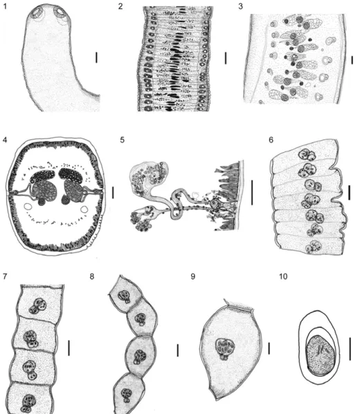
Diagnosis - Strobila apolytic becoming thinner in the terminal portion. Segments numerous, acraspedote, with external segmentation apparent in gravid and pre-gravid segments. Scolex with four suckers, simple, un-armed. Large ventral osmoregulatory canals. Double re-productive structures, including the following: bilateral genital atrium, two compact medullary dorsal testes, two lateral piriform cirrus pouches, two ovaries and two vitelline glands. Genital atrium lightly muscular. Cirri pouches thin not divided by a septum. Cirri with termi-nal spines. Vas deferens arise from testis loops twice, enters in medially to the cirrus pouch. External seminal vesicle absent. Paruterine organs paired. Two paruterine capsules, united basally, not surrounded by an outer en-velope structure. Eggs with three membranes.
Etymology - The genus has been named after the late Reinalda M Lanfredi, PhD, in recognition of her valuable contributions to Brazilian helminthology and helminth systematics.
Emended key to the genera of the Nematotaeniidae Lühe, 1910.
1. Single group of reproductive organs (1 testis, 1 cir-rus, 1 ovary and 1 vitelline gland) ……… 3
2. Double group of reproductive organs (2 testes, 2 cirri, 2 ovaries and 2 vitelline glands) …… Lanfrediella
3a. Two paruterine capsules or organs per segment … 4 3b. More than two paruterine capsules or organs per segment ………. 5
4a. Paruterine capsules surrounded by a second enve-lope ……… Bitegmen Jones, 1987 4b. Paruterine capsules not surrounded by a second envelope ……… Cylindrotaenia Jewell, 1916
5a. Paruterine capsules in row of two-six pairs of or- gans; four-12 paruterine capsules clustered in the pos- terior half of the segment … Distoichometra Dickey, 1921
5b. Paruterine organs not paired; five-150 paruter-ine capsules or paruterparuter-ine organs distributed randomly throughout the segment ……… Nematotaenia Lühe, 1899
Lanfrediella amphicirrus gen. nov. sp. nov. (Figs 1-33)
thin-walled piriform, cirrus pouches, not divided by a septum, 40.05 ± 1.50 (36.84-41.57) × 18.00 ± 1.05 (15.78-19.47) (Figs 4, 5, 11-13); genital atrium lightly muscu-lar, cirri straight, with terminal spines, 16.54 ± 0.97 (15.52-18.15) in length (Figs 24, 26, 27); two ovaries, spherical to oval, situated in the middle of the segment, 48.24 ± 4.56 (38.90-55.19) × 38.47 ± 6.30 (30.51-50.00); two vitelline glands, oval, dorsolateral to the ovaries, 30.38 ± 4.74 (24.00-38.90) in diameter (Fig. 11); uter-us dorsal or dorsolateral to the ovaries (Figs 4, 5, 11); pregravid segments 80.76 ± 8.42 (76.13-98.73) × 853.41 ± 36.63 (789.42-894.73); paruterine complex 96.05 ± 15.76 (65.78-110.52) × 34.29 ± 5.79 (28.31-47.36); gravid segments 259.67 ± 65.08 (191.56-374.68) × 251.95 ± 63.16 (200.00-388.96); two paruterine capsules, oval
to spherical, 67.20 ± 8.29 (51.95-77.92) × 34.29 ± 5.79 (28.31-47.36) (Figs 6-9); seven-10 oncospheres per cap-sule and 15-20 per segment; oncospheres with an oval outer envelope 30.15 ± 3.18 (26.31-33.94) × 16.45 ± 1.61 (13.94-18.68), embryophore 18.83 ± 5.88 (17.30-22.36) × 13.73 ± 1.73 (12.76-15.52) and internal hooks, 7.85 ± 1.07 (6.57-9.89) in length (Fig. 10).
The arrangement of the cirrus pouches was recon-structed from the 116 serial longitudinal dorsoventral sections of mature strobila segments. This 3D recon-struction revealed the equidistant distribution of the cir-rus pouches that line each lateral margin of the mature segment of the strobila, between opposing pouches. This reconstruction confirms the presence of two sets of re-productive structures in each proglottid (Figs 14, 15).
Type host - R. marina (Linnaeus, 1758) (Amphibia: Bufonidae), Cane Toad, Giant Toad.
Site of infection -Small intestine.
Type-locality - Belém, PA (01º28’03”S 48º20’18”W).
Type data and depository - Holotype, deposited in the Helminthological Collection of the Oswaldo Cruz Institute (CHIOC) of the Oswaldo Cruz Foundation in Rio de Janeiro, Brazil, under catalogues CHIOC 37314a (holotype) and CHIOC 37314b, 35706 (paratype).
Molecular data -A partial sequence (1,253 nucleotides) of the 18S rRNA gene was obtained from L. amphicirrus
gen. nov. sp. nov. and added to GenBank under accession HM185494. A comparison with D. bufonis identified a number of sites with potential for phylogenetic differen-tiation, including 18 substitutions and 15 indels, restricted to the 5’ half of the alignment (not shown).
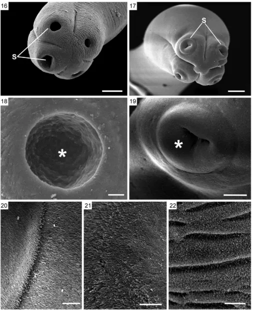
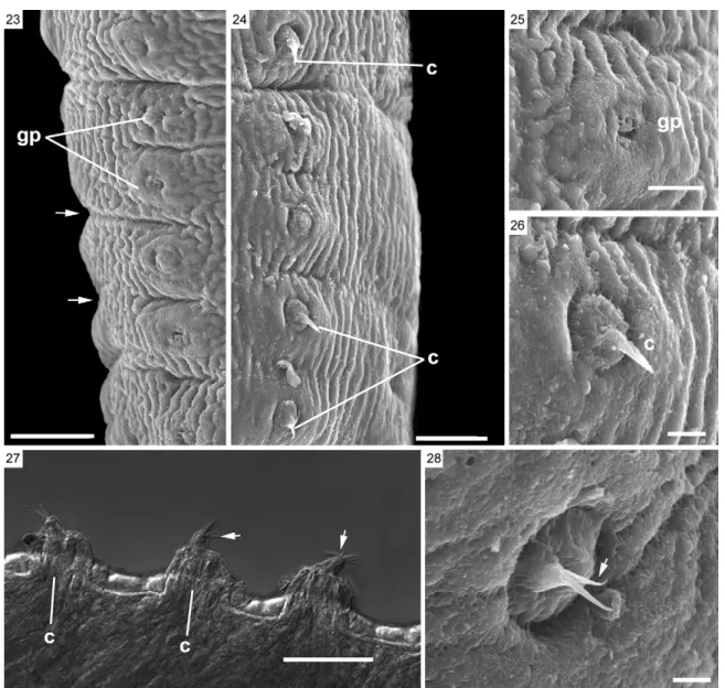
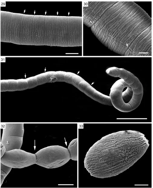
SEM of L. amphicirrus gen. nov. sp. nov. (Figs 16-26, 28-33) - SEM revealed that the scolex has simple suckers and no apical organ (Figs 16-19). The microtriches ob-served throughout the surface of the parasite are delicate, very short and filiform (Figs 20-22). The body presents distinct segmentation only in its most posterior portion. Two genital pores were observed in the mature segments
(Figs 23-25). Some mature segments have partially ex-troverted, conical cirri with spine-like structures located distally (Figs 24, 26, 28). Genital pores could not be ob-served in the gravid segments (Figs 30-33).
Host-parasite data - Prevalence: 45%.
Etymology - The species name is derived from the Greek prefix amphi-, meaning both, and thus refers to the double cirri and other reproductive structures.
DISCUSSION
The new genus Lanfrediella was allocated to the Nematotaeniidae family due to the presence of diagnos-tic morphological traits described for this family by Jew-ell (1916), Hsü (1935), Douglas (1958), Jones (1987) and Khalil et al. (1994). These traits include few testes per
segment (2 or rarely 3), a cylindrical form, acraspedote segments with segmentation evident only in the posteri-or region of the body, the presence of conical paruterine organs immediately adjacent to the uterus, a distinct pat-tern of paruterine capsules (which are thin-walled that bear eggs formation developing at the anterior surface of uterus) and cylindrical strobila. All nematotaeniids are found in amphibian and reptile hosts.
Khalil et al. (1994) proposed a key to the family Nematotaeniidae that uses characteristics that are visible in the gravid and pregravid segments to distinguish the genera Nematotaenia, Cylindrotaenia, Distoichometra and Bitegmen. These genera can be differentiated pri-marily by the number and arrangement of the paruterine organs. Lanfrediella gen. nov. can be differentiated eas-ily from Nematotaenia and Distoichometra. While the
new genus has only two paruterine capsules per gravid segment, Nematotaenia and Distoichometra have more than two capsules (Hsü 1935, Douglas 1958, Yamaguti 1959, Jones 1987, Khalil et al. 1994).
Bitegmen has only two paruterine capsules per seg-ment, although each capsule is surrounded by an outer enveloping structure (Jones 1987, Khalil et al. 1994). The sperm duct does not loop within the cirrus pouch, the an-terior portion of the genital atrium is surrounded by mus-cular tissue and a septum separates the distal portions of the cirrus pouch. In contrast, mature and gravid segments of Lanfrediella gen. nov. lack all of these traits.
According to Jones (1987) and Khalil et al. (1994), the mature segments of Cylindrotaenia (Jewell 1916) have two
paired paruterine organs that are united basally and grav-id segments with two paruterine capsules, a feature also seen in Lanfrediella gen. nov. However, the new genus differs conspicuously from Cylindrotaenia in that it has two of each reproductive structure (cirri, cirrus pouches, testes, ovaries and vitelline glands) in each segment.
L. amphicirrus gen. nov. sp. nov. is the first cyclo-phyllidean tapeworm to be found in Amazonian R. ma-rina. Specimens of Cylindrotaenia americana have been collected from Rhinella toads in southern Brazil, includ-ing the states of Paraná (Stumpf 1982), São Paulo (Jones 1987) and Rio Grande do Sul (Santos & Amato 2010).
The comparison of Lanfrediella gen. nov. with oth-er nematotaeniid genoth-era using classical techniques was
hampered by the difficulties of observing the internal or-gans as described by Jones (1987), showing the necessity for the use of different tools to improve the classification
This is the first study of cestodes that used His-toresin, which simplified the diagnosis and observation of internal structures, such as testes, ovaries, vitelline glands, sperm ducts and cirrus pouches. Based on these results, this technique is recommended for use in future studies of these organisms. Embedding in Historesin preserved the tegument and other structures, which be-come retracted in specimens embedded in paraffin.
Reconstruction of complex internal structures from serial histological sections is a complex and time-con-suming task (Fiala 2005); however, in the present study, 3D reconstruction facilitated the visualisation of these structures in the helminths, which are difficult to
ob-serve using standard techniques. As the segmentation of the body is difficult to observe in mature segments, this technique may become an extremely useful tool for the analysis of internal structures in nematotaeniids and the development of a more systematic approach to the tax-onomy of these helminths.
This is also the first study of a nematotaeniid para-site using SEM, which revealed morphological details of structures such as the scolex, suckers, genital pores, cirri, microtriches and segmentation of the strobila, which are representative of the genus. The use of SEM improved upon the results obtained with light micros-copy in these analyses. The use of SEM also permitted the detection of two genital pores per segment and thus appears to be a valuable tool for analysing the external morphology of cestodes.
The molecular database for cestodes is still very limited and most of the nucleotide sequences available in GenBank as of December 2010 were from species of medical or veterinary interest. The only available sequences from nematotaeniids are short fragments of the 18S rRNA gene of D. bufonis obtained by Mariaux (1998). Our molecular analysis results represent an im-portant complement to the data available for the Nemato-taeniidae, which will be valuable for future phylogenetic analyses and species classifications through the addition of new taxa and genes.
ACKNOWLEDGEMENTS
To late Reinalda Marisa Lanfredi (in memoriam) and Djane Clarys Baia da Silva, for their valuable assistance, to Dr Edilson Matos (Laboratório de Pesquisa Carlos Azevedo/ ISPA/UFRA), Dr Hilton Tulio Costi (Laboratory of Scan-ning Electron Microscopy/MPEG), for his technical support with the SEM analyses, to Dr Wanderley de Souza, head of the Hertha Meyer Cell Ultrastructure Laboratory of UFRJ, for access to SEM, to Dr Stephen Ferrari, for English revi-sion, and the anonymous referees, for valuable comments and suggestions.
REFERENCES
Barton DP 1997. Introduced animals and their parasites: the cane toad
Bufo marinus in Australia. Aust J Ecol29: 316-324.
Douglas LT 1958. The taxonomy of nematotaeniidae cestodes. J Par-asitol4: 261-273.
Espínola-Novelo JF, Guillén-Hernandez S 2008. Helminth parasites in Chaunus and Cranopis valliceps (Anura: Bufonidae) from La-gunas Yalahau, Yucatan, Mexico. J Parasitol94: 672-674.
Espinoza-Jiménez A, Garcia-Sarabia L, Osorio-Sarabia D, León-Règagnon V 2007. Checklist of helminth parasites of the cane toad Bufo marinus (Anura: Bufonidae) from Mexico. J Parasi-tol93: 937-944.
Fiala JC 2005. Reconstruct: a free editor for serial section micros-copy. J Microsc 218: 52-61.
Hall TA 1999. BioEdit: a user-friendly biological sequence alignment editor and analysis program for Windows 95/98/NT. Nucl Acids Symp Ser 41: 95-98.
Hsü HF 1935. Contributions à l’étude des cestodes de Chine. Pev S Zoole42: 447-570.
Jewell ME 1916. Cylindrotaenia americana nov. spec. from the crick-et frog. J Parasitol2: 186-196.
Jones MK 1987. A taxonomic revision of Nematotaeniidae Lühe, 1910 (Cestoda: Cyclophylidea). Syst Parasitol10: 165-245.
Khalil LF, Jones MK, Bray RA 1994. Keys to the cestodes parasites of vertebrates, CAB International, St Albans, 751 pp.
Mariaux J 1998. A molecular phylogeny of the Eucestoda. J Para-sitol84: 114-124.
Santos JN, Giese EG, Maldonado Jr A, Lanfredi RM 2008. A new species of Oswaldocruzia (Molineidae: Nematoda) in Chaunus marinus (Amphibian: Bufonidae) (Linnaeus, 1758) from Brazil.
J Parasitol 94: 264-268.
Santos JN, Melo FTV, Nascimento LCS, Nascimento DEB, Giese EG, Furtado AP 2011. Rhabdias paraensis sp. nov.: a parasite of the lungs of Rhinella marina (Amphibia: Bufonidae) from Brazilian Amazonia. Mem Inst Oswaldo Cruz106: 433-440.
Santos VTG, Amato SB 2010. Helminth fauna of Rhinella fernandezae
(Anura: Bufonidae) from the Rio Grande do Sul coastland, Brazil: analysis of the parasite community. J Parasitol96: 823-826.
Skeriková A, Hypsa V, Scholz T 2001. Phylogenetic analysis of Eu-ropean species of Proteocephalus (Cestoda: Proteocephalidea): compatibility of molecular an morphological data and parasite-host coevolution. Int J Parasitol31: 1121-1128.
Stumpf IVK 1982. Ciclo evolutivo de Cylindrotaenia americana Jew-ell, 1916 (Cyclophyllidea: Nematotaeniidae) em Bufo ictericus
Spix, 1824. Acta Biol Paran10/11: 31-39.