U m in ho | 2 01 2
Ana Marta Gomes Duarte
Identification of genes and the signal transduction
pathways involved in the regulation of acetic
acid-induced programmed cell death
A n a M a rt a G o m es D u a rt e I d e n ti fi ca ti o n o f g e n e s a n d t h e s ig n a l t ra n sd u ct io n p a th w a ys in vo lv e d in t h e r e g u la ti o n o f a ce ti c a ci d -in d u ce d p ro g ra m m e d c e ll
Dissertação de Mestrado
Mestrado em Genética Molecular
Trabalho realizado sob a orientação de
Orientadora: Professora
Orientadora: Doutora Susana Chaves
Doutora Maria João Sousa
Identification of genes and the signal transduction
pathways involved in the regulation of acetic
Nome: Ana Marta Gomes Duarte
Endereço eletrónico: [email protected] Telefone: 961244992
Nº do Bilhete de Identidade: 13561270
Título da Tese de Mestrado:
Identification of genes and the signal transduction pathways involved in the regulation of acetic acid-induced programmed cell death
Orientadores:
Professora Doutora Maria João Sousa Doutora Susana Alexandra Rodrigues Chaves
Instituições de Acolhimento:
Centro de Biologia Molecular Ambiental (CBMA)
Ano de Conclusão: 2012
Designação do Mestrado:
Mestrado em Genética Molecular
1. É AUTORIZADA A REPRODUÇÃO INTEGRAL DESTA TESE, APENAS PARA EFEITOS DE INVESTIGAÇÃO, MEDIANTE DECLARAÇÃO ESCRITA DO INTERESSADO, QUE A TAL SE COMPROMETE.
Universidade do Minho, 31 de Outubro de 2012
_____________________________________________ Ana Marta Gomes Duarte
iii
Esta tese é o resultado de um trabalho que se tornou possível graças a um grupo de pessoas. Assim sendo, gostaria de agradecer aqueles que me ajudaram e tornar possível a concretização deste trabalho:
Agradeço as minhas orientadoras, Professora Doutora Maria João Sousa e Drª. Susana Chaves, pela excecional orientação científica, apoio prestado, partilha de conhecimentos e simpatia. Muito obrigado.
Agradeço à Professora Doutora Manuela Côrte-Real pelo acolhimento neste grupo de investigação e simpatia.
A todos os meus colegas do laboratório pelo excelente ambiente de trabalho, por toda a ajuda prestada e companheirismo.
Ao meu pai e irmãs, por todo o apoio e compreensão que demonstraram, pelos muitos fins-de-semana em que estive ausente.
À memória da minha mãe, graças a ela sou a pessoa que me tornei hoje, os teus ensinamentos tornaram-me uma pessoa lutadora.
Ao meu namorado Filipe, pelo amor e conforto nos momentos de desânimo, por me fazeres acreditar sempre que seria capaz. Obrigado por estes anos ao meu lado. Para ti um beijo muito especial, sem ti não seria capaz.
A todos os docentes e funcionários do Departamento de Biologia.
Este trabalho foi desenvolvido e financiado pelo projecto FCT, Portugal Grant PTDC/AGR-ALI/102608/2008.
v
Abstract
After many years of research, S. cerevisiae was accepted as a powerful model that allows increasing our comprehension about the underlying mechanisms of apoptosis in more complex and less accessible organisms. So, to better understand these apoptotic mechanisms we performed a functional analysis, at whole-genome scale, with the Euroscarf mutants collection. This analysis reveled 2159 resistant mutants and 391 mutants more sensitive to acetic acid induced cell death than the parental strain BY4741. The results obtained contribute to further characterize acetic acid-induced programmed cell death (PCD), and provide information on new putative targets for its control.
Most of the studies on apoptosis in yeast have been centered in the identification of apoptotic markers, however less is known about the signal transduction pathways that induce apoptosis. Cells possess a network of signal transduction pathways, which allow them to respond to different stimulus, implying several changes in genetic expression. Sfl1p is a transcription factor (TF) involved in repression of flocculation-related genes, and activation of stress responsive genes. We studied, cell death induced by acetic acid in yeast strains deleted in SFL1 and in genes potentially regulated by Sfl1p (AQY2,
FMP42, FMP45, SUC2, HSP30, HSP104, NNF2, FLO1 FLO8, YMR173W-a, YJR11W e YCR006C). The results obtained suggest that Sfl1p and the genes under its regulation,
share a role in the mediation of acetic acid-induced apoptosis. Slf1p harbors 3 domains characteristic of the c-myc oncoprotein, a transcription factor with an important role in apoptosis induction and often found mutated in cancer cells. Our results showing that Sfl1p is also involved in the regulation of apoptosis in yeast suggest that these domains can have a conserved function in apoptosis regulation across kingdoms.
We also studied the involvement of genes regulated by Rlm1p on cell death induced by acetic acid. This TF coordinates an adaptive transcriptional response to the stress induced in the cell wall. Our results show that the genes that confer stability to the cell wall, confers sensitivity to acetic acid, when mutated. On the other hand, the genes involved in the cell wall formation, confers resistance, when mutated.
vii
Resumo
Depois de muitos anos de pesquisa, a S. cerevisiae foi aceite como um poderoso modelo que permitiu aumentar a compreensão dos mecanismos subjacentes à apoptose, em organismos mais complexos e menos acessíveis. Assim, para melhor compreensão dos mecanismos apoptóticos, realizámos uma análise funcional, à escala do genoma, com a coleção de mutantes da EUROSCARF. Esta análise revelou 2159 mutantes resistentes e 391 mutantes mais sensíveis à morte induzida por ácido acético do que a estirpe parental BY4741. Os resultados obtidos contribuem para uma melhor caraterização da PCD induzida por ácido acético e fornecem informação sobre hipotéticos alvos para o seu controlo.
A maioria dos estudos sobre apoptose em levedura têm-se centrado na identificação de marcadores apoptóticos, no entanto pouco é conhecido sobre as vias de transdução de sinais que induzem apoptose. As células possuem uma rede de vias de transdução de sinais que lhes permitem responder a diferentes estímulos, implicando grandes mudanças na sua expressão genética. Sfl1p é um fator de transcrição (FT) envolvido na repressão de genes relacionados com a floculação e na ativação de genes de resposta ao stress. Estudamos os genes potencialmente regulados pelo Sfl1p na presença de ácido acético (AQY2, FMP42, FMP45, SUC2,
HSP30, HSP104, NNF2, FLO1 FLO8, YMR173W-a, YJR11W e YCR006C). Os
resultados obtidos indicam que Sfl1p, e os seus genes alvo têm um papel na regulação na apoptose induzida por ácido acético. A proteína Slf1 contém 3 domínios característicos da oncoproteína c-myc, um fator de transcrição com um papel importante na indução de apoptose e muitas vezes alterado em células cancerígenas. Os nossos resultados sugerem que estes domínios podem ter uma função conservada na regulação da apoptose em leveduras.
Estudámos também o envolvimento dos genes regulados pelo Rlm1p, na morte celular induzida por ácido acético. Este FT coordena uma resposta de transcrição adaptativa, ao stress provocado na parede celular. Os nossos resultados mostram que os genes que conferem estabilidade à parede celular, quando mutados, conferem sensibilidade ao acido acético. Por outro lado, os genes envolvidos na formação da parede celular, quando mutados, conferem resistência.
Agradecimentos ... iii Abstract ……….v Resumo ………..………..……….vii Index ……….ix Abbreviations ... xi 1. Introduction ... 1 1.1. Cell death ... 3 1.2. Apoptosis ... 3
1.2.1. The extrinsic apoptotic pathway ... 4
1.2.2. The intrinsic apoptotic pathway ... 6
1.2.2.1. The BCL-2 family ... 8
1.2.2.2. Pro-apoptotic proteins released from mitochondria ... 9
1.2.2.3. Caspases ... 11
1.3. Apoptosis and diseases ... 13
1.4. The S. cerevisiae model ... 14
1.5. Apoptosis in the yeast S. cerevisiae ... 15
1.6. Acetic Acid ... 18
1.7. Genetic expression of transcription factors involved in apoptosis ………...20
1.7.1. The transcription factor SFL1p ... 22
1.7.1.1. Regulation of SFL1p ... 23
1.7.1.2. Sfl1p homology in mammals ... 25
1.7.2. The transcription factor Rlm1p ... 26
2. Objetives ... 31
3. Material and methods ... 35
3.1. Yeast strains ... 37
3.3.1. PI staining ... 38
3.3.2. ROS Production ... 39
3.3.3. DAPI / Chromatin Condensation ... 39
3.4. Screening of the EUROSCARF deletion mutant collection ... 39
4. Results ... 41
4.1. Part I ... 43
4.1.1. Genes whose deletion causes sensitivity to acetic acid-induced cell death ... 45
4.1.2. Genes whose deleted causes resistance to acetic acid-induced cell death ... 50
4.2. Part II ... 53
4.2.1. Sfl1p and the response to acetic acid ... 55
4.2.2. Role of the catalytic subunits of PKA in acetic acid-induced cell death ... 58
4.2.3. Identification of the downstream targets of Sfl1p involved in programmed cell death ... 62
4.2.3.1. Characterization of the role of genes under Sfl1p regulation in acetic acid-induced cell death ... 67
4.2.3.2. Cell death markers ... 70
4.2.3.3. Alignment Mycp with Sfl1p ... 75
4.3. Part III ... 77
4.3.1. Optimization of screening conditions ... 79
4.3.2. Functional categories significantly enriched in the data set of resistant strains ... 83
4.3.3. Functional categories significantly enriched in the data set of sensitive strains ... 84
5. Discussion ... 87
xi
Abbreviations
AIF - Apoptosis-inducing Factor
Apaf-1 - Apoptotic Protease Activating Factor-1 ATP - Adenosine Triphosphate
C.F.U. - Colony forming units
c-FLIP - Cellular-FLICE (FADD-like IL-1β-converting enzyme)-inhibitory Protein
DAPI - 4,6-Diamino-2-phenyl-indole
dihydrochlorid
DD - Death Domain
DED - Death Effector Domain DHE - Dihydroethidium
DISC - Death Inducing Signaling Complex DNA - Deoxyribonucleic Acid
DR - Death Receptors Endo G - Endonuclease G ER - Endoplasmatic Reticulum
FADD - Fas-Associated Death Domain H2O2 – Hydrogen Peroxide
HtrA2/Omi - High Temperature Requirement Protein A2 IAPs - Inhibitors of Apoptosis Proteins
MAPK - Mitogen-activated Protein Kinases
MOMP - Mitochondrial Outer Membrane Permeabilization PCD – Programmed Cell Death
PI - Propidium Iodide
ROS - Reactive Oxygen Species
SC Gal - Synthetic Complete Galactose medium
Smac/Diablo - Second Mitochondria-derived Activator of Caspases/Direct Inhibitor of
Apoptosis Protein (IAP)-Binding Protein With Low Pi
TNF-R - Tumor-Necrosis Factor Receptor TRADD - TNF-R-Associated Death Domain
1
3
1.1. Cell death
Cell death plays an important role in the maintenance of tissue homeostasis and in the development of organisms (Judah et al., 1965). There are different types of cell death, and their classification has undergone significant evolution. The first descriptions of programmed cell death (PCD) mechanisms date back to the mid-1960s, but the term was first used by Lockshin and Williams to describe a type of cell death that was not accidental (Lockshin and Williams, 1965). In 1972, Kerr and coworkers implemented the term apoptosis to define a new pattern of cell death, a genetically controlled sequence of steps that lead to specific morphological and biochemical changes (Kerr et al., 1972). Apoptosis was later considered a synonym of PCD and cell death classified into apoptosis and necrosis. For a long time, necrosis has been considered an accidental cell death mechanism. It is now clear that necrosis can occur in a regulated manner, and that necrotic cell death has a prominent role in multiple physiological and pathological settings. The term ‘necroptosis’ has recently been used as a synonym of regulated necrosis. However, since necrosis may also be regulated and other forms of cell death exist, this classification was abandoned. Recently, the Nomenclature Committee on Cell Death proposed a functional classification of cell death which includes extrinsic apoptosis, caspase-dependent or -independent intrinsic apoptosis, regulated necrosis, autophagic cell death and mitotic catastrophe (Galluzzi et al, 2012).
1.2. Apoptosis
Apoptosis is the best characterized form of programmed cell death. It was originally defined based on morphological and biochemical features found in mammalian cells. The morphological appearance includes chromatin condensation, nuclear fragmentation and cell shrinkage. Biochemical features include high molecular weight DNA fragmentation, phosphatidyl serine externalization and proteolytic cleavage of a number of intracellular substrates (Cohen et a.l, 1994; Martin and Green, 1995). The process of apoptosis ensures the quick removal of cells without rupture of the plasma membrane,
4
thus preventing inflammation (Ballard and Holt, 1968; Bertolaccini and Olivero, 2002). Several diseases associated with severe human pathologies (cancer and neurodegenerative disorders) can be linked to poor regulation of apoptosis. In human adults, 50 to 70 billion cells are eradicated by this process every day (Matsuyama et al., 1999), and therefore it is not surprising that apoptosis de-regulation can contribute to several diseases. The identification of components of the different apoptotic pathways and understanding the mechanisms underlying their regulation is critical to the development of new strategies of prevention and treatment against those diseases.
Apoptosis is mediated by intrinsic and extrinsic mechanisms (Hengartner, 2000). The extrinsic pathway or death receptor pathway (such as TNF receptor-1) is defined as mitochondria-independent, although mitochondria can be involved in the amplification of the death signal. This pathway involves the activation of receptors in the plasma membrane through binding of ligands that trigger a proteolytic process. The second mechanism, the intrinsic or mitochondrial pathway, involves the permeabilization of the mitochondrial outer membrane allowing the release of proapoptotic proteins into the cytosol. The two pathways differ in the initiator caspases that transmit the signal, but later converge at the level of activation of the same caspases. These proteases are responsible for morphological and biochemical alteration typical of apoptosis, and for the rapid clearance of the dying cell (Leist and Jäättelä, 2001; Riedl and Salvesen, 2007; Ow et al., 2008).
1.2.1. The extrinsic apoptotic pathway
The extrinsic pathway involves the activation of receptors in the membrane through binding of ligands that trigger a proteolytic cascade responsible for the characteristic morphological features of apoptosis. Surface death receptors (DR) are characterized by the presence of an intracellular death domain (DD), a stretch of approximately 80 amino acids (Boldin et al., 1995; Chinnaiyan et al., 1995). To date, six human DD-containing receptors have been identified: TNF-R1 (p55/p60 TNF-R), CD95 (Fas, APO- 1), death receptor
5
3 (DR3, TRAMP), TRAIL-R1 (DR4), TRAIL-R2 (DR5), and DR6 (TNFRSF21). These receptors are activated by their respective ligands: TNF, CD95L (FasL/APO-1L), TL1A, TRAIL (Apo2L) (Friesen et al., 1996). The DD plays a crucial role in signaling induced by these receptors, as it enables the recruitment of proteins that themselves contain DDs (Ashkenazi and Dixit, 1999).
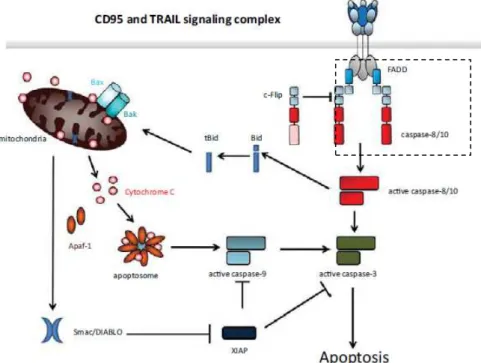
The most prominent and decisive integrators of death receptor signaling are the proteins known as Fas-associated DD (FADD or MORT1) and TNFR-associated DD (TRADD) (Ashkenazi and Dixit, 1998; Yeh et al., 1998). FADD and TRADD do not exert any enzymatic function, but form a bridge between proteins, in this case between receptor and signaling effector proteins. These have their own DD and are recruited to the DD of the activated death receptors. These adapter proteins also have a death-effector domain (DED), with which the DED of procaspase-8 can interact to form the Death Inducing Signaling Complex (DISC). The DISC is formed by FADD and caspase-8. Homotypic interaction of the DEDs of FADD and caspase-8 (Caspase-8 is present in the cytosol as a proenzyme) results in dimerization of caspase- 8, inducing a conformational change that allows caspase-8 to become enzymatically active. It then proteolytically activates the downstream effector caspase-3 (Leist and Jäättelä, 2001). The process leading to the activation of caspase-8 it is identical to that of caspase-10. This pathway is illustrated Figure 1.
The proteolysis of effector caspase substrates is responsible for the characteristic biochemical and morphological hallmarks of apoptosis, proteolysis of vital cellular proteins, including structural components, but also of other proteins such as the inhibitor of caspase-activated DNAse, and cleavage of nuclear DNA (Ding and Yin, 2004).
6
Figure 1 - Schematic representation the extrinsic apoptotic pathway. Binding of CD95 or
TRAIL to their respective receptors leads to receptor trimerization and formation of DISC. The FADD is recruited to the DISC where the DD of both interact. Subsequently, procaspases -8 and -10 are recruited to interact with FADD via the DEDs. cFLIP can compete with caspase-8 for binding to FADD. DISC-activated caspase-8 and -10 starts a caspase cascade by cleavage of caspase-3, and also initiate the mitochondrial apoptosis pathway (adapted from Kantari and Walczak, 2011).
DISC can be inhibited by the antiapoptotic factor FLICE-like inhibitory protein (cFLIP), a caspase-8 inhibitor, leading to inactivation of DISC (Hengartner, 1997; Lawen, 2003). cFLIP is structurally similar to caspase-8 and -10, and contains two N-terminal DEDs. However, unlike cysteine proteases, it lacks a cysteine in what otherwise would be its active center, and thus cFLIP lacks enzymatic activity as a protease. Three different splice variants of cFLIP may exert apotosis inhibitory effects: cFLIPL, cFLIPS, and cFLIPR (Irmler el al, 1997; Van Parijs et al, 1999).
1.2.2. The intrinsic apoptotic pathway
The intrinsic pathway is activated mainly by non-receptor stimuli, such as DNA damage, endoplasmic reticulum stress, metabolic stress, UV radiation or
7
growth-factor deprivation. As mentioned, the central event in the intrinsic pathway is mitochondrial outer membrane permeabilization (MOMP), which allows the release of proapoptotic proteins from the mitochondrial intermembrane space into the cytosol, such as cytochrome c (cyt c), Apoptosis-inducing factor (AIF), Second Mitochondria-derived Activator of Caspases/Direct Inhibitor of Apoptosis Protein (IAP)-Binding Protein With Low Pi (Smac/Diablo) and High Temperature Requirement protein A2 (HtrA2/Omi) (Gulbins et al., 2003). In the cytosol, cyt c binds to apoptotic protease-activating factor-1 (Apaf-1) and ATP/dATP, forming a large complex known as the apoptosome, a molecular platform which promotes the proteolytic maturation of caspase-9 (Cain et al., 2002). When caspase-9 is activated, it activates caspases-3 and -7. These are subject to a number of controls, for example from proteins that bind and inactivate caspases (Inhibitors of Apoptosis, IAPS). Smac/DIABLO and HtrA2/Omi relieve caspase inhibition.
Figure 2 - Schematic representation, of two signaling pathways leading to apoptosis
(extrinsic and intrinsic pathways) in mammalian cells (Reed and Green, 2011).
The intrinsic and extrinsic pathways are not completely independent; in some cells activation of caspase 8 results in activation of the mitochondrial
8
pathway (figure 2). In this case, among other things, caspase 8 cleaves the BH3-only protein BID, generating a truncated fragment known as truncated BID (tBID) that can permeabilize the mitochondrion resulting in MOMP (Favaloro et
al., 2012).
1.2.2.1. The BCL-2 family
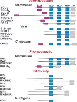
BCL-2 (B-cell leukemia/lymphoma-2) was the first protein of this family to be discovered and thus lends its name to the entire protein family. Since then, all the proteins of the family that have been discovered are related to BCL-2 by sequence homology, containing at least one BCL-2 homology (BH) domain in their structure as well as an involvement in apoptosis control (Tsujimoto, 1998). This family contains proteins that induce or prevent MOMP and consequently apoptosis. It is sub-divided into anti-apoptotic proteins (which contain all four BH domains (BCL-2, BCL-XL, BCL-W, MCL-1, and A1)), pro-apoptotic multidomain proteins (BAX and BAK) and pro-apoptotic BH3-only proteins (BID, PUMA, NOXA, BIM, BAD, and BIK), represented in figure 4 (these are called BH3-only proteins because of Bcl-2 homology regions, they share only the third) (Fletcher and Huang, 2006; Willis et al, 2007; Youle and Strasser, 2008; Brenner and Mak, 2009).
Figure 3 - Bcl-2 family members
can be subdivided into three categories according to their function and structure: anti-apoptotic, such as BCL-2, BCL-XL, BCL-W, MCL-1, and A1 (BFL-1); pro-apoptotic, such as BAX, BAK, and BOK (Mtd); and the BH3-only proteins, Bid, Bad, and Bim (Gross A. et al, 1999).
9
Pro-apoptotic proteins, BAX and BAK, are essential effectors of apoptotic signaling in the mitochondrion, when their activated form induces MOMP, which allows the release of IMS proteins (Mcdonnell et al., 1999). The main steps for BAX activation are translocation to the mitochondrion, conformational change, insertion into the mitochondrial membrane, oligomerization and pore formation. To date, two models describing the interaction between BCL-2 proteins that lead to BAX and BAK activation have been reported: in the indirect model, BAX and BAK are sequestered and held inactive by anti-apoptotic BCL-2 proteins. The binding of pro-apoptotic BH3-only proteins to these anti-apoptotic BCL-2 proteins triggers the release of BAX and BAK. The direct model proposes that BAX and BAK are activated by direct binding of pro-apoptotic BH3-only proteins, called activators, such as BID, BIM or PUMA (Willis et al., 2007; Brenner and Mak, 2009). BID is activated by proteolytic cleavage to generate t-BID, which translocates to mitochondria (Mcdonnell et al., 1999; Yin, 2006; Zaltsman et al., 2010). BIM and BAD are activated by dephosphorylation, whereas PUMA and NOXA are transcriptionally regulated by p53 (Youle and Strasser, 2008). Studies using models of combined deletion of BAX and BAK show that there is no MOMP in the absence of both proteins.
Expression of the anti-apoptotic BCL-2 family proteins allows the cell to survive a wide variety of attacks that might induce apoptosis. These proteins inhibit cell death by binding to pro-apoptotic proteins inhibiting the processes described previously. Perhaps their most important function is to bind and sequester the activator BH3-only proteins to prevent their interaction and activation of BAX and BAK (Letai et al., 2002). The fate of the cell is therefore determined by the balance between the intracellular levels and/or activities of the anti-apoptotic BCL-2 family members and the pro-apoptotic BH3-only proteins (Brenner and Mak, 2009).
1.2.2.2. Pro-apoptotic proteins released from mitochondria
Mitochondria are essential organelles that exist in dynamic networks, and often change their localization and shape during stress conditions (Giannattasio
10
et al., 2005). The action of pro-apoptotic proteins, BAX and BAK, when in the
activated form induces MOMP, which allows the release of IMS proteins, such as cyt c, Smac/DIABLO, HtrA2/Omi and AIF to the cytosol (Gulbins et al., 2003; Armstrong, 2006).
Cyt c was the first mitochondrial protein with an apoptotic function identified, and established the general importance of mitochondria in apoptosis, represented schematicly in figure 5 (Liu et al., 1996; Cai et al., 1998). When in the cytosol, cyt c binds to Apaf-1 and forms the apoptosome together with deoxyadenosine triphosphate (dATP) (Zou et al., 1997). The apoptosome activates caspase-9, (Ow et al., 2008) which mediates activation of caspase-3 and -7 and the execution of apoptosis (Zou et al., 1999; Acehan et al., 2002).
Figure 4 - Schematic representation of the actions of BAX and BAK on the OMM. These
cause the permeability of the mitochondria membrane and induce the release of the proteins from the mitochondrial intermembrane space.
Other proteins that accompany cyt c during MOMP include SMAC/Diablo and Omi/HtrA2, both of which assist in caspase activation by antagonizing IAPs. This will be discussed in detail in the next section (Wu et al., 2000; Du et al., 2000; Verhagen et al., 2000).
AIF exists in the IMM and appears to play a role in mitochondrial complex I assembly or function. Once MOMP has occurred in response to apoptotic stimuli, AIF is also released from mitochondria and is translocated to the nucleus. When translocated into the nucleus, AIF induces DNA fragmentation
11
and chromatin condensation (Candé et al., 2002). The contribution of AIF to cell death depends on the cell-type and apoptotic stimulus, and is only seen when caspases are inhibited or not activated, because it functions in a caspase-independent manner (Wissing et al., 2004).
1.2.2.3. Caspases
In 1992, two groups identified a human protease responsible for activating the precursor of interleukin-1β (interleukin-1β converting enzyme) (ICE). Later, it was found that one of the key genes that regulate apoptosis in C. elegans (CED3) shows homology with ICE (Alnemri et al., 1996). These publications initiated a search over the ensuing years for mammalian ICE homologs that should govern cell death. Today these proteases are known as caspases (standing for cysteine dependent aspartate-specific protease) (Thornberry et al., 1992).
Of the eleven caspases in humans, seven are known to be involved in apoptosis, three are involved primarily in pro-inflammatory cytokine activation and one is involved in keratinocyte differentiation, figure 5. Thus caspases can be divided into initiators caspases (caspases-2, -8, -9 and -10) and executioners caspases (caspases-3, -6, and -7) (Budihardjo et al., 1999).
Figure 5 - Schematic representation of human caspases: activation, specificity, and
12
Caspase-9 is activated by the apoptosome, which then activates caspase-3 and -7. These proteases are responsible for the cleavage of many cellular proteins, which results in the phenotypic hallmarks of apoptosis (Stennicke and Salvesen, 1998). These hallmarks include cutting of DNA into small fragments, condensation of chromatin in the nucleus, dissipation of the mitochondrial membrane potential, and redistribution of phosphatidylserine (Liu et al., 1997; Enari et al., 1998).
Cells also contain natural inhibitors of caspases. IAPs were first identified in baculovirus but were subsequently found in human cells (XIAP, c-IAP1, and c-IAP2) (Deveraux and Reed, 1999; Miller, 1999). IAPs are a family of apoptosis-suppressing proteins that contain at least one copy of a conserved domain called baculoviral IAP repeat (BIR), which represents the defining characteristic of the family (LaCasse et al., 1998; Miller, 1999). This family of proteins inhibits caspases directly, blocking apoptosis. The best-characterized endogenous caspase inhibitor is the X-linked inhibitor of apoptosis protein (XIAP) (Deveraux el al., 1999). Activated caspases-3, -7 and -9 are potently inhibited by XIAP (figure 6) (Fuentes-Prior and Salvesen, 2004; Salvesen and Riedl, 2007; Ow et al., 2008), but this inhibition can be relieved by the action of IAP antagonists, like SMAC/Diablo and serine protease HtrA2/Omi, through the IAP-binding motif (IBM) that disrupts IAP (Suzuki et al., 2004; Brenner and Mak, 2009). Thus XIAP operates both within the intrinsic pathway, downstream of Apaf-1 and at the point of convergence of several apoptosis pathways, where caspases-3 and -7 operate as executioners of the cell death program. In cells that express high levels of XIAP, direct activation of caspase-3 by caspase-8 is blocked so that these cells require the mitochondrial changes induced by cleavage of BID and its pro-apoptotic activity.
13
Figure 6 - Events that occur downstream of mitochondrial outer membrane
permeabilization and their effects on cytosolic components.
1.3. Apoptosis and diseases
Based on its role in maintaining tissue homeostasis, it is not surprising that alterations in apoptosis play an important role in diseases development. Alterations in the upstream regulators of these pathways are the most common alterations in cancer cells. Disruption of the balance between cell death and proliferation is considered a major factor in the growth of tumors or their regression during therapy. This balance can be disrupted in two ways: by increasing proliferation and/or decreasing apoptosis.
A variety of alterations in the different BCL-2 family members have been described, illustrating the importance of these proteins in cancer development. BCL-2 has been found overexpressed in a variety of cancers. BAX and BAK mutations are frequent in tumours. Various BH3 protein alterations have also been implicated in cancer development; as an example, Bid-deficient mice are prone to develop a form of chronic myelomonocytic leukemia (Zinkel et al., 2005), as well as diffuse large B-cell lymphoma. The possibility to target Bcl-2 family member proteins to induce apoptosis in cancer cells has been studied, and particular attention has been given to BH3 only proteins in the design of drugs that would mimic their pro-apoptotic functions. Some of these are currently being tested in phase I/II clinical trials (Esposti, 2010; Placzek et al., 2010; Kelly and Strasser, 2011). Antisense oligonucleotides targeting BCL-2
14
have also been developed and in one case have reached the phase III clinical trial for patients with chronic lymphocytic leukemia.
Caspases are the final effectors of both extrinsic and intrinsic apoptosis; interfering with their function impairs these pathways, leading to a survival advantage for cancer cells. Caspase alterations are frequent in a variety of tumours (Olsson and Zhivotovsky, 2011). The altered caspase function can also be a consequence of modified expression of their specific inhibitors. As an example, cFLIP which competes with caspase 8 for FADD binding, thus preventing its activation, is often elevated in tumours, while its down-regulation can sensitize tumour cells to therapy. Among caspase inhibitors, an important role is played by IAPs. Indeed alterations of IAPs also are found in a variety of human cancers (Favaloro et al., 2012).
1.4. The S. cerevisiae model
In 1996, S. cerevisiae became the first eukaryotic organism to have a fully sequenced genome (Dujon, 1996; Goffeau et al., 1996), thus leading to the creation of several widely accessible databases. After the complete sequencing of the genome, a search for homologies in databases to uncover potential regulators of apoptosis was performed. It was questioned why yeast, an organism composed of a single cell, would undergo a suicide program. Several authors argue that despite the fact that yeast is a unicellular organism, apoptosis could provide an evolutionary advantage at the colony level. Yeasts in the wild exist in multicellular colonies and not as individuals, in which apoptosis may be a mechanism that saves and releases nutrients to the healthier cells, and apoptosis is like a mechanism of self-preservation of the colony as a whole (Gourlay and Ayscough, 2006). So the possibility of a single cell organism to undergo a programmed death program is becoming widely accepted. Indeed, during the last 13 years, many studies have reported the existence of programmed cell death in yeast.
15
The recognition of a mitochondria-mediated apoptotic pathway in yeast, showing similarities with the mammalian intrinsic pathway was of particular interest. S. cerevisiae has characteristics like a short generation time (90-120 min), simple and inexpensive culturing, ease and safety of handling, ability to grow at different temperatures, easy manipulation of mitochondrial respiration, a good characterization of many of its genes (thanks to its responsiveness deletion genes), gene marking or mutations and easy genetic manipulation. Another very important characteristic is its distinctive ability to survive without mitochondrial respiration, which makes them a powerful model to study the involvement of mitochondria in cell death (Pereira et al., 2008). Because of these and other advantageous features, S. cerevisiae proved to be a valuable model organism in which several intracellular processes could be characterized in great detail. Thus the S. cerevisiae model has become one of the most studied models systems to many researchers in the field of molecular and cellular biology.
1.5. Apoptosis in the yeast S. cerevisiae
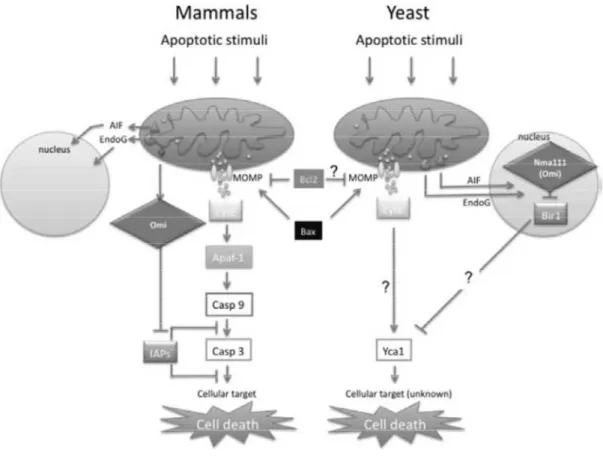
The apoptosis pathways described previously are from vertebrate organisms; however, other pathways with homologous proteins exist in invertebrates. S. cerevisiae PCD shares many morphological and biochemical features with apoptosis in mammalian cells, although there are some differences (figure 7). The first observation that there is apoptosis mechanism in
S. cerevisiae was made in the Cdc48S565G mutant, a temperature-sensitive mutant. When incubated above the restrictive temperature, these cells showed an apoptotic phenotype with characteristics like DNA damage, phosphatidylserine exposure on the outer leaflet of the plasma membrane, chromatin condensation and fragmentation, ROS production and release of cyt
c (Madeo et al., 1997; Braun et al., 2006). Several genetic studies contributed to
understand the mechanisms of cell death in yeast. Some genes involved in metazoan cell death have been confirmed as apoptotic regulators in yeast. Key events of apoptosis in mammalian cells also occur in S. cerevisiae; for example, cyt c is also translocated from yeast mitochondria into the cytosol. The yeast
16
genome also harbors a gene, called NMA111, homologous to vertebrate HtrA2/Omi mitochondrial serine protease (Vande et al., 2008). Nuc1p is the yeast homolog of metazoan endonuclease G (EndoG), exerting its pro-death action upon exit from mitochondria and translocation to the nucleus (Büttner et
al., 2007). The AIF is a highly conserved protein from yeast to human, which
after apoptosis induction translocates to the nucleus, where it participates in apoptotic chromatinolysis.
As in mammalian cells, a IAP has been identified in yeast, termed Bir1p (Uren et al., 1999). Yeast orthologues of mammalian ANT and VDAC have also been identified, AAC1/2/3 and POR1, respectively (Wissing et al., 2004; Ludovico et al., 2005), as has a nuclease (TAT-D) that is apparently involved in DNA degradation during apoptosis. Recently, yeast homologues of mitochondrial fission factors such as Dnm1p (Drp-1 homologue), Mdv1/Net2 and Fis1p were reported to also regulate yeast PCD. Yeast suicide proteins 1, - 2 (Ysp1p and Ysp2p), are also required for mitochondrial fragmentation induced by PCD (Pozniakovsky et al., 2005; Sokolov et al., 2006). Mitochondrial fragmentation has been described in yeast, after acetic acid treatment, leading to the formation of the typical punctate pattern (Fannjiang et al., 2004). Yeast internal NADH dehydrogenase (NDI1) is the homolog of metazoan AMID, and seems to also be involved in apoptosis.
The yeast protein Yor197w, with structural homology with mammalian caspases, is called Yeast Caspase-1 (YCA1) (Madeo et al., 2002). Overexpression of YCA1 in combination with oxidative stress efficiently triggered yeast cell death, accompanied by common apoptotic features. YCA1 belongs to the family of metacaspases, proteases that have a caspase-like fold (Uren et al., 2000). Yca1p overexpression enhances apoptotic-like death of the cells, whereas its knockout reduces cell death in response to several stimuli (Madeo et al., 2002b; Silva et al., 2005).
Regulators such as Apaf-1 and most members of the Bcl-2 family of proteins seem to be absent in yeast (Jin and Reed, 2002; Leist and Jäättelä, 2001). Moreover, only a yeast BH3-only protein was identified so far, Ybh3p. Ybh3p translocates to the mitochondria and is capable of mediating the
17
mitochondrial apoptosis pathway (Büttner et al., 2011). Although the yeast genome does not appear to contain very evident orthologs of the mammalian BCL-2 family genes, expression of pro-apoptotic BAX in yeast leads to apoptotic cell death (Ligr et al., 1998; Priault et al., 1999). This can be prevented by co-expression of antiapoptotic BCL-2 and BCL-XL, suggesting that the function of Bcl-2 family proteins is potentially conserved in yeast and that it can function in yeast in an analogous manner to its role in mammals (Hanada et al., 1995).
In addition to the release of mitochondrial proteins, dissipation of the mitochondrial membrane potential also causes the loss of cell homeostasis via generation of reactive oxygen species (ROS). In S. cerevisiae, ROS accumulation is evident in almost every apoptotic scenario. Various studies have identified mitochondria as the major site of ROS production, and implicate ROS as a component of the apoptotic cascade. A Rho0 strain (lacking mitochondrial DNA) has been shown to display an increased resistance to many apoptotic stimuli. For some stimuli, the higher resistance was accompanied by a decrease in ROS levels (Pereira et al., 2008). Therefore, like in mammalian cells, mitochondria in yeasts play a key role in the apoptotic process.
Several assays for apoptosis detection are routinely used in yeast, and include assessment of viability (CFU), ROS accumulation (DHE), cell integrity (Propidium iodide (PI) staining), chromatin condensation (DAPI staining), DNA fragmentation (TUNEL assay -Terminal dUTP nick-end labeling) and exposure of phosphatidylserine at the outer surface of the plasma membrane (Annexin-V staining) (Carmona-Gutierrez et al., 2010).
18
Figure 7 - Components of apoptotic pathways are conserved from yeast to mammals
(Reed and Green, 2011).
1.6. Acetic Acid
Apoptosis in yeast can be induced by a variety of compounds and conditions, including hydrogen peroxide, acetic acid, amiodarone, hyperosmotic stress, and aging. Ludovico et al., in 2001, showed that acetic acid in low concentrations (20–80 mM) induces PCD in S. cerevisiae cells, which display chromatin condensation and DNA fragmentation. At higher concentrations (above 120 mM), acetic acid induces cell morphological changes typical of necrosis (Ludovico et al., 2001). Later in 2002, Ludovico and collaborators showed a mitochondria-dependent pathway implicated in cell death induced by acetic acid. Translocation of cyt c to the cytosol and ROS production was also observed in yeast cells treated with acetic acid (Ludovico et al., 2002).
When an inhibitory concentration of a weak acid is added to an exponentially growing yeast culture, this acid enters the cell in the undissociated
19
form by simple diffusion. In a growth medium with a pH equal or below pKa, the acetic acid (pKa=4.7) in the undissociated form (RCOOH) prevails. An undissociated form enters the yeast cells by simple diffusion through the plasma membrane (Casal et al., 1996). When inside the cell (where the pH is usually close to neutrality), the chemical dissociation of the weak acid occurs, leading to the release of protons (H+) and of the respective counter ion (RCOO-) and accumulation of protons and acetate in the cell interior figure 8. The S.
cerevisiae response to weak acids depends on the side chain of R group
(R-COOH) (Mira et al, 2010).
Figure 8 - Recovery of intracellular pH requires stimulation of the activity of plasma
membrane, which couples ATP hydrolysis with proton extrusion.
This undissociated form of the acid, due to its electric charge, is not able to cross the hydrophobic lipid plasma membrane bilayer and accumulates in the cell interior. This will lead to intracellular acidification, anion accumulation and inhibition of cell metabolic activity. It also has an impact on the lipid organization and function of cellular membranes, consistent with its strong propensity to become more inhibitory as it becomes more hydrophobic (Stratford and Anslow 1996; Piper et al., 1998).
It has also been described that acetic acid also enters the cell in the undissociated form by simple diffusion, mediated by the aquaglyceroporin
20
Fps1p. Studies demonstrated that deletion of FPS1 (gene that encodes an aquaglyceroporin channel) abolishes the accumulation of undissociated acetic acid in the cell and leads to resistance to acetic acid (Mollapour and Piper, 2007). The recovery of intracellular pH requires the stimulation of the activity of plasma membrane Pma1p (PM-H+-ATPase), which couples ATP hydrolysis with proton extrusion, figure 9. Acetic acid has been extensively used as an inducer of apoptosis. In yeast, acetic acid-induced apoptosis is among the best-characterized apoptotic pathways.
Figure 9 - Entry of undissociated acid into the cell through the Fps1p channel
(Mollapour and Piper, 2008).
1.7. Genetic expression of transcription factors involved in
apoptosis
To date, most studies regarding yeast apoptosis have focused on the identification of apoptotic markers. However, little is known about the signal transduction pathways that induce apoptosis. Cells possess a network of signal transduction pathways that enable them to respond to different stimuli, which implies strong changes in gene expression. Signal integration occurs at several levels of transduction, including transcriptional control of gene expression, translational regulation, and posttranslational modifications.
Initiation of transcript
regulate gene expression. Transcription initiation begins with recruitment of RNA polymerase to a specific locus upstream of the gene known as the promoter. Transcription factors (TFs) are proteins that b
can activate or repress transcription depending where they bind relatively to the transcription start site of the target gene, and are thus classified as acti
repressors (Fulton et al.
activate or repress the transcription of target genes typically in response to an environmental or cellular trigger (Browning
Figure 10 - Various studies in
differentially induced in response to different stress
binding to genes encoding other transcriptional regulators. Lines with arrows depict binding o
to the gene encoding another regulator. Circles with arrows depict binding of a regulator to the promoter region of its own gene. (A) Circle divided into functional categories based on the functions of the target gene. (B) Representative cycle of transcription factors involved in stress response
The basic principles of transcriptional regulation are similar between prokaryotes and eukaryotes (Lee
transcription initiation in eukaryo
related to the genome size as it forms an impediment to the binding of T (Kornberg, 1974; Richmond
promoter DNA sequences and inhibit transcription by steric hindrance of RNA A
21
Initiation of transcription is arguably the most important control point to regulate gene expression. Transcription initiation begins with recruitment of RNA polymerase to a specific locus upstream of the gene known as the promoter. Transcription factors (TFs) are proteins that bind to the promoter and can activate or repress transcription depending where they bind relatively to the transcription start site of the target gene, and are thus classified as acti
et al., 2009). This regulation of transcription initiation can
activate or repress the transcription of target genes typically in response to an environmental or cellular trigger (Browning et al., 2004).
Various studies in S. cerevisiae have led to the identification of genes which are differentially induced in response to different stresses. Representation network of transcriptional regulators binding to genes encoding other transcriptional regulators. Lines with arrows depict binding o
to the gene encoding another regulator. Circles with arrows depict binding of a regulator to the promoter region of its own gene. (A) Circle divided into functional categories based on the functions of the target
e of transcription factors involved in stress response (Lee
The basic principles of transcriptional regulation are similar between ukaryotes (Lee et al, 2002). Despite these similarities, transcription initiation in eukaryotes is considerably more complex, and is related to the genome size as it forms an impediment to the binding of T (Kornberg, 1974; Richmond et al., 1984). Prokaryotic repressor proteins bind promoter DNA sequences and inhibit transcription by steric hindrance of RNA
B
ion is arguably the most important control point to regulate gene expression. Transcription initiation begins with recruitment of RNA polymerase to a specific locus upstream of the gene known as the ind to the promoter and can activate or repress transcription depending where they bind relatively to the transcription start site of the target gene, and are thus classified as activators or on initiation can activate or repress the transcription of target genes typically in response to an
have led to the identification of genes which are etwork of transcriptional regulators binding to genes encoding other transcriptional regulators. Lines with arrows depict binding of a regulator to the gene encoding another regulator. Circles with arrows depict binding of a regulator to the promoter region of its own gene. (A) Circle divided into functional categories based on the functions of the target
Lee et al., 2002).
The basic principles of transcriptional regulation are similar between ). Despite these similarities, tes is considerably more complex, and is related to the genome size as it forms an impediment to the binding of TFs 1984). Prokaryotic repressor proteins bind to promoter DNA sequences and inhibit transcription by steric hindrance of RNA
22
polymerase. In eukaryotic cells, repression does not occur simply by binding of repressor proteins. Eukaryotic DNA is tightly wrapped around histones, forming nucleosomes, the basic units of chromatin, which becomes limiting for the binding of TFs (Richmond et al., 2003). Chromatin modifier complexes are required that either displace or evict nucleosomes or covalently modify histones to loosen their interactions with DNA (Galeote et al., 2007). TFs overcome the chromatin barrier to access DNA through interactions with a host of coregulators that modify the chromatin state.
It is believed that transcriptional activity is in some cases correlated with histone acetylation. Thus, chromatin modifiers can also function as co-repressors by effecting a more closed chromatin conformation. Given the drastic changes in the integrity of DNA and the state of chromatin compaction during apoptosis, histone modifications may play a functional role in promoting these changes. Methylation of sequences in promoter regions is commonly observed during tumor progression to inactivate genes whose products are important for processes such as DNA repair, cell-cycle regulation, cell adhesion, angiogenesis and apoptosis (Miranda et al., 2007). The stress transcription factors are interesting models, and their characterization can lead to the identification of new components of stress signaling pathways in yeast.
1.7.1. The transcription factor SFL1p
In the yeast S. cerevisiae, the global transcriptional regulator Ssn6 (Cyc8)-Tup1 was the first transcriptional co-repressor to be described (Keleher et al., 1992; Tzamarias and Struhl, 1994). Ssn6(Cyc8)-Tup1 is recruited to promoters via interactions with DNA-binding proteins, each of which represses genes in a specific biological pathway and inhibits the transcription of a diverse set of genes under a variety of stress conditions (Keleher et al., 1992). This complex is composed of one Cyc8 subunit and four Tup1 subunits (Tzamarias et al., 1994). Tup1 bears the transcriptional repression activity of the co-repressor complex, exerting its function via two distinct mechanisms. One model suggests that Tup1 controls nucleosome positioning so as to mask DNA targets for
23
activators or transcription factors (Grunstein, 1990; Edmondson et al., 1996; Watson et al., 2000). A second model suggests that Tup1 directly inhibits the function by interacting with subunits of the RNA polymerase II holoenzyme,
such as Sin4, Srb10/11, Med3, Hrs1 and Srb7 (Kuchin et al., 1998; Gromoller 2000; Papamichos-Chronakis et al., 2000). Studies show that mutations in
components of Pol II holoenzyme alleviate the repression by Tup1 (Balciunas and Ronne, 1995).
Sfl1p was first described as a transcriptional repressor but it can also act as an activator; it is, involved in repression of flocculation-related genes, and activation of stress responsive genes. Steven Conlan and Dimitris Tzamarias in 2001 showed that Sfl1p interacts directly with Ssn6p. In vivo repression data suggest that Sfl1p inhibits transcription by recruiting Ssn6p-Tup1p via a specific domain in the Sfl1 protein. Components of specific RNA polymerase II sub-complexes, Sin4p and Srb10p, are necessary for the Ssn6p-Tup1p repression activity of Sfl1 function. Sfl1p interacts with Tpk2p, a cAMP-dependent subunit that negatively regulates Slf1p function. This interaction of Sfl1p with DNA is thus regulated by Tpk2p, which is involved in the regulation of Sfl1p recruitment to some Ssn6p-regulated genes (Conlan et al., 2001).
1.7.1.1. Regulation of SFL1p
In eukaryotic cells, the secondary messenger cyclic adenosine monophosphate (cAMP) is produced in response to extracellular stimuli (D'Souza and Heitman, 2001). The central role of this signaling pathway in S.
cerevisiae is nutrient sensing and regulation of diverse biological processes
including growth, metabolism, stress resistance, and entry into either meiosis or pseudohyphal diferentiation (D'Souza and Heitman, 2001). The cyclic AMP-dependent signaling transduction pathway is a multienzyme cascade that regulates diverse biological processes. Specific connection of appropriate G-protein receptors followed by adenylate cyclase activation leads to the production of cyclic AMP. Cyclic AMP then binds to cytoplasmic protein kinase
24
A (PKA), which consists of a single regulatory subunit encoded by the BCY1 gene and three catalytic subunits, Tpk1p, Tpk2p and Tpk3p, figure 11.
Figure 11 - Schematic representation Sfl1p repression by isoform Tpk2p.
When cAMP levels increase, it binds to the regulatory subunits and induces a conformational change that causes dissociation of the tetramer into dimeric regulatory subunits. These catalytic subunits are enzymatically active and phosphorylate target substrates that include metabolic enzymes and transcription factors (Sfl1p), which elicit alterations in cell cycle progression and stress responses (D'Souza and Heitman, 2001). Tpk1p has been implicated in the branched chain amino acid biosynthesis pathway, mitochondrial iron homeostasis and mtDNA stability. Tpk2p has been shown to influence iron uptake, trehalase synthesis, water homeostasis, pseudohyphal growth and negative regulation of Sfl1 protein (figure 11). Tpk3p is a regulator of mitochondrial function. Its overexpression has been shown to inhibit growth, and deletion of TPK3 is sufficient to prevent the production of ROS, as this PKA subunit regulates mitochondrial function (Leadsham et al., 2010).
Previous studies in yeast have established links between Ras signaling and mitochondrial function, via cAMP/PKA. The cAMP pathway is the most explored signaling pathway controlled by Ras proteins; it affects a large number of genes, some of which are important for the defence against oxidative stress. In yeast, Ras/cAMP/PKA signaling also controls cellular processes that include
25
cell growth and proliferation, making this pathway a good candidate to integrate environmental signaling with mitochondrial regulation (Hlavata et al., 2008).
1.7.1.2. Sfl1p homology in mammals
Sfl1p encodes a 767-amino acid transcription factor, which has two domains significantly homologous to Myc protein (Fujita et al., 1989). The proto-oncogene c-Myc encodes a transcription factor that plays a biological role through the modulation of genes in multiple cellular processes like cell growth, proliferation, differentiation and apoptosis (Askew et al,. 1991; Evan et al., 1992). Deregulated expression of this oncogene is associated with a wide range of human cancers. This deregulated expression causes uncontrolled cell proliferation, which characterizes most, if not all, human cancer cells (Klefstrom
et al., 2002).
Expression of c-MYC sensitizes cells to mechanistically diverse pro-apoptotic insults, including DNA damage, death receptor signaling, hypoxia, genotoxic stress, and nutrient deprivation (Askew et al., 1991; Evan et al., 1992; Klefstrom et al., 1994; Alarcon et al., 1996; Hueber et al., 1997). There are two discrete pro-apoptotic effector pathways mediating this sensitization: stabilization of p53 through the ARF (Active Response Factor)/MDM2 (Mouse Double Minute-2) pathway, which serves as a sentinel for genotoxic damage, and release of cyt c from mitochondria into the cytosol, possibly through activation of the pro-apoptotic molecule BAX by a mechanism that is independent of both Fas-FasL and DNA damage pro-apoptotic pathways (figure 12) (Juin et al., 1999). Studies investigated a possible physical interaction between c-Myc protein and the Bax promoter using an immunoprecipitation assay. It was found that c-MYC strongly binds to the BAX promoter region, contributing to BAX expression. c-MYC is a transactivator of BAX, based on the presence of four CACGTG motifs located in the BAX gene (Mitchell et al., 2000). Activated BAX within the mitochondrial membrane leads to apoptosis, through the mechanisms described previously. It remains to be established if there are other mitochondrial factors, such as AIF, released during c-MYC-induced apoptosis, and their involvement in this process.
26
Figure 12 - Expression of c-MYC triggers two discrete proapoptotic effector pathways:
the stabilization of p53 through the ARF/MDM2 pathway, which serves as a sentinel for genotoxic damage, and the triggers the release of cyt c from mitochondria into the cytosol, possibly through activation of the expression of the pro-apoptotic molecule BAX (Pelengaris et al., 2002).
Survival signals, which serve to block c-MYC, include signaling via IGF1R (Insulin-like Growth Factor-1 Receptor). Activation of the IGF-1 receptor triggers a survival-signal, routing through Ras, PI3-kinase, serine/threonine kinase PKB/Akt and subsequent phosphorylation of the pro-apoptotic protein BAD. Phosphorylated BAD is sequestered and inactivated by cytosolic 14-3-3 proteins. Functionally, this inactivates BAD, which cannot antagonize BCL-2 (Kauffmann-Zeh et al., 1997; Evan and Littlewood 1998).
1.7.2. The transcription factor Rlm1p
Four essential mitogen-activated protein kinase (MAPK) cascades respond to different external signals in yeast (figure 13). The mating pathway is activated by pheromones and induces cell-cycle arrest and the morphological
27
changes required for mating (Gustin et al., 1998). The Kss1 vegetative growth pathway may be activated by cell wall stress or changes in osmolarity (Lee and Elion, 1999; Cullen et al., 2000). The invasive growth pathway is activated by starvation. The high osmolarity glycerol (HOG) pathway increases intracellular glycerol levels in response to hypertonic stress. The cell wall integrity pathway (CWI) is activated by hypotonic stress, heat shock, or impaired cell wall synthesis.
Figure 13 - Overview of the MAPKinase pathways in yeast (Qi M. and Elion EA. 2005).
The cell wall of S. cerevisiae is an external envelope that protects it against different environmental conditions. The adaptive response of yeast to cell wall stress is mainly mediated by the CWI pathway (Levin et al., 2005; Fuchs and Mylonakis, 2009; Kim and Levin, 2011).
Two membrane proteins, namely Mid2 and Wsc1, act as the main sensors of the CWI pathway. These, when activated, interact with the guanine nucleotide exchange factor Rom2, activating the GTPase Rho1, which then interacts and activates Pkc1. Pkc1 then activates a downstream MAP kinase
28
cascade comprising three protein kinases, MAPKKK (Bck1), MAPKK (Mkk1/ Mkk2), and finally the MAPK (Mpk1/Slt2) (figure 14) (Kim and Levin, 2011).
Mpk1/Slt2 targets the transcription factor complex SBF (SCB-binding factor) and Rlm1p. SBF is a complex of two proteins, Swi4 and Swi6, which is involved in the regulation of the yeast cell cycle and polarized growth, especially during the transition from G1 to S phase, via transcriptional activation of genes such as CLN1, CLN2, PCL1 and PCL2 (Fong et al., 2008; Chiu et al., 2011). Rlm1p is a MADS-box transcription factor that promotes the expression of cell wall maintenance proteins (Watanabe et al., 1997; Heinisch et al., 1999; Jung et
al., 2002; Garcia et al., 2004; Fuchs and Mylonakis, 2009). Thus the final
consequence of the activation of the CWI pathway by cell wall stress is the induction of an adaptive transcriptional response (Jung and Levin, 1999; Lagorce et al., 2003; García et al., 2009). The elements of the yeast transcriptional machinery working in concert with Rlm1p for transcriptional activation upon cell wall stress and the molecular mechanisms involved in this process are completely unknown. Under cell wall stress conditions, Slt2p phosphorylates Rlm1p and the SWI/SNF recruited complex is targeted to the
Figure 14 - Schematic
overview of the cell wall integrity pathway.
29
promoters of CWI-responsive genes, altering the nucleosome positioning at the promoter, facilitating the binding of Rlm1p to sites previously occluded by nucleosomes (Kasten et al., 2011). Finally, binding of Pol II stimulates transcription initiation. S. cerevisiae SWI/SNF has 11 subunits: Arp7, Arp9, Snf2, Snf5, Snf6, Snf11, Snf12, Swi1, Swi3, Swp82 and Taf14 (Yudkovsky et
31
33
This work aimed to identify genes involved in the regulation of acetic acid-induced apoptosis, and involved three approaches:
The first part of this work aimed to identify, at a whole-genome scale, the genes required for sensitivity/resistance phenotypes under acetic acid-induced apoptotic conditions (400 mM acetic acid, at pH 3.0) in S. cerevisiae, by screening the EUROSCARF haploid mutant collection (http://web.unifrankfurt. de/fb15/mikro/euroscarf/). A set of genes involved in resistant and sensitive phenotypes were clustered according to biological function (MIPS Functional Catalogue) and known physical and genetic interactions (STRING Protein-Protein Interactions).
In the second part, the aim was to identify regulators and downstream targets of Sfl1p involved in acetic acid-induced apoptosis. Deletion mutants in genes regulated by Sfl1p were tested for their sensitivity/resistance to acetic acid-induced cell death and cell death markers were assessed in mutants displaying altered resistance.
The third part was identification the downstream targets of Rlm1p involved in acetic acid-induced apoptosis. Deletion mutants defective in genes regulated by Rlm1p were tested for their sensitivity/resistance to acetic acid.
35
37
3.1. Yeast strains
In this study microorganisms used were the parental strain of S. cerevisiae BY4741 (MATa, his3∆1, leu2∆0, met15∆0, ura3∆0) and the respective EUROSCARF collection of derived deletion mutant strains, containing all the non-essential open reading frames replaced by the KanMX cassette.
Figure 15 - Strategy for deletion of genes used in the construction of the EUROSCARF
mutant collection (Saccharomyces Genome Deletion Project).
3.2. Growth conditions and treatments
Yeast cells were grown on YPDA medium (1% yeast extract, 1% Bacto-peptone, 2% glucose and 2% agar) plates for 2 days at 30 ºC. After growth on YPDA, cells were then inoculated in 10 ml of YPD medium until early exponential phase (OD640nm = 0.5 - 0.7) at 30°C in a shaker at 200 rpm. Thereafter cells were harvested, suspended in Erlenmeyers with 10 ml YPD medium adjusted to pH 3.0 with HCl and 120mM of acetic acid (with a ratio of flask volume/medium of 5:1), and incubated for up to 220 min at 30°C, with agitation (200 rpm). At specific time intervals (0, 60, 120, 180, 200 and 220 min), 100цl of cells were collected, ressuspended in water, and serial dilutions were plated on YPDA. After 2 days of incubation at 30 °C, cell viability was
38
measured as a percentage of Colony Forming Units (CFU). The percentage of viable cells was estimated, considering 100% survival the number of CFU obtained in time 0, by the formula:
% min
0 100
For semi-quantitative viability assays, 10µL of cell suspensions in water with the dilution rate of 10-1 were spotted onto YPDA plates. After 2 days of incubation at 30 °C, photographs of the plates were taken with ChemiDoc XRS (BioRad).
Cell viability assays were also performed in galactose medium. In thses assays, after growth on YPDA the strains regulated by RLM1 were inoculated into 10 ml of Synthetic Complete Galactose medium (SC Gal- 2% galactose, 0.67% yeast nitrogen base without aminoacids, 0.14% Dropout mixture lacking 0.008% histidine, 0.04% leucine, 0.008% tryptophan and 0.008% Uracil). SC Gal treatment medium was adjusted to pH 3.0 with HCl and contained 100 mM acetic acid.
3.3. Analysis of apoptotic markers
3.3.1. PI staining
Detection of the integrity of the cell plasma membrane was assessed by flow cytometry using propidium iodide (PI). Cells were treated with acetic acid as described above, and, after specific time intervals (0, 60, 120, 180, 200 and 220 minutes), 100 ul of cells were collected by centrifugation, washed in deionised water, resuspended in 500 uL of phosphate buffered saline (PBS) (80 mM Na2HPO4, 20 mM NaH2PO4 and 100 mM NaCl) and stained with PI (1
µg/ml) (Sigma). Afterwards, samples were incubated for 10 min at room temperature in the dark. Finally, fluorescence was detected in an Epics® XL™ (Beckman Coulter) flow cytometer, where 30,000 cells from each sample were analyzed. Cells with red fluorescence (FL-3 (488/620 nm)) were considered to contain plasma membrane disruption.