UNCORRECTED
PROOF
Nanomedicine: Nanotechnology, Biology, and Medicine xxx (2017) xxx-xxx
Contents lists available at ScienceDirect
Nanomedicine: Nanotechnology, Biology, and Medicine
journal homepage: www.elsevier.comImmobilization of bacteriophage in wound-dressing nanostructure
Frederico Nogueira
a, b, Natia Karumidze
c, Ia Kusradze
c, Marina Goderdzishvili
c, Pilar Teixeira
d, Isabel C. Gouveia
b,⁎ aCICS-UBI – Health Sciences Research Centre, University of Beira Interior, PortugalbFibEnTech – Fiber Materials and Environmental Technologies, University of Beira Interior, Portugal cG. Eliava Institute of Bacteriophages, Microbiology and Virology, Tbilisi, Georgia
dInstitute for Biotechnology and Bioengineering (IBB), Portugal
A R T I C L E I N F O Article history: Received 25 March 2017 Accepted 9 August 2017 Available online xxx Key words: Bacteriophages Antimicrobial agents Surface immobilization Electrospinning Pseudomonas aeruginosa A B S T R A C T
Opportunistic bacteria that cause life-threatening infections are still a central problem associated with a healthcare setting. Bacteriophage capsid immobilization on nanostructured polymers maximizes its tail expo-sure and looks promising in applications toward skin-infections as alternative to antibiotics standardly used. The main goal of this work was to investigate the covalent immobilization of vB_Pae_Kakheti25 bacterio-phage capsid on polycaprolactone (PCL) nanofibers (non-woven textile), as a potential effective antimicro-bial, laundry resistant and non-toxic dressing for biomedical use. Surface analyses showed that the immobi-lization of vB_Pae_Kakheti25 bacteriophage capsid on PCL nanofibres oriented bacteriophage tails to inter-act with binter-acteria. Furthermore, antimicrobial assays showed a very effective 6 log binter-acterial reduction, which was equivalent to 99.9999%, after immediate and 2 hours of contact, even following 25 washing cycles (due to covalent bond). The activity of PCL-vB_Pae_Kakheti25 against P. aeruginosa was immediate and its re-duction was complete.
© 2017.
The skin of patients with inflammatory skin-diseases alongside with chronic or burn wounds and exit-sites of catheters is partic-ularly susceptible to infection by different microorganisms. Oppor-tunistic pathogens are the cause of skin diseases, infections, and the inability of chronic wounds to heal.1–4They are capable of produc-ing virulence factors, includproduc-ing enzymes that promote tissue invasion and extracellular polymers, which form the biofilm that contributes to the perpetuation of skin inflammation, even in normal-appearing skin. Fortunately, the majority of our resident skin microorganisms are non-pathogenic and many of these contribute to maintaining health.1
Accordingly, skin-disease/injury management demands an integrated approach aimed not only at diminishing infection but also at regulat-ing the skin microbiome.2,5
P. aeruginosa is the most common infectious agent among Pseudomonas spp. As a versatile and opportunistic microorganism it can colonize the skin, soft tissue, gastrointestinal tract, armpits, eye and ear.6–8P. aeruginosa is the agent responsible for the most com-mon infections under hospital settings, through catheter and ventila-tor contaminations leading to nosocomial infections, such as pneumo-nia, urinary tract and wound burn infections, as well as bacteremia, especially in patients with diabetes or immunodeficiency.8The major
Acknowledgments: The authors acknowledge the Fundação para a Ciência e Tecnologia (FCT) for the PhD grant SFRH/BD/91444/2012 and Programa Operacional Capital Humano (POCH) and European Union for co-funding the work.
⁎Corresponding author at: FibEnTech R&D Fiber Materials and Environmental
Technologies, University of Beira Interior, 6201-001 Covilhã, Portugal.
Email address: [email protected] (I.C. Gouveia)
concerns about the control of nosocomial infections vary from the problems of drug safety associated with a high human toxicity, the long-term and large scale application of broad-spectrum antibiotic drugs, to the increased resistance to conventional therapies. These in-fections tend to chronicity and may fail to be treated with almost any combination of antibiotics, showing mortalities up to 61%.9The
com-bination therapy to fight P. aeruginosa infections is very difficult to achieve, due to the compromised immune system of the majority of in-fected patients, and the intrinsic resistance of microorganisms to vari-ous antibiotics.10
Recent research has been conducted on the three groups of natu-rally occurring antimicrobials as novel alternatives to antibiotics: bac-teriophages, bacterial cell wall hydrolases (BCWHs), and antimicro-bial peptides (AMPs).11 Among them, bacteriophages are the most
highly specific toward both Gram-positive and Gram-negative bac-teria and they are also highly efficient and relatively cost-effective. In contrast, AMPs have a broad-spectrum against bacteria and fun-gus, low level of induced resistance, but may cause toxicity at high doses in order to be efficient, and are more costly to produce.11BCWH has limitations toward Gram-negative bacteria, as a result of the pres-ence of the outer membrane, and important Gram-positive pathogens like S. aureus are already resistant to lysozymes. To overcome the changing tide of nosocomial diseases and increasing reports of mi-croorganism-acquired resistances, recently the United States, Canada and European countries have started to take a close interest in bac-teriophage-based therapies,12,13 in which they act without
mecha-nism-based host toxicity.
The bacteriophage vB_Pae_Kakheti25 has a potent activity against P. aeruginosa and appears as an alternative approach to con
https://doi.org/10.1016/j.nano.2017.08.008 1549-9634/© 2017.
UNCORRECTED
PROOF
ventional treatments, especially as an antimicrobial agent for dressing.It is representative of siphoviral family, has dsDNA as its genome, icosahedral capsid, and long non-contractile tail. vB_Pae_Kakheti25 undergoes a lytic cycle through which it self-replicates and lyses a broad range of P. aeruginosa strains in order to spread copies of itself. Polycaprolactone (PCL), an hydrophobic polyester, can be ex-plored as a substrate for skin regeneration due to its high elasticity and slow biodegradability.14,15Furthermore, it is not broken down by en-zymes and microorganisms.14These features seem worthy of dressing applications.16
The textile and medical industries continue to look for eco-friendly processes that may replace the currently used toxic textile chemicals and the use of antibiotics, respectively.17–19The demand for medici-nal products alternative to antibiotics has increased considerably and our proposed application seems promising due to its versatility, low content of impurities, antistatic properties and good mechanical prop-erties. Furthermore, as PCL is unfavorable for the development of mi-croorganisms, its shelf life and users' health status are also ensured.
In this work, in order to eliminate the growth of P. aeruginosa, PCL electrospun nanofibers were threaded and then vB_-Pae_Kakheti25 bacteriophages were covalently immobilized by their capsid via acid-amine reactions, forming amide linkages. The effect of anti-P. aeruginosa activity of PCL-vB_Pae_Kakheti25 dressing was evaluated under various parameters, so as to produce appropriate ap-plications toward skin-infections, and aiming to further highlight the potential of phage as the “antibiotic” of the millennium by minimizing bacterial resistance and preserving skin-microbiome.
Methods
Materials
Polycaprolactone nanostructure (PCL), average Mn 45,000 (Sigma), was functionalized in a vB_Pae-Kakheti25 bacteriophage so-lution. PA25 (DSM 25642) clinical isolates of Pseudomonas aerug-inosa from the Eliava culture collection were used for isolation and subsequent growth of vB_Pae-Kakheti25 bacteriophages. P. aerugi-nosa was grown on Brain Heart Infusion (BHI) agar and then on Brain Heart Infusion (BHI) broth (Sigma) at 30 °C and in shaker at 200 rpm. Methods
Electrospinning
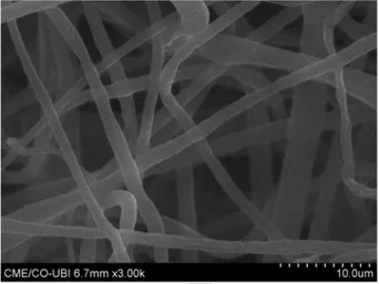
Nanofibers were produced by NanoSpider (Elmarco s.r.o. Liberec, Czech Republic). PCL 15% (w/v) was dissolved in a mixture of ab-solute ethanol/chloroform (65:35 vol.%) to prepare a homogeneous solution. Different ratios of ethanol/chloroform solvents were used in order to optimize the final nanostructure. The final concentration of PCL and ratio of solvents were set according to the homogene-ity of resulting nanofibers, their easy-detachment from polypropy-lene-coated collecting electrode and tensile strength. The electrospin-ning process was done under the following experimental conditions: RH ≈65%, temperature ≈25 °C, electric voltage ≈80 kV, distance be-tween electrodes = 8.98 cm, and electrode spin = 7 r/min (44 Hz). Figure 1 shows the representative images of these nanofibers. Tensile strength assays
Tensile strength of electrospun PCL was evaluated with resis-tance-to-rupture assays. These assays were performed in a Dy
Figure 1. Polycaprolactone (PCL) 15% (w/V) electrospun fibers. Tendency of forming
layers of curlicues. 3000× magnification.
namometer (Thwing-Albert Instrument Co.) according to Standard EN ISO 2062, at 20 ± 2 °C, under 60% relative humidity. Samples were strip-cut 1×5 cm, with an average thickness of 50.33 μm, grammage of 18.6 g/m2, and placed between dynamometer tweezers. A defined pre-tension was set in the beginning, and the test ended with the rup-ture of samples. Seven replicates were used.
Protein structure modeling – I-TASSER method
The sequence of amino acids (UniProtKB) of the Major capsid pro-tein referred to as H6WTZ9–1 and Major tail tube propro-tein referred to as H6WU05-1 of vB_Pae-Kakheti25 bacteriophage were the follow-ing, respectively: MALSDLAVYSEYAYSAFSETLRQQVDLFN- TATGGAIMLQSAAHQGDFSDVAFFAKVTGGLVRRRNAYGS- GTVAEKVLKHLVDTSVKVAAGTPPVRLDPGQFRWIQQNPE- VAGAAMGQQLAVDTMADMLNVGLGSVYSALSQVSDVVY- DATANTDAADKLPTWNNLNNGQAKFGDQSSQIAAWIMH- STPMHKLYGSNLTNGERLFTYGTVNVVRDPFGKLLVMTD- SPNLFAAGTPNVYHILGLVPGGVLIGQNNDFDANEETKNG- DENIIRTYQAEWSYNIGVKGFAWDKANGGKSPTDAALFTST-NWDKYATSHKDLAGVVVKTN; MVCEIAKIDSNITGLAFAEEECLKQLPTTPVWYGLEPNSYS- DFGGELSTVARAPIDPSRQNKKGTITDLDASGGFNADFTK- TNLARILQGFFFADARELPSTQPLNGASVALTGVTAID- STYAAASGLGVFGADMLVYATGFANAANNGLKTVVSA- TAAGVVVAETLIDETPPAGAKLECVGRQLAAADANIAVT- GNVVSLIVTAGDFTTMPELFAGRWVFVGGDATANRFAN- NVGYARIKSVAAKALVFDDVTWQAVNETGTGK- SIRLFVGTVIKNEKTPALIKRRSYQIERTLGEGLNGTQCEYLE- GAVPNEFTLNVPQADKLNADLSFVACDNTYRSGDPGDEQK- AGTRVPAPGEDAYNTSSDVYRIKMAVHDAASSNPAALF- GYVSEANVSINNNVTPNKAVGVLGAFDTSAGNFEVGGSI- TAYFTTVAAVKAVRANADVGLSVISAAKNAGFVFDIPLL- GLGGGRLNVEKDAPITVPLEPAGAENANGYTMLYEVF-SYLPNLAMPD.
For the simulation, these sequences were threaded from Protein Data Bank library, where multiple template alignments were gener-ated. Then, the continuous fragments were excised, reassembed, clus-tered and this full process was repeated. I-TASSER used the most sig-nificant templates with the lowest energy.20–23The predicted struc-tures were analyzed for polarity and ligand binding sites.
UNCORRECTED
PROOF
NinhydrinIn order to ascertain the presence of free amines on Pae-Kakheti25 capsid, i.e. the estimation of the quantity of vB_-Pae-Kakheti25 that PCL nanofibers were able to crosslink, ninhydrin assays were done. Ninhydrin hydrates with amino groups, producing purpurous Ruhemann's cromophore (λmax 570 nm). This colorimet-ric assay was performed after drawing a calibration with known con-centrations of L-cysteine. Each sample was added 2 mL of 108 vB_-Pae-Kakheti25/ml and 1 ml of 8% Ninhydrin (acetone). ddH2O was
used for controls. Test tubes were placed in water bath at 100 °C for 10 minutes and then cooled down to room temperature before they were read at 570 nm.
PCL substrate preparation Nanofiber functionalization
After being formed with electrospinning, non-woven nanofibrous PCL was washed for 4 hours with distilled water, at 40 °C, 110 rpm, and dried afterwards at 30 °C until used, in order to remove any surface residues and impurities. Samples were stored in the desic-cator until use. For the activation of the surface, samples were sub-mersed in acidic solution (pH 5.5) for 60 minutes at 40 °C. Soon af-terwards they were washed in deionized water. After the activation process, these were submitted to the functionalization process by ex-haustion with vB_Pae-Kakheti25 bacteriophages via their amine com-pounds. For the exhaustion functionalization process, a MOI of 0.1 vB_Pae-Kakheti25 bacteriophages was prepared. Each sample was immersed in a bioactive solution at a liquor ratio of 1:50 in the beakers of the Ahiba datacolor dyeing machine and submitted to 40 °C for 60 minutes. Finally, samples were washed in deionized water and stored in the fridge.
Washing cycles assay – resistance to laundry
Laundry operations were performed before any characterization, so as to assess PCL-vB_Pae-Kakheti25 covalent bonding resistance. The methodology was adapted from the international standard EN ISO 105-C06:2010, as follows: (i) washing in ddH2O throughout 5 and 25
washing cycles, at 40 °C for 30 minutes each, and (ii) washing in a 4 g/L solution of AATCC 1993 Standard Reference Detergent WOB throughout 5 and 25 washing cycles at 40 °C during 30 minutes each.
Fourier transform infrared spectroscopy (FT-IR)
The chemical arrangement of PCL nanostructure and PCL-vB_-Pae-Kakheti25 was analyzed after three rinses (no washing cycles), 5 and 25 washing cycles with Thermo-Nicolet is10 spectrophotometer. Samples were scanned 64 times, with a spatial frequency resolution of 4 cm−1.
Antimicrobial activity assessment
PCL-vB_Pae-Kakheti25 substrates were tested according to Japan-ese Industrial Standard JIS L 1902:2002, designed to test all fabric samples tailored to healthcare industry. Pseudomonas aeruginosa was the selected agent once it is responsible for the most common in-fections under hospital settings, with mortalities up to 61%.9Briefly, the inoculum of P. aeruginosa was adjusted to 1–5 × 10⁶bacteria/ mL, of which 200 μl were inoculated on 0.4 g PCL-vB_Pae-Kakheti25 circle samples. Following an incubation of 2 hours, antimicrobial activity was calculated in order to determine bacterial
growth inhibition (% of Inhibition) at 2 hours:
Where C is the average value of Colony Forming Units (CFU) of controls, and A represents the average value of CFU of PCL-vB_-Pae-Kakheti25.
Growth kinetics of infected bacteria
One-step growth experiments were performed in order to evaluate the latent phase and burst size of vB_Pae-Kakheti25, according to.24
Transmission electron microscopy (TEM)
The vB_Pae-Kakheti25 life cycle together with its host P. aerug-inosa was monitored by TEM (HITACHI HT7700) at 0, 15, 30, 60 and 120 minutes. Furthermore, the bonding frequency of vB_-Pae-Kakheti25 on PCL nanofibers was also visualized. Samples were stained with 2% uranyl acetate during 5 minutes, and framed on a thin bar copper web covered with formvar. Various magnitudes were used at an accelerating voltage of 80 KV.
Cytotoxicity Activity Assay
The cytotoxicity of PCL-vB_Pae-Kakheti25 was performed ac-cording to,25in two different assays: perspiration extract assay and di-rect contact assay. In the first assay, 2 g of PCL-vB_Pae-Kakheti25 samples were immersed for 24 h at 37 °C in PBS. The resulting per-spiration extract was filtered (0.2um) and was then blended with cells, at concentrations of 4.4, 6.6, 9.9, 14.8, 22.2, and 33.3%. The second assay, direct contact assay, consisted in placing at the center of each well a PCL-vB_Pae-Kakheti25 sample disc that covered 1/10 of its area. Fibroblasts 3 T3 were used to seed each well at a concentration of 2 × 10⁴cells/well. In the final part of both assays incubation was conducted at 37 °C for 24 h, under a humidified atmosphere contain-ing 5% CO2. MTS viability assay was performed. Solvent, positive
and negative controls were included. Statistical analysis
Statistics software SPSS 21.0 was used to calculate one-way ANOVA and Tukey post-hoc tests for establishing multiple comparisons between all samples. P-values below 0.05 were consid-ered statistically significant.
Results
Sample characterization Electrospun fibers
Nanofibers were produced with an average thickness of 1.57 nm ± 0.26 Figure 1.
Tensile strength assays
Tensile tests evaluated the mechanical properties of developed electrospun samples. Elastic modulus was observed for electrospun PCL nanofibers (10.64 Mpa ± 2.99) with an elongation of 13.95% ± 2.43. Results between samples were not significantly differ-ent, which means it demonstrated reproducibility.
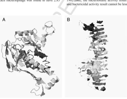
Protein structure modeling
The predicted secondary structure of Major capsid protein was arranged in globular domains, forming hexamers, comprised of alpha and beta structures. Major tube protein was arranged in β-helix, ac
UNCORRECTED
PROOF
cording to the I-TASSER algorithm.16–19Furthermore, thebioinfor-matics modeling of these structures were generated using the SPICKER program based on pair-wise structure similarity. C-scores varied from −1.72 to −1.38. In Figure 2, A and B, it is presented the structure predictions of Major capsid protein and Major tube pro-tein, respectively. Non-polar side chains remained uncharged at phys-iological pH and were incapable of participating in hydrogen bond-ing. On the other hand, in Major tube protein, seven polar leucine-15,47,88,112,178,385, and 464 were identified. Furthermore four polar asparagine191,259,277, and 336 were detected, as well as two polar glutamine-11 and 303. All of them contained an α-amino group protonated at –NH3+under biological conditions. These are
im-portant for cross-linking to outer sheath proteins around Major tube protein. Furthermore leucine-507, methionine-509, proline-510 and aspartate-511 constitute an acidic terminal region, important to bind and be capped by the spike protein.
The first subunit of the Major capsid protein presents polar ly-sine-38 and glycine-45. Furthermore it presents an RGD se-quence-44,45,46; aspartate-82,98, valine-85, and glycine-90,112 in the second subunit; aspartate-144,148, leucine-132, valine-135, ser-ine-137 in the third subunit; aspartate-157,174, serine-177,186, me-thionine-184,189, histidine-185 in the fourth subunit; aspartate-224, serine-225, valine-236, glycine-245, glutamate-259, asparagine-263 in the fifth subunit; glycine-284 in the sixth subunit. Under physiological conditions (pH 7.4) lysine, arginine, histidine side chains were posi-tively charged –NH3+and important for covalent bonding to PCL
sur-face.
Ninhydrin
The free amines on vB_Pae-Kakheti25 capsid were quantified with ninhydrin reagent. A calibration curve (r2= 0.99) was performed with known concentrations of L-cysteine (which contains only one amino group), and finally, the concentration of amines in bacteriophage so-lution was determined. Each bacteriophage was found to have 2.85 picogram of free amines.
Fourier transform infrared spectroscopy (FT-IR)
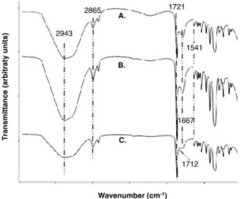
The FT-IR spectrum of unmodified nanofilm is shown in Figure 3, A. This spectrum displays the expected representative peaks of polycaprolactone, namely C-H, related with CH2stretching vibrations
at 2943 cm−1and 2865 cm−1, and with carbonyl adsorption at 1721 cm−1.26
The covalent bonds between PCL and vB_Pae-Kakheti25 on nanofibers, were also assessed by FT-IR after 5 and 25 washing cy-cles. IR spectra showed PCL surface with carboxylic acid after its acti-vation at 1712 cm−1, and vB_Pae-Kakheti25 grafted on nanofibers, as
denoted by the increase of the peaks owed to covalent bonds formed with PCL surface, namely at 1667 cm−1and 1541 cm−1, assigned to Amide I and II, respectively, as well as the decrease of free carboxylic acid at 1711 cm−1, as shown in Figure 3, B and C.
Antimicrobial assays
Quantitative method (suspension)
In order to evaluate the antimicrobial effectiveness of PCL-vB_-Pae-Kakheti25, the adsorption test was performed to quantify the abil-ity of nanofibers functionalized with bacteriophages to prevent bac-terial growth Figure 4. The results showed that there were marked reductions in Pseudomonas aeruginosa at T0 h and T2 h. PCL acti-vated nanofibers showed outstanding microbial inhibition, as shown in Figure 4. We could observe that PCL-vB_Pae-Kakheti25 eliminated 6 log of Pseudomonas aeruginosa at 0 and 2 hours, which was equiva-lent to 99.9999% reduction.
After 5 and 25 washing cycles with WOB at 40 °C, the same 6 log elimination of Pseudomonas aeruginosa was observed, either at 0 or at 2-hour. These results proved that covalent bonding between PCL and the bacteriophage capsid was stable and strong enough to resist to several washings.
In order to know whether the samples behaved as bacteriosta-tic or bactericidal, according to Japanese Industrial Standard JIS L 1902:2002, the bacteriostatic activity result can be not less than 2.0 and bactericidal activity result cannot be less than 0. Briefly, bacterio
Figure 2. Major capsid protein (A) and Major tube protein (B) modeled structures generated by SPICKER program16–19based on pair-wise similarity. C-scores varied from −1.72 to
UNCORRECTED
PROOF
Figure 3. IR spectra of unmodified PCL (A) and PCL-vB_Pae_Kakheti25 after (B) 5washing-cycles and (C) 25 washing-cycles.
static activity = log10 of the average of bacterial colonies on the con-trol samples at T24 h - log10 of the average of bacterial colonies on the functionalized samples at T24 h; and bactericidal activity = log10 of the average number of bacterial colonies on the control samples at T0 - log10 of the average of bacterial colonies on the functionalized samples at T24 h.
The calculated values from these equations yielded the following values: after three rinses, bacteriostatic activity was 0.94 and bacte-ricidal activity was 1.63; after 5 washing cycles, bacteriostatic activ-ity was 0 and bactericidal activactiv-ity was 0.98; after 25 washing cycles, bacteriostatic activity was 0 and bactericidal activity was 0. These re-sults state that PCL-vB_Pae-Kakheti25 has a bactericidal effect after 3 rinses, 5 and 25 washing cycles, which means that the immobilization of vB_Pae-Kakheti25 on PCL lead to death of P. aeruginosa and do not interfere in the development and growth of bacteria.
Growth kinetics of infected bacteria
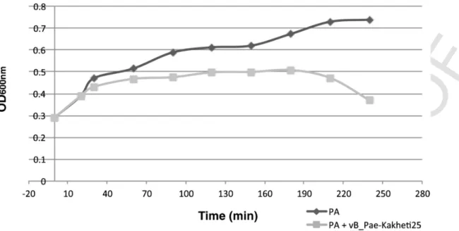
This assay aimed to study the growth kinetics of the infected host cells, using a control (uninfected bacteria), at the dressing change tem-perature of 29.9 °C Figure 5.
At this temperature the infected P. aeruginosa showed lower ab-sorbance values than those obtained by the non-infected P. aerugi
nosa from 20 minutes after the beginning of incubation. This indicated that vB_Pae-Kakheti25 had been using the bacterial machinery for its benefit (latent period). A decrease in bacterial growth rate after 120 minutes were also observed, which indicated the onset of cell lysis phase, resulting from bacteriophage infection.
Transmission electron microscopy (TEM)
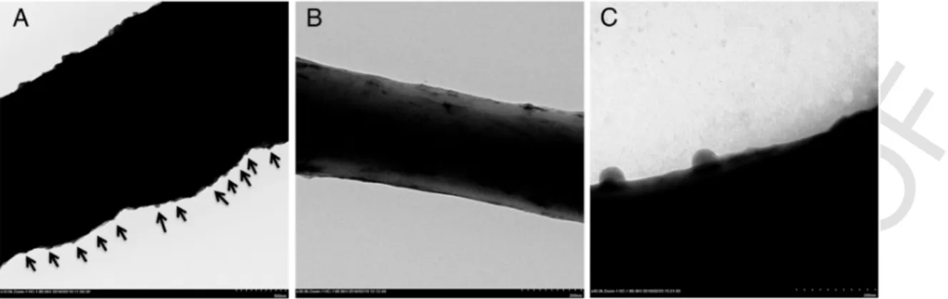
The interaction between vB_Pae-Kakheti25 tail and P. aeruginosa membrane is shown in Figure 6, A and B. The vB_Pae-Kakheti25 life cycle inside P. aeruginosa cells was also monitored at 0, 20, 30, and 120 minutes, as shown in Figure 6, C-F. vB_Pae-Kakheti25 bacte-riophage fiercely degraded P. aeruginosa membranes, especially after 20 minutes (Figure 6, D-F). Furthermore, the spacing of the bonding of vB_Pae-Kakheti25 on PCL after 25 washing cycles was also vi-sualized, as shown in Figure 7. The frequency of covalent bonds of vB_Pae-Kakheti25 on PCL nanofibers averaged 118 nm, as shown in Figure 7, A.
Tumoral activity assay
PCL-vB_Pae-Kakheti25 did not show any cytotoxic effect over mouse fibroblast cell line BALB/c 3 T3 Figure 8. Both perspiration extract assay and direct contact assay showed values that did not ranged beyond 30% from controls. Only an alteration under or over 30% in comparison with controls would be considered cytotoxic or pro-tumorigenic, respectively.21
Discussion
As part of natural defenses of ecosystem, the mode of action of bacteriophages is very specific and relies on infecting exclusively their host bacteria, leaving the rest of microbiota and human tis-sues intact. In this investigation, the capsid of the bacteriophage vB_-Pae-Kakheti25 was covalently cross-linked to polycaprolactone (PCL) nanofibrous dressing, in order to maximize vB_Pae-Kakheti25 tail exposure, evaluate its stability, and its antimicrobial effect over Pseudomonas aeruginosa bacteria. The electrospun nanofibrous struc-tures offer larger area of contact and a smoother surface, which dimin-ishes roughness and scratching.27
A nanofibrous dressing with an average thickness of 1.57 nm was produced by electrospinning. For the covalent immobilization of bac-teriophage capsid onto PCL nanofibers, the first step was the forma-tion of reactive acid groups on PCL, namely ester groups (−COO-) that were hydrolyzed to carboxylic acid groups from ester under acidic solution (pH 5.5) via the reverse of Fischer esterification.
Figure 4. Percentage of microbial inhibition at 0 hours (A) and 24 hours (B) – all of these values were obtained by comparison with results of native PCL. *Statistically significant
UNCORRECTED
PROOF
Figure 5. One-step growth kinetics of P. aeruginosa strain and P. aeruginosa infected with the bacteriophage vB_Pae-Kakheti25 at 29.9 °C.Figure 6. Adsorption of vB_Pae-Kakheti25 to P. aeruginosa membrane at 50000× magnification, (A) and (B). vB_Pae-Kakheti25 life cycle infecting P. aeruginosa monitored at 0
(C), 20 (D), 30 (E), and 120 minutes (F). 6000× and 25,000× magnifications.
Amino groups of vB_Pae-Kakheti25 capsid – 2.85 picogram of free amines per bacteriophage – reacted as soon as they contacted the car-boxylic acids of PCL, via acid-amine reactions, forming amide link-ages. vB_Pae-Kakheti25 bacteriophage contains an icosahedral head structure that is composed of many copies of Major capsid protein (UniProtKB) with 325 amino acid length and lysine, arginine, histi-dine side chains that were positively charged under physiologic pH. Also vB_Pae-Kakheti25 presents a tail that recognizes surface recep
tors of P. aeruginosa. Mechanical properties of PCL nanofibers showed high elasticity and were not significantly altered in any treat-ments in this study, which means PCL structure was not degraded dur-ing the experiment.
Fourier Transform Infrared Spectroscopy (FT-IR) and Transmis-sion Electron Microscopy (TEM) provided evidence for the proposed bacteriophage immobilization aligned in a head-to-tail orientation. FT-IR spectra confirmed that bacteriophage capsid was covalently
UNCORRECTED
PROOF
Figure 7. The bonding frequency of vB_Pae-Kakheti25 to PCL single nanofibre (A), 3D visualization (B), and vB_Pae-Kakheti25 adsorption arrangement (C). 10,000×, 30,000×and 40,000× magnifications.
Figure 8. Perspiration extract assay and direct contact assay against mouse fibroblast cell line BALB/c 3 T3. immobilized on PCL surface, by the comparison of peak height
pro-portions (1667 cm−1, 1541 cm−1and 1711 cm−1) between PCL and PCL grafted with vB_Pae-Kakheti25. Furthermore, spectra showed the covalent cross-link between PCL and vB_Pae-Kakheti25 to be multiple washing-resistant – 5 and 25 washing cycles – and conse-quently stable, because the aforementioned peaks height proportions were maintained. In accordance with these previous data, TEM im-ages showed vB_Pae-Kakheti25 immobilized on PCL nanofibers and aligned in a head-to-tail orientation after it had been washed 25 times. Immobilized bacteriophages were also spaced ≈118 nm apart on both sides.
Antimicrobial assays of vB_Pae-Kakheti25-bearing PCL showed that it acted as soon as it contacted with P. aeruginosa, with an-timicrobial reducing rates of 99.9999% (6 log), p < 0.05. In addi-tion, PCL-vB_Pae-Kakheti25 was identically active after 25 washing cycles, either at immediate contact, or at 2-hour contact. These re-sults were higher than the 5 log reduction observed by Huang et al. for silver-chitosan.28 The low natural adhesion of PCL29 due to its hydrophobic character - contact angle of 109.9° ± 5.0°29 – may also have facilitated the contact of P. aeruginosa with the bacteriophage
tails. H. Yang and Y. Deng30also showed that a hydrophobic surface
reduced adhesion capacity of PCL to bacteria.30
Although Karumidze et al24 studied the effect of Pae-Kakheti25 on P. aeruginosa, the immobilization of vB_-Pae-Kakheti25 on whichever surface has not been reported yet. Since a vB_Pae-Kakheti25 multiplicity of infection of 0.1 led to a general growth inhibition of P. aeruginosa in BHI agar plates,24it would be assured that a surface functionalized with the same amount of vB_-Pae-Kakheti25 would also kill P. aeruginosa, especially due to its maximized tail exposure and the larger area of contact of electrospun nanofibers. On the other hand, when in solution, vB_Pae_Kakheti25 bacteriophages aggregated together by generating bonds between their tails, and consequently decreasing their accessibility to interact with P. aeruginosa, as also proposed by Langlet et al.31
The growth kinetics was studied at 29.9 °C, which is the temper-ature of the dressing change procedure. The P. aeruginosa was ob-served to halt its exponential growth as it became infected by vB_-Pae-Kakheti25 bacteriophage. This demonstrated that as soon as P. aeruginosa was infected, vB_Pae-Kakheti25 used its machinery for self-replication, the so-called latent period. The lysis phase began when bacteria started to have a decrease in bacterial growth rate at
UNCORRECTED
PROOF
120 minutes, where P. aeruginosa mainly undergone the release ofre-cently-formed bacteriophages.
Our samples were adequately tested on mouse fibroblast cell line in order to ascertain whether they were cytotoxic. The perspiration ex-tract of samples showed an average cell growth inhibition of 5.3% and the direct contact assay showed an average growth enhancement of 0.33%. A variation of growth of more than 30% in comparison with the controls is regarded as cell-toxic effect.32Therefore, the cytotox-icity assay for PCL-vB_Pae-Kakheti25 showed less than 30% of cel-lular viability reduction, making our invention a safe agent for use in contact with skin.
While there are a few commercial antimicrobial applications, they present limitations. Dermasilk® is a silk-based material with Si-QAC (Silane quaternary ammonium compounds) combining the smooth-ness of silk with the antimicrobial properties.2,33Another example is Padycare® a micromesh polyamide/lycra containing woven silver fil-aments.34Similarly, TheraBond® is a silver fabric dressing designed to optimize wound fluid management without compromising the an-timicrobial properties.2,3 In vitro studies of these garments demon-strated a significant decrease in P. aeruginosa. Even though there is no evidence for skin sensitization by bulk silver, a possible systemic absorption of silver particles because of a disrupt skin barrier has to be considered, while Si-QAC is reported to cause bacterial resis-tance and show evidence of skin sensitization.2,33,35For this reason, the relatively lower incidence of adverse reactions of natural products has been intensively investigated.4,33,35Overall, antimicrobial textiles
have found uses in hygienic rather than medical field especially due to possible cytotoxic effect and sometimes low effectiveness.4,35 Antimi-crobial textiles have been designed to address activity against a broad spectrum of microorganisms33,35,36but there remain open questions
re-garding toxicity on human health, which is causing a paradigm change to move on to highly specific agents toward pathogenic bacteria. Like-wise, the rising flow of strains resistant to last-resort antibiotics rekin-dles interest in novel approaches.11,35–37
Phage immobilization strategies on organic and inorganic surfaces have been recently reported37–45: while in some cases electrostatic binding showed phage instability and detachment,46covalent attach-ment offers a much stronger bond.47Still, for antimicrobial surfaces
designed to utilize the inherent binding specificity and lytic activity of phage toward the bacterial host, tailed phage should be immobilized through the capsid and filamentous phage with their bacterial-captur-ing pole exposed.44–46
Therefore, in this work we surface-anchored vB_Pae-Kakheti25 bacteriophages to develop highly specific antibacterial non-woven textiles/dressings especially – but not limited to – for skin associated infections, as a skin microbiome regulator. Such textiles could also find application as outside catheter dressings to provide adequate fix-ation and inhibition of bacterial adhesion during catheter lifetime. To authors' knowledge this is the first investigation exploiting phages as antibacterial agents for textiles. The closest approach that one can con-sider related has been reported by Mao et al.,48where a non-lytic fil-amentous phage (M13) was engineered to express negatively-charged glutamates further glutaraldehyde-crosslinked upon extrusion of fibers and Kevlar coatings with the ability to attract Ag+in a silverization
process. The engineered phage was the basis of the fiber and coating but silver was responsible for the antibacterial activity since non-lytic phage was used. Our strategy, in contrary, immobilized lytic phage onto non-woven textile as antibacterial agent.
This study showed that vB_Pae-Kakheti25 covalent immobiliza-tion on PCL nanofibers with a head-to-tail orientaimmobiliza-tion added antimi-crobial properties to the proposed dressing. It presented a miantimi-crobial reduction of 6 log, even after 25 wash cycles, comparatively with the
non-modified nanostructure after 2 h of incubation. Its activity was immediate and P. aeruginosa reduction was complete.
On the contrary, when in solution, vB_Pae_Kakheti25 bacterio-phage aggregated and decreased its availability to interact with P. aeruginosa.
References
1. J. Oh, A.L. Byrd, C. Deming, S. Conlan, et al., Biogeography and individuality shape function in the human skin metagenome, Nature 514 (2014) 59–64. 2. C. Lopes, D. Silva, L. Delgado, O. Correia, A. Moreira, Functional textiles for
atopic dermatitis: a systematic review and meta-analysis, Pediatr Allergy Im-munol 24 (2013) 603–613.
3. J.G. Powers, L.M. Morton, T.J. Phillips, Dressings for chronic wounds, Dermatol Ther 3 (2013) 197–206.
4. J. Ring, A. Alomar, T. Bieber, et al., Guidelines for treatment of atopic eczema (atopic dermatitis) part II, J Eur Acad Dermatol Venereol 26 (2012) 1176–1193. 5. A.M. Misic, S.E. Gardner, E.A. Grice, The wound microbiome: modern
ap-proaches to examining the role of microorganisms in impaired chronic wound healing, Adv Wound Care 3 (7) (2014) 502–510.
6. K. Gjødsbøl, J.J. Christensen, T. Karlsmark, et al., Multiple bacterial species re-side in chronic wounds: a longitudinal study, Int Wound J 3 (3) (2006) 225–231. 7. F. Nogueira, L. Granadeiro, C. Mouro, I.C. Gouveia, Antimicrobial and antioxi-dant surface modification toward a new silk-fibroin (SF)-l-cysteine material for skin disease management, App Surf Sci 364 (2016) 552–559.
8. L.M. Weiner, A.K. Webb, B. Limbago, M.A. Dudeck, J. Patel, A.J. Kallen, et al., Antimicrobial-resistant pathogens associated with healthcare-associated infec-tions: summary of data reported to the National Healthcare Safety Network at the Centers for Disease Control and Prevention, 2011-2014, Infect Control Hosp Epi-demiol 37 (11) (2016) 1288–1301.
9. K.A. Traugott, K. Echevarria, P. Maxwell, K. Green, J.S. Lewis II, Monotherapy or combination therapy? The Pseudomonas Aeruginosa Conundrum, Pharma-cotherapy 31 (6) (2011) 598–608.
10. D.L. Stevens, Infections of the skin, muscle and soft tissues, in: D.L. Kasper, E. Braunwald, A.S. Fauci, et al. (Eds.), Harrison's Principles of Internal Medicine, 16 ed., McGraw-Hill, New York, 2006, p. 740.
11. A. Parisien, B. Allain, J. Zhang, R. Mandeville, C.Q. Lan, Novel alternatives to antibiotics: bacteriophages, bacterial cell wall hydrolases, and antimicrobial pep-tides, J Appl Microbiol 104 (2008) 1–13.
12. J.S. Soothill, Bacteriophage prevents destruction of skin grafts by Pseudomonas aeruginosa, Burns 20 (3) (1994) 209–211.
13. C.S. McVay, M. Velásquez, J.A. Fralick, Phage therapy of Pseudomonas aerugi-nosa infection in a mouse burn wound model, Antimicrob Agents Chemother 51 (6) (2007) 1934–1938.
14. B.D. Ratner, Biomaterials science: an introduction to materials in medicine, 2nd ed., Elsevier Academic Press, Amsterdam, Boston, 2004854.
15. B.D. Ulery, L.S. Nair, C.T. Laurencin, Biomedical applications of biodegradable polymers, J Polym Sci B 49 (12) (2011) 832–864.
16. Y. Ikada, H. Tsuji, Biodegradable polyesters for medical and ecological applica-tions, Macromol Rapid Commun 21 (3) (2000) 117–132.
17. V. Ilić, Z. Šaponjić, V. Vodnik, R. Molina, S. Dimitrijević, P. Jovančić, et al., An-tifungal efficiency of corona pretreated polyester and polyamide fabrics loaded with ag nanoparticles, J Mater Sci 44 (2009) 3983–3990.
18. S.H. Lim, S.M. Hudson, Review of chitosan and its derivatives as antimicrobial agents and their uses as textile chemicals, J Macromol Sci Polym Rev 43 (2) (2003) 223–269.
19. S. Reardon, Antibiotic alternatives rev up bacterial arms race, Nature 521 (2015) 402–403.
20. J. Yang, R. Yan, A. Roy, D. Xu, J. Poisson, Y. Zhang, The I-TASSER suite: pro-tein structure and function prediction, Nat Methods 12 (2015) 7–8.
21. J. Yang, Y. Zhang, I-TASSER server: new development for protein structure and function predictions, Nucleic Acids Res 43 (2015) W174–W181.
22. A. Roy, A. Kucukural, Y. Zhang, I-TASSER: a unified platform for automated protein structure and function prediction, Nat Protoc 5 (2010) 725–738. 23. Y. Zhang, I-TASSER server for protein 3D structure prediction, BMC
Bioinfor-matics 9 (2008) 40.
24. N. Karumidze, J.A. Thomas, N. Kvatadze, M. Goderdzishvili, K.W. Hakala, S.T. Weintraub, et al., Characterization of lytic Pseudomonas aeruginosa bacterio-phages via biological properties and genomic sequences, Appl Microbiol Biotech-nol 94 (2012) 1609.
25. ISO/TC 194 biological and clinical evaluation of medical devices, in: http://www. iso.org/iso/catalogue_detail.htm%3Fcsnumber=36406.
26. G. Socrates, Infrared and Raman characteristic group frequencies: tables and charts, John Wiley & Sons, Ltd., 2004.
UNCORRECTED
PROOF
27. Y. Gao, Y.B. Truong, Y. Zhu, I.L. Kyratzis, Electrospun antibacterial nanofibers: production, activity, and in vivo applications, J Appl Polym Sci 131 (18) (2014) 1–13.
28. L. Huang, T. Dai, Y. Xuan, G.P. Tegos, M.R. Hamblin, Synergistic combination of chitosan acetate with nanoparticle silver as a topical antimicrobial: efficacy against bacterial burn infections, Antimicrob Agents Chemother 55 (7) (2011) 3432–3438.
29. F. Nogueira, I.C. Gouveia, Amino acid based material for the complementary therapy of decubitus ulcers, J Microbiol Biotechnol 26 (11) (2017).
30. H. Yang, Y. Deng, Preparation and physical properties of superhydrophobic pa-pers, J Colloid Interface Sci 325 (2) (2008) 588–593.
31. J. Langlet, F. Gaboriaud, J.F.L. Duval, C. Gantzer, Aggregation and surface prop-erties of F-specific RNA phages: implication for membrane filtration processes, Water Res 42 (2008) 2769–2777.
32. E. Borenfreund, O. Borrero, In vitro cytotoxicity assays. Potential alternatives to the Draize ocular allergy test, Cell Biol Toxicol 1 (1984) 55–65.
33. L. Windler, M. Height, B. Nowack, Comparative evaluation of antimicrobials for textile applications, Environ Int 53 (2013) 62–73.
34. A. Gauger, S. Fischer, M. Mempel, et al., Efficacy and functionality of sil-ver-coated textiles in patients with atopic eczema, J Eur Acad Dermatol Venereol 20 (2006) 534–541.
35. Y. Gao, R. Cranston, Recent advances in antimicrobial treatments of textiles, Text Res J 78 (1) (2008) 68–72.B.
36. M. Pedrosa, C. Mouro, F. Nogueira, J. Vaz, I.C. Gouveia, Comparison of the an-tibacterial activity of modified-cotton with Magainin I and LL-37 with potential as wound-dressings, J Appl Polym Sci 131 (21) (2014) 40997, [1-8]. 37. J.J. Barra, Bacteriophage adhering to mucus provide a non–host-derived
immu-nity, PNAS 110 (2013) 10771–10776.
38. H. Anany, W. Chen, R. Pelton, M.W. Griffiths, Biocontrol of listeria mono-cyto-genes and E. coli o157:H7 in meat using phage immobilized on modified cellulose membranes, Appl Environ Microbiol 77 (18) (2011) 6379–6387.
39. R. Cademartiri, H. Anany, I. Gross, R. Bhayani, M. Griffiths, M.A. Brook, Immo-bilization of bacteriophages on modified silica particles, Biomaterials 31 (2010) 1904–1910.
40. P.J. Yoo, K.T. Nam, J. Qi, S.-K. Lee, J. Park, A.M. Belcher, et al., Spontaneous assembly of viruses on multilayered polymer surfaces, Nat Mater 5 (2006) 234–240.
41. N. Tawil, E. Sacher, R. Mandeville, M. Meunier, Strategies for the immobiliza-tion of bacteriophages on gold surfaces monitored by surface Plasmon resonance and surface morphology, J Phys Chem C 117 (13) (2013) 6686–6691. 42. S.K. Arya, A. Singh, R. Naidoo, P. Wu, M.T. McDermott, S. Evoy, Chemically
immobilized T4-bacteriophage for specific Escherichia coli detection using sur-face plasmon resonance, Analyst 136 (3) (2011) 486–492.
43. W. Salalha, J. Kuhn, Y. Dror, E. Zussman, Encapsulation of bacteria and viruses in electrospun nanofibres, Nanotechnology 17 (2006) 4675–4681.
44. A.K. Trillinga, M.M. Harmsenc, V.J.B. Ruigrokd, H. Zuilhofb, J. Beekwildera, The effect of uniform capture molecule orientation on biosensor sensitivity: de-pendence on analyte properties, Biosens Bioelectron 40 (1) (2013) 219–226. 45. J.-W. Lee, J. Song, M.P. Hwang, K.H. Lee, Nanoscale bacteriophage biosensors
beyond vphage display, Int J Nanomedicine 8 (2013) 3917–3925.
46. Z. Hosseinidoust, A. Olsson, N. Tufenkji, Going viral: designing bioactive sur-faces with bacteriophage, Colloids Surf B Biointersur-faces 124 (2014) 2–16. 47. H.A. Pearson, G.S. Sahukhal, M.O. Elasri, M.W. Urban, Phage-bacterium war on
polymeric surfaces: can surface-anchored bacteriophages eliminate microbial in-fections?, Biomacromolecules 14 (2013) 1257–1261.
48. J.Y. Mao, A.M. Belcher, K.J. Van Vliet, Genetically engineered phage fibers and coatings for antibacterial applications, Adv Funct Mater 20 (2010) 209–214.