The use of hydroxyapatite for arthrodesis in dogs and cats: a clinical study
[Utilização de hidroxiapatita para artrodeses em cães e gatos: ensaio clínico]
F.A. Dórea Neto1,2, J.G. Padilha Filho2 L.A. Santos4, A.P. Oriá1,2, J.C. Canola3 S.A. Stefanes2, E. Regonato2
1
União Metropolitana de Educação e Cultura Av. Luiz Tarquínio Pontes, 600 42700-000 - Lauro de Freitas, BA
2
Aluno de pós-graduação - FCAV-UNESP – Jaboticabal, SP
3
Faculdade de Ciências Agrárias e Veterinária - UNESP – Jaboticabal, SP
4
Universidade Federal do Rio Grande do Sul – Porto Alegre, RS
ABSTRACT
Twenty-five arthrodeses were performed in four cats and 17 dogs using synthetic hydroxyapatite as fresh autogenous graft cancellous bone substitute. Arthrodesis was performed in the carpal joint in eight cases, in the tarsal joint in 10, in the elbow joint in six, and in the knee joint in one case. The mean radiographic follow-up time was 30 days in one animal, 45 days in another animal and 60 days in the 19 remaining cases. Bone union was observed in 24 arthrodeses. Non-union of one elbow arthrodesis was due to failure of stabilization. Restoration of limb functionality was classified as good to excellent in 22 cases. Hydroxyapatite was able to promote bone growth and is suitable for using in routine surgical procedures for small animals.
Keywords: dog, cat, bone fusion, carpal, tarsal, stifle, elbow
RESUMO
Realizaram-se 25 artrodeses em 21 casos, quatro em gatos e 17 em cães, utilizando hidroxiapatita sintética como substituto ao enxerto ósseo autógeno esponjoso fresco, sendo oito na articulação do carpo, 10 na articulação do tarso, seis na do cotovelo e uma na do joelho. As avaliações radiográficas foram realizadas aos 30 dias em um animal, aos 45 dias em outro e aos 60 dias nos 19 casos restantes. Visibilizou-se união óssea em 24 artrodeses e a não-união em um cotovelo foi atribuída a falha na estabilização. O retorno à função do membro foi classificado de bom a excelente em 22 casos. A hidroxiapatita foi capaz de viabilizar o crescimento ósseo e mostrou-se factível para utilização na prática cirúrgica rotineira em pequenos animais.
Palavras-chave: cão, gato, fusão óssea, carpo, tarso, joelho, cotovelo
INTRODUCTION
Arthrodesis is defined as the surgical fixation of a joint in order to permit fusion of the articular surfaces through the proliferation of bone cells (Penwick, 1987; Lesser, 1993; Turner and Lipowitz, 1996). Arthrodesis has been well documented as a treatment option for comminuted joint fractures, irreparable injuries
Recebido em 19 de julho de 2006 Aceito em 2 de abril de 2007 E-mail: [email protected]
Since their introduction in the beginning of the 1980s, calcium phosphate ceramics, especially hydroxyapatite, have been used for the remodeling and reconstruction of bone defects. The properties of biocompatibility, bioactivity and osteoconduction of bioceramics permit their implantation into the bone site without inducing an immune response. These materials are able to bind directly to bone tissue, promoting bone growth along its surface (Legeros, 1991). However, these synthetic bone substitutes are not commonly employed in veterinary surgery (Fitch et al., 1997).
The objective of the present study was to clinically and radiographically evaluate the use of hydroxyapatite as a graft in joints submitted to arthrodesis.
MATERIAL AND METHODS
Twenty-five arthrodeses were performed in 21 animals, four cats and 17 dogs, for the treatment of severe joint disorders using synthetic hydroxyapatite as a fresh autogenous graft cancellous for defect filling between opposite articular sides.
Medical records as well as data regarding surgical treatment (arthrodesis) in cats and dogs with severe joint injuries, pain and instability, were obtained from January 2001 to December 2002. The data included clinical signs, etiology of the injury, concurrent problems, extent and site of the injuries, time elapsed until surgical stabilization of the joint, stabilization methods and postoperative management, duration of hospitalization, cultures and antibiotic therapy, healing period and complications. The interval between the occurrence of the injury and surgery ranged from 24h in some cases of shearing injury to 3 months in cases of osteoarthrosis.
Clinical reassessment comprised complete clinical and orthopedic examination and was performed whenever possible. The presence of any gait abnormality and the presence or absence of pain, crepitation and articular edema were evaluated.
The age of the animals, including 12 (57.1%) males and nine (42.9%) females, ranged from 4 months to 10 years. Six (28.6%) of the 21
animals were younger than one year, 14 (66.7%) were from 1 to 6 years and one animal (4.7%) was over 10 years old. The weight of the animals ranged from 2 to 35kg, with 12 (57.1%) animals weighing less than 10kg, seven (33.4%) between 10 and 20kg and two (9.5%) more than 20kg.
Arthrodesis was performed in the carpal joint in eight (32%) cases, in the tarsal joint in 10 (40%), in the elbow in six (24%), and in the knee joint in one (4%).
The injuries presented by the animals included radial agenesis in two (9.5%); fracture of the calcaneus in three (14.3%); luxation due to shearing injury in seven (33.3%); fracture caused by a firearm projectile in one (4.5%); closed luxation, open luxation and osteoarthritis in two animals each (9.5% each); contracture of the carpal flexor tendons caused by fracture of the radius in one (4.5%); and fracture of the olecranon in one (4.5%).
Bilateral carpal arthrodesis was performed in one animal (Table 1, case 2). Two animals presented radial agenesis and were submitted to carpal and elbow arthrodesis (Table 1, cases 4 and 5). Three animals underwent pantarsal arthrodesis due to the extent of the injuries (Table 1, cases 12, 15 and 17).
The joints were prepared by debridement of articular cartilage using curettes and an electrical milling machine coupled to a high-rotation micro-rectifier, hand-held saws and bone files. The contour of the joint was preserved in all cases, except for the knee joint in which the femoral condyles and proximal portion of the tibia were sawed off, forming a platform to provide better coaptation of the two bones. After joint preparation, the defect produced by curettage was completely filled with granule hydroxyapatite (<177µm)1 (Fig. 1).
The joints were positioned at angles predetermined with a goniometer, ranging from 175 to 180o for carpal joints, from 125 to 145o for tarsal joints, from 130 to 150o for elbow joints and an angle of 140o for knee joint.
1
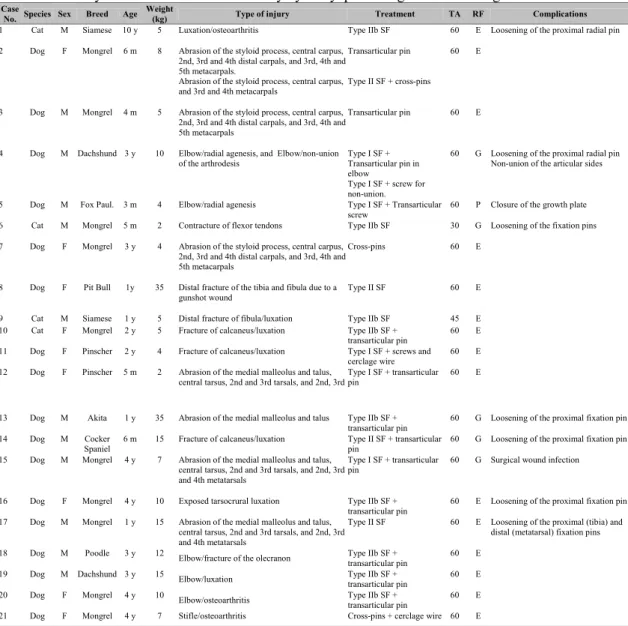
Table 1. Summary data: arthrodesis with treat by hydroxyapatite as graft bone in dogs and cats Case
No. Species Sex Breed Age Weight
(kg) Type of injury Treatment TA RF Complications
1 Cat M Siamese 10 y 5 Luxation/osteoarthritis Type IIb SF 60 E Loosening of the proximal radial pin
2 Dog F Mongrel 6 m 8 Abrasion of the styloid process, central carpus, 2nd, 3rd and 4th distal carpals, and 3rd, 4th and 5th metacarpals.
Abrasion of the styloid process, central carpus, and 3rd and 4th metacarpals
Transarticular pin
Type II SF + cross-pins
60 E
3 Dog M Mongrel 4 m 5 Abrasion of the styloid process, central carpus, 2nd, 3rd and 4th distal carpals, and 3rd, 4th and 5th metacarpals
Transarticular pin 60 E
4 Dog M Dachshund 3 y 10 Elbow/radial agenesis, and Elbow/non-union of the arthrodesis
Type I SF + Transarticular pin in elbow
Type I SF + screw for non-union.
60 G Loosening of the proximal radial pin Non-union of the articular sides
5 Dog M Fox Paul. 3 m 4 Elbow/radial agenesis Type I SF + Transarticular screw
60 P Closure of the growth plate
6 Cat M Mongrel 5 m 2 Contracture of flexor tendons Type IIb SF 30 G Loosening of the fixation pins
7 Dog F Mongrel 3 y 4 Abrasion of the styloid process, central carpus, 2nd, 3rd and 4th distal carpals, and 3rd, 4th and 5th metacarpals
Cross-pins 60 E
8 Dog F Pit Bull 1y 35 Distal fracture of the tibia and fibula due to a gunshot wound
Type II SF 60 E
9 Cat M Siamese 1 y 5 Distal fracture of fibula/luxation Type IIb SF 45 E 10 Cat F Mongrel 2 y 5 Fracture of calcaneus/luxation Type IIb SF +
transarticular pin
60 E
11 Dog F Pinscher 2 y 4 Fracture of calcaneus/luxation Type I SF + screws and cerclage wire
60 E
12 Dog F Pinscher 5 m 2 Abrasion of the medial malleolus and talus, central tarsus, 2nd and 3rd tarsals, and 2nd, 3rd
Type I SF + transarticular pin
60 E
13 Dog M Akita 1 y 35 Abrasion of the medial malleolus and talus Type IIb SF + transarticular pin
60 G Loosening of the proximal fixation pin
14 Dog M Cocker
Spaniel
6 m 15 Fracture of calcaneus/luxation Type II SF + transarticular pin
60 G Loosening of the proximal fixation pin
15 Dog M Mongrel 4 y 7 Abrasion of the medial malleolus and talus, central tarsus, 2nd and 3rd tarsals, and 2nd, 3rd and 4th metatarsals
Type I SF + transarticular pin
60 G Surgical wound infection
16 Dog F Mongrel 4 y 10 Exposed tarsocrural luxation Type IIb SF + transarticular pin
60 E Loosening of the proximal fixation pin
17 Dog M Mongrel 1 y 15 Abrasion of the medial malleolus and talus, central tarsus, 2nd and 3rd tarsals, and 2nd, 3rd and 4th metatarsals
Type II SF 60 E Loosening of the proximal (tibia) and distal (metatarsal) fixation pins
18 Dog M Poodle 3 y 12
Elbow/fracture of the olecranon Type IIb SF + transarticular pin
60 E
19 Dog M Dachshund 3 y 15
Elbow/luxation Type IIb SF +
transarticular pin
60 E
20 Dog F Mongrel 4 y 10
Elbow/osteoarthritis Type IIb SF + transarticular pin 60 E
21 Dog F Mongrel 4 y 7 Stifle/osteoarthritis Cross-pins + cerclage wire 60 E
M: male; F: female; SF: skeletal fixator; TA: time of radiographic assessment (days) after the surgical procedure; RF: restoration of limb functionality (E: excellent; G: good; P: poor).
The joints were stabilized using pins, cerclage wires, screws and types I, II and IIb external fixators combined or not with pins, transarticular screws and cerclage wires as separately listed in Table 1.
The bone fixation pins of the external fixators were connected to polymethylmethacrylate bars. During the postoperative period, padded bandages were used to protect the limb and fixators. Three (14.3%) animals undergoing carpal arthrodesis also received an indirect pedicled skin flap on the lateral chest due to injuries caused by shearing trauma. The owners were advised to keep the animals in confinement until removal of the implants.
Empirical antibiotic therapy was adopted after surgery and consisted of the subcutaneous administration of 2.2mg/kg sodium ceftiofur2 every 24h for 6 days. In cases of bone exposure, 25mg/kg metronidazole3 was additionally administered orally every 12h for 5 days. This procedure was also adopted for cases in which signs of infection or osteomyelitis persisted until the results of cultures and sensitivity tests permitted the most appropriate choice of antibiotic in each case. Additionally,
1.0mg/kg ketoprofen4 was administered
2
Excenel, Schering Plough Veterinária 3 Flagil, Searle
subcutaneously every 24h for 3 days and 0.01mg/kg buprenorphine5 was injected intramuscularly every 12h for 3 consecutive days.
The animals were clinically and radiographically assessed immediately after surgery and on the 60th day; however, two animals were evaluated less than 60 days (case 6 on the 30th day and case 9 on the 45th day). The following aspects were analyzed: arthrodesis stability, loosening of the metal implants, presence of infection, exudate or signs of sequestration of the implanted bioceramic and bone union.
RESULTS AND DISCUSSION
The results obtained for each case are individually illustrated in Table 1. No formation of fistulas or a purulent exudate was observed during follow-up of the animals. During the immediate postoperative period, intense edema was noted in the extremity of the limb and around the pins, which persisted on average until day 8. Hot and cold compresses were administered as coadjuvant therapy to the anti-inflammatory drugs.
With respect to the degree of lameness, the animals were non-weightbearing on the operated limb on days 1st to 2nd after surgery, presented intermittent weightbearing lameness between days 4th and 6th, and continuous weightbearing until removal of the implants. Three animals (Table 1, cases 6, 17 and 21) were no longer weightbearing due to loosening of the metal implants during the evaluation period.
The bone fixation methods were well tolerated by the animals, with rapid restoration of limb functionality. Serous secretion was observed in most animals receiving external fixators, especially in the proximal pins, by postoperative on day 30th in case 6, day 45th in case 9 and day 60th in the other cases.
Radiographically, bone union was noted after 30 days in case 6, after 45 days in case 9 (Fig. 2) and
after 60 days in the other animals submitted to
arthrodesis with hydroxyapatite, except for one animal (Table 1, case 4). In this case, the non-union was due to instability of the bone fixation. A new arthrodesis was performed in this animal using a type I external fixator with hydroxyapatite and bone union was observed upon reassessment after 60 days (Fig. 3). In the two animals evaluated 30 days
5 Tengesic, Schering Plough
after surgery (Table 1, cases 7 and 9), bone union was already present after this period.
No formation of fistulas or a purulent exudate that could be associated with the presence of hydroxyapatite was observed. Radiographic analysis revealed migration or excess of the biomaterial applied during the surgical procedure in soft tissues.
Figure 1. Photographic image of the surgical procedure of elbow arthrodesis. Hydroxyapatite was used to fill the bone defect.
Figure 2.Radiographic images of traumatic luxation of the tarsocrural joint in a cat. (A) Forty-five days after tarsocrural arthrodesis. (B) Stable union of the arthrodesis (arrows).
Evaluation of the bone fixation methods showed loosening of the pins of some external fixators and transarticular pins. However, there was no need to replace any fixation pin. Premature closure of the growth plates was observed in growing animals. In animal 5, which presented radial agenesis and functional impotence of the affected limb, the procedure only permitted the use of the limb as support due to limb shortening as a result of premature closure of the growth plate.
In this study, the relative outcomes of the 25 arthrodeses, in terms of limb functionality after removal of the fixation systems, were excellent in 68% of the cases, good in 20% and poor in 12%.
Arthrodesis is indicated in most cases as a limb saving method due to the impossibility of correction of alterations resulting from degenerative processes, for luxations in which functional reduction was not possible and for traumatic injuries that compromise limb functionality. Arthrodesis is also recommended for processes that involve chronic pain, instability and functional loss, restoring limb functionality (Penwick, 1987; Lesser, 1993; Johnson, 1995; Moak et al., 2000). Shanil et al. (2006) reported the successful use of type II external fixator that spans the tarsometatarsal joint in four dogs. This technique enables the patient to bear weight on the limb immediately after surgery until bony fusion is achieved. These authors reported the successful use of type II external fixator that spans the tarsometatarsal joint in four dogs.
According to Toombs (1992), the frequent use of ESFs in small animal orthopedics can be attributed to the improvement of fixator pin mechanics, insertion techniques of the implants and biomechanical characteristics of the different configurations. When ESFs are used as the exclusive method for stabilization in arthrodesis, these fixators should be strong enough to withstand folding, torsion, sliding and axial forces (Nieves, 2002). This author also emphasized that ESF devices should be used during the process of arthrodesis to evaluate the joint and to promote alignment and juxtaposition of the articular surfaces. According to Decamp et al. (2002), type I or type II ESFs can be used for the stabilization of tarsal shearing injuries for 4-6
weeks as an adjuvant or as the primary fixation, reducing or eliminating the orthopedic implant at the site of open luxation.
The lack of bone union in case 4 was due to instability of the bone fixation. Possible causes of arthrodesis failure, associated with other complications, include inadequate reduction, persistence of articular cartilage, failure in the application of the bone graft, infection, failure of fixation, diaphyseal fractures and early migration of the implants (Lesser, 1993; Johnson, 1995; Piermattei and Flo, 1997).
In the present study, the arthrodeses was evaluated immediately after surgery and on the 60th day. Bone fusion was observed on the 60th day in all but one case. The time to bone fusion in arthrodeses is variable, ranging from 4 to 8 weeks (Johnson, 1995). The times to bone fusion obtained in the present study were similar to those reported in the literature for experimental carpal arthrodesis in dogs using a fresh autogenous graft cancellous (Johnson and Belenger, 1980; Grumadas, 1987). This finding might be attributed to the osteotransductive capacity of hydroxyapatite which implies the simultaneous biodegradation and replacement of this material with new bone (Carrodeguas et al., 1999).
In the seven cases with shearing injuries and in the two cases with open luxations, no bone abnormalities that could be associated with infection were observed and the process of bone union occurred normally within a period similar to that observed for arthrodeses performed in the other animals described in this study.
osteoconductivity. These materials have been proven to be efficient as bone substitutes (Carrodeguas et al., 1999; Nicolazo et al., 2003).
Calcium phosphate is prepared in powder, granule or block form. The hydroxyapatite used in the present cases in the form of granules migrated outside the joint at the time of implantation, absorbing humidity and modifying its physical conformation to a more pasty consistency. The adjacent soft tissues were affected as can been seen on the radiographs. In animals in which the arthrodeses were redone, hydroxyapatite was present in the soft tissues scattered around the joints. Driessens et al. (1998) also reported that hydroxyapatite granules may migrate outside the implantation site reaching the soft tissues leading to adverse reactions and even expulsion of the material.
Hydroxyapatite was chosen for filling of the articular space created by curettage based on its frequent application in medicine and dentistry and because it is one of the most biocompatible materials known (Blokhuis et al., 2000). The biocompatibility of hydroxyapatite is attributed to its hexagonal crystal structure and similarity to the mineral phase of bone tissue, in addition to the porosity of the bioceramic that influences its osteoconductivity by serving as a framework for the migration of blood vessels and deposition of new bone (Blokhuis et al., 2000). A similar phenomenon has also been reported by Steveson (1993) and Parker (1995) for bone grafts. In experimental tarsocrural arthrodesis using hydroxyapatite as a bone graft substitute, articular fusion was observed within 30 days, i.e., 15 days more than for the control group without a fresh autogenous graft cancellous (Dórea Neto, 2002).
Despite the extensive and frequent use of hydroxyapatite in medicine and dentistry, in veterinary medicine its application is restricted almost exclusively to experimental procedures in which the material is implanted at various sites but only for assessment of its biocompatibility. These studies have also reported satisfactory results in terms of the incorporation and osteointegration of hydroxyapatite and bone growth between the ceramic and host bone (Daculsi et al., 1989; Fuller et al., 1996; Delécrin et al., 1997; Dórea Neto, 2002). A scanning electron microscopy study of experimental
tarsocrural arthrodesis has demonstrated the presence of bone-bioceramic integration and the absence of gap formation (Dórea Neto, 2002). Histologically, there was active remodeling of host bone accompanied by the presence of giant cells, multinuclear osteoclasts and basophilic osteoblasts. In addition, absorption of hydroxyapatite was observed parallel to the formation of new bone, a finding that, according to this author, encourages the use of hydroxyapatite in arthrodeses.
CONCLUSION
The clinical and radiographic results regarding bone union of the arthrodeses obtained with the use of hydroxyapatite indicate this material to be a promising bone substitute in small animal surgical practice.
ACKNOWLEDGMENTS
The authors thank Fundação de Amparo a Pesquisa do Estado de São Paulo (FAPESP) for financial support (grants 00/007715-5 and 00/12655-1).
REFERENCES
BLOKHUIS, T.J.; TERMAAT, M.F; DEN BOER F.C. et al. Properties of calcium phosphate ceramics in relation to their in vivo behavior. J. Trauma, v.48, p.179-186, 2000.
CARRODEGUAS, R.G., SANTOS, L.A., OLIVEIRA, L.C. et al. Cimentos de alfa-fosfato tricálcico de fraguado doble. Rev. CENIC – Cien. Quim., v.30, p.153-158, 1999.
CHARREIÈRE, E.; LAMAITRE J.; ZYSSET, PH. Hydroxyapatite cement scaffolds with controlled macroporosity: fabrication protocol and mechanical properties.Biomaterials, v.24, p.809-817, 2002.
DACULSI, G.; PASSUTI, N.; MARTIN, S. et al. Comparative study of bioactive calcium phosphate ceramics after implantation in spongy bone in dogs. Histologic, ultrastructural and electron probe microanalysis. Rev. Chir. Orthop., v.75, p.65-71, 1989.
DECAMP, C. E. External skeletal fixation.
Proceedings… Western Veterinary Conference
2002. Accessed February 15, 2006. Online. Available at
DELECRIN, J.; AGUADO, E.; NGUYEN, J.N. et al. Influence of local environment on incorporation of ceramic for lumbar fusion. Comparison of laminar and intertransverse sites in a canine model.
Spine, v.22, p.1683-1689, 1997.
DÓREA NETO, F.A. Avaliação da hidroxiapatita
em artrodeses experimentais e eme ensaio clínico.
2003. 71f. Dissertação (Mestrado) – Faculdade de Ciências Agrárias e Veterinárias, Universidade Estadual Paulista, Jaboticabal, SP.
DRIESSENS, F.C.M.; BOLTONG, M.G.; DE MAEYER, E.A.P. et al. In:_____LEGEROS R.Z.,
LEGEROS J.P. (Eds). Bioceramics, New York:
World Scientific Publishing, 1998. v.11, p.231-233.
FITCH, R.; NEWMAN-GAGE, H.; SINIBALDI, K.R. Bone autografts and allografts in dogs. Comp.
Cont. Educ. Pract. Vet., v.19, p.558-575, 1997.
FULLER, D.A.; STEVENSON, S.; EMETY, S.E. The effects of internal fixation on calcium carbonate. Ceramic anterior spinal fusion in dogs.
Spine, v.21, p.2131-2136, 1996.
GRUMADAS, C.E.S. Pan-artrodese do carpo na
correção de instabilidade articular provocada experimentalmente por neurectomia do radial em
caninos. 1987. 77f. Dissertação (Mestrado) –
Universidade Federal de Santa Maria, Santa Maria, RS.
JOHNSON, K.A. Arthrodesis.
In:____OLMSTEAD, M.L. Small animal
orthopedics. 3.ed, Philadelphia: Mosby, 1995.
p.503-529.
JOHNSON, K.A.; BELENGER, C.R. The effects of autologous bone grafting on bone healing after carpal arthrodesis in the dog. Vet. Rec., v.107, p.126-132, 1980.
LEGEROS, R.Z. Calcium phosphate in oral biology
and medicine. In:____ MYERS, H.M. Monographs
in oral science. Basel: Karger, 1991. p 210.
LESSER, A.S. Arthrodesis. In:____SLATTER, D.
Textbook of small animal surgery. 2.ed.,
Philadelphia: Saunders, 1993. v.2, p.1888-1900.
MOAK, P.C., LEWIS, D.D.; ROE, S.C. et al. Arthrodesis of the elbow in three cats. Vet. Comp.
Orthop. Traumatol.,v.13, p.149-153, 2000.
NIEVES, M. A. Tarsal luxation. Proceedings…
Western Veterinary Conference 2002. Disponível em Feb. 15, 2006. <http:// www.vin.com/Members/Proceedings>. Acessado em 15 fev.2006.
NICOLAZO, C.; GAUTIER, H.; BRANDÃO, M.J. et al. Compactibility study of calcium phosphate biomaterials.Biomaterials, v.24, p.255-262, 2003.
PARKER, R.B. Injertos ósseos en cirurgía de pequeños animales. Waltham Focus, v.5, p.90-99, 1995.
PENWICK, R.C. Arthrodesis. Vet. Clin. N. Am.:
Small Anim. Pract., v.17, p.811-819, 1987.
PIERMATTEI, D.L. Approach to distal radius and carpus through a dorsal incision.In: ___. An atlas of surgical approaches to the bones and joints of the dog and cat. 3.ed. Philadelphia: Saunders, 1993. p.204-205.
PIERMATEI, D. L., FLO, G. L. Arthrology. In:___
Handbook of small animal orthopedics and fracture
repair. 3.ed. Philadelphia: Saunders. 1997.
p.170-200.
SHANIL, J.; WESHURUN, Y.; SHAHAR, R. et al. Arthrodesis of the tarsometatarsal, using type II ESF with acrylic connecting bars in four dogs.Vet.
Comp. Orthop. Traumatol., v.19, p.61-63, 2006.
STEVESON, S. Bone grafting. In:___SLATTER,
D. Textbook of small animal surgery. 2.ed,
Philadelphia: Saunders, 1993. v.2, p.1694-1703
TOOMBS, J.P. Transarticular application of external skeletal fixation. Vet. Clin. N. Am.: Small.
Anim. Pract., v.22, p.181-194, 1992.
TROSTEL, C.T.; RADASCH, R.M. Tarsocrural arthrodesis: a clinical report using a circular external fixator. Vet. Comp. Orthop. Traumatol.,
v.11, p.193-196, 1998.
TURNER, T.M.; LIPOWITZ, A .J. Artrodese. In:___BOJRAB, J. M. Técnicas atuais em cirurgia
de pequenos animais. 3.ed., São Paulo: Roca, 1996.