1
The Role of Endoscopy, Biomarkers and
Imagiology in the Clinical Management
of Inflammatory Bowel Disease Patients
Contributo da Endoscopia, Biomarcadores e
Imagiologia na evolução clinica dos doentes
com Doença Inflamatória IntestinaI
Susana Isabel Oliveira Lopes
2018
3
Dissertação de candidatura ao grau de Doutor apresentada à Faculdade
de Medicina da Universidade do Porto, no âmbito do Programa Doutoral
em Medicina.
This PhD thesis has been submitted in fulfilment of the requirements
for the PhD degree in Medicine at the Medical School of the University
of Porto.
5
Contributo da Endoscopia, Biomarcadores e Imagiologia na
Evolução Clínica dos Doentes com Doença Inflamatória IntestinaI
The Role of Endoscopy, Biomarkers and Imagiology in the Clinical
Management of Inflammatory Bowel Disease Patients
7
Artigo 48º, & 3º
A Faculdade não responde pelas doutrinas expendidas na dissertação.
Regulamento da Faculdade de Medicina do Porto, Lei nr. 19337, de 29
de Janeiro de 1931.
9
Júri
Presidente:
Doutora Maria Amélia Duarte Ferreira
Professora Catedrática da Faculdade de Medicina da Universidade do Porto
Vogais:
Doutora Isabel Maria Amorim Pereira Ramos
Professora Catedrática da Faculdade de Medicina da Universidade do Porto
Doutora Maria de Fátima Machado Henriques Carneiro
Professora Catedrática da Faculdade de Medicina da Universidade do Porto
Doutor Mário Jorge Dinis Ribeiro
Professor Catedrático Convidado da Faculdade de Medicina da Universidade do
Porto
Doutora Carla Rolanda Rocha Gonçalves
Professora Associada Convidada da Escola de Medicina da Universidade do
Minho
Doutor Fernando José Magro Dias
Professor Associado Convidado da Faculdade de Medicina da Universidade do
Porto e Orientador da Tese
Doutora Paula Maria Ferreira Brinca Borralho Nunes
Professora Auxiliar Convidada da Faculdade de Medicina da Universidade de
Lisboa
Doutor Paulo André Vinagreiro Freire
Assistente Convidado e Especialista na Área da Faculdade de Medicina da
Universidade de Coimbra
11
Professores Catedráticos
Maria Amélia Duarte Ferreira
José Agostinho Marques Lopes
Patrício Manuel Vieira Araújo Soares Silva
Alberto Manuel Barros da Silva
José Manuel Lopes Teixeira Amarante
José Henrique Dias Pinto de Barros
Maria Fátima Machado Henriques Carneiro
Isabel Maria Amorim Pereira Ramos
Deolinda Maria Valente Alves Lima Teixeira
Maria Dulce Cordeiro Madeira
Altamiro Manuel Rodrigues Costa Pereira
José Carlos Neves da Cunha Areias
Manuel Jesus Falcão Pestana Vasconcelos
João Francisco Montenegro Andrade Lima Bernardes
Maria Leonor Martins Soares David
Rui Manuel Lopes Nunes
José Eduardo Torres Eckenroth Guimarães
Francisco Fernando Rocha Gonçalves
José Manuel Pereira Dias de Castro Lopes
António Albino Coelho Marques Abrantes Teixeira
Joaquim Adelino Correia Ferreira Leite Moreira
Raquel Ângela Silva Soares Lino
12
Professores Catedráticos Jubilados e Aposentados
Alexandre Alberto Guerra Sousa Pinto
Álvaro Jerónimo Leal Machado de Aguiar
António Augusto Lopes Vaz
António Carlos de Freitas Ribeiro Saraiva
António Carvalho Almeida Coimbra
António Fernandes Oliveira Barbosa Ribeiro Braga
António José Pacheco Palha
António Manuel Sampaio de Araújo Teixeira
Belmiro dos Santos Patrício
Cândido Alves Hipólito Reis
Carlos Rodrigo Magalhães Ramalhão
Cassiano Pena de Abreu e Lima
Eduardo Jorge Cunha Rodrigues Pereira
Fernando Tavarela Veloso
Henrique José Ferreira Gonçalves Lecour de Menezes
Jorge Manuel Mergulhão Castro Tavares
José Carvalho de Oliveira
José Fernando Barros Castro Correia
José Luís Medina Vieira
José Manuel Costa Mesquita Guimarães
Levi Eugénio Ribeiro Guerra
Luís Alberto Martins Gomes de Almeida
Manuel Alberto Coimbra Sobrinho Simões
Manuel António Caldeira Pais Clemente
Manuel Augusto Cardoso de Oliveira
Manuel Machado Rodrigues Gomes
Manuel Maria Paula Barbosa
Maria da Conceição Fernandes Marques Magalhães
Maria Isabel Amorim de Azevedo
Serafim Correia Pinto Guimarães
Valdemar Miguel Botelho dos Santos Cardoso
Walter Friedrich Alfred Osswald
13
Acknowledgements
My first words are dedicated to the Supervisor of this work, Professor
Fernando Magro, to whom I would like to acknowledge the support, help
and guidance throughout my PhD thesis and related research. He taught
me the fundamental steps in research and provided me the insights and
the sense of feasibility and adequacy in research methodology.
A very special word to Patricia. We walked side by side in this adventure
and she made the job easier, by being always there. I thank her the
hours dedicated to this project, the encouragement and all the motivation,
loyalty and altruism shown. This journey made possible to create
bonds of friendship and partnership, far beyond the trainee-mentor
pathway we have shared.
A special word to Joana Afonso, from Farmacology and Therapeutics
Department at Porto Medical School, for her enthusiasm, competence
and friendship. Without her commitment and permanent availability it
would not have been possible to conduct this project.
I am thankful for the continuous support and friendship across these years
that Professor Fatima Carneiro, Director of the Pathology Department
provided me, along with the all the help from Dr Joanne Lopes in so many
important moments of this work.
My gratitude is also expressed to the Directors of the Department
of Imagiology, Professor Isabel Ramos, and Department of Clinical
Pathology, Professor Tiago Guimarães, for their involvement in this
project. They allowed some of the work here included to be developed
in their Departments and managed to give a precious support through
the involvement of Dr Rui Cunha (from the Imagiology Department),
Dr Joana Sobrinho Simões and the technician Silvia Conde (from the
Clinical Pathology Department). They in fact enabled me to conduct all
the steps needed to achieve relevant clinical information with proper
methodology and pace.
A special thank to many others involved in the clinical and research
work included in this thesis, not only for the lively discussions, doubts
14
and agreements that we had but also the kindness and good will of
after hours working with me, namely, Eduardo Pinto, Rui Gaspar and
Rodrigo Liberal.
To Professor Cláudia Camila Dias, from the Health Information and
Decision Sciences Department at Porto Medical School, for participating
and contributing on the discussion of the results.
To all medical staff, nurses and administrative officers of the
Gastroenterology Department of Centro Hospitalar São João, who with
their continuous help, understanding and enthusiasm, encouraged
me to move on with this thesis. Thank you for taking care of all my
requests and needs, so important to get along with these projects.
To Chief Nurse Sonia Barros, for all the commitment in making possible
everything to happen in so many busy endoscopy days.
A special word to GEDII for providing some of the funding for this
project.
To my closest friends for just being there for me, with all the support,
joy and encouragement that made possible many accomplishments.
To my Parents, for their example, for the trust, patience and love they
were always able to give me.
To Guilherme… for being here, there and everywhere, just to make my
dreams come true.
15
Abbreviations
Anti- TNFα Anti-tumor necrosis factor alfa
BD Bowel damage
CD Crohn´s Disease
CDAI Crohn´s Disease Activity Index
CECDAI Capsule Endoscopy Crohn´s Disease Activity Index
CMV Cytomegalovirus
CTE Computed Tomography Enterography
CU Colite Ulcerosa
DAE Device Assisted Enteroscopy
DAMP Damage-Associated Molecular Pattern
DC Doença de Crohn
DEB Dilatação Endoscópica com Balão
DII Doença Inflamatória Intestinal
DNA Deoxyribonucleic acid
EBD Endoscopic Balloon Dilation
EBV Epstein-Barr virus
ECCO European Crohn´s and Colitis Organization
ESGE European Society of Gastrointestinal Endoscopy
FC Fecal calprotectin
FL Fecal lactoferrin
HSSV Human Herpes simplex virus
IBD Inflammatory Bowel Disease
MaRIA Magnetic Resonance Index of Activity
MH Mucosal Healing
MNV Murine norovirus
MRE Magnetic Resonance Enterography
mSES-CD modified Simple Endoscopic Score for Crohn’s disease
NK-cells Natural killer cells
PCR Polymerase Chain Reaction
PICE Pan-Intestinal Capsule Endoscopy
16
PTLD Post-Transplant Lymphoproliferative disease
SB Small Bowel
SES-CD Simple Edoscopic Score for Crohn´s Disease
SICUS Small Intestine Contrast Ultrasonography
STRIDE Selecting Therapeutics Targets in Inflammatory Bowel Disease
TAC Tomografia Axial Computorizada
UCEIS Ulcerative Colitis Endoscopic Index of Severity
UC Ulcerative Colitis
17
List of Publications
Along with the specific studies designed and conducted for the present
Thesis, the author was also actively involved in several other projects
that were related with this thesis, and which will also be presented here.
The list of publications conducted for this thesis is hereby presented:
1. Lopes S, Andrade P, Conde S, Liberal R, Dias CC, Fernandes S, Pinheiro
J, S. Simões J, Carneiro F, Magro F, Macedo G. Looking into Enteric
Virome in Patients With IBD: Defining Guilty or Innocence?
Inflamm Bowel Dis 2017;Aug 23 (8):1278-1284.
2. Lopes S, Andrade P, Afonso J, Rodrigues-Pinto E, Dias CC, Macedo
G, Magro F. Correlation Between Calprotectin and Modified
Rutgeerts Score. Inflamm Bowel Dis 2016; 22:2173-2181.
3. Lopes S, Andrade P, Rodrigues-Pinto E, Afonso J, Macedo G, Magro
F. Fecal Markers Levels as Predictors of the Need for
En-doscopic Balloon Dilation in Crohn´S Disease Patients with
Anastomotic Strictures. World J Gastroenterol 2017 September
21; 23 (35): 6482-6490.
4. Lopes S, Rodrigues-Pinto E, Andrade P, Afonso J, Baron TH, Magro
F, Macedo G. Endoscopic Balloon Dilation of Crohn’s Disease
Strictures – Safety, Efficacy and Clinical Impact. World J
Gas-troenterol 2017 November 7; 23 (41): 7397-7406.
5. Lopes S, Andrade P, Afonso J, Cunha R, Rodrigues-Pinto E, Ramos
I, Macedo G, Magro F. Monitoring Crohn´s Disease Activity:
Endoscopy, Fecal Markers and CT Enterography. In press in
Therap Adv Gastroenterol 2018.
6. Lopes S, Andrade P, Cunha R, Magro F. Transmural Healing in
Crohn´s Disease: Beyond Mural Findings. Dig Liver Dis 2018 Jan;
50 (1): 103-104.
18
7. Santos-Antunes J, Cardoso H, Lopes S, Marques M, Nunes ACR,
Macedo G. Capsule Enteroscopy is Useful for the
Therapeu-tic Management of Crohn´s Disease. World J Gastroenterol
2015 November 28; 21 (44): 12660-12666.
The other projects that resulted in related publications are:
1. Magro F, Lopes J, Borralho P, Lopes S, Coelho R, Cotter J, Dias de
Castro F, de Sousa HT, Salgado M, Andrade P, Vieira AI, Figueiredo P,
Caldeira P, Sousa A, Duarte MA, Avila F, Silva J, Moleiro J, Mendes S,
Giestas S, Ministro P, Sousa P, Gonçalves AR, Gonçalves B, Oliveira A,
Rosa I, Rodrigues M, Chagas C, Dias CC, Afonso J, Geboes K, Carneiro
F; Portuguese IBD Study Group (GEDII).Comparison of different
histological indexes in the assessment of ulcerative colitis
activity and their accuracy regarding endoscopic outcomes
and faecal calprotectin levels.
Accepted for publication in Gut
2018.
2. Magro F, Lopes S, Coelho R, Cotter J, Dias de Castro F, Tavares de
Sousa H, Salgado M, Andrade P, Vieira AI, Figueiredo P, Caldeira P,
Sousa A, Duarte MA, Ávila F, Silva J, Moleiro J, Mendes S, Giestas S,
Ministro P, Sousa P, Gonçalves R, Gonçalves B, Oliveira A, Chagas C,
Torres J, Dias CC, Lopes J, Borralho P, Afonso J, Geboes K, Carneiro F;
Portuguese IBD Study Group [GEDII]. Accuracy of Faecal
Calpro-tectin and Neutrophil Gelatinase B-associated Lipocalin in
Evaluating Subclinical Inflammation in UlceRaTIVE Colitis
- the ACERTIVE study. J Crohns Colitis. 2017 Apr 1;11(4):435-444.
3. Magro F, Dias CC, Coelho R, Santos PM, Fernandes S, Caetano C,
Rodrigues Â, Portela F, Oliveira A, Ministro P, Cancela E, Vieira
AI, Barosa R, Cotter J, Carvalho P, Cremers I, Trabulo D, Caldeira
P, Antunes A, Rosa I, Moleiro J, Peixe P, Herculano R, Gonçalves
R, Gonçalves B, Tavares Sousa H, Contente L, Morna H, Lopes
S. Impact of Early Surgery and Immunosuppression on
Crohn’s Disease Disabling Outcomes.
19
4. Magro F, Lopes SI, Lopes J, Portela F, Cotter J, Lopes S, Moreira MJ,
Lago P, Peixe P, Albuquerque A, Rodrigues S, Silva MR, Monteiro P,
Lopes C, Monteiro L, Macedo G, Veloso L, Camila C, Afonso J, Geboes
K, Carneiro F; Portuguese IBD group [GEDII]. Histological Outcomes
and Predictive Value of Faecal Markers in Moderately to
Se-verely Active Ulcerative Colitis Patients Receiving Infliximab.
J Crohns Colitis. 2016 Dec;10(12):1407-1416.
5. Rodrigues-Pinto E, Cardoso H, Rosa B, Santos-Antunes J, Rodrigues S,
Marques M, Lopes S, Albuquerque A, Carvalho P, Moreira M, Cotter J,
Macedo G. Development of a predictive model of Crohn’s
di-sease proximal small bowel involvement in capsule endoscopy
evaluation. Endosc Int Open. 2016 Jun;4(6):E631-6.
6. Albuquerque A, Cardoso H, Marques M, Rodrigues S, Vilas-Boas F,
Lopes S, Dias CC, Macedo G. Predictive factors of small bowel
patency in Crohn’s disease patients. Rev Esp Enferm Dig. 2016
Feb;108(2):65-70.
7. Magro F, Santos-Antunes J, Albuquerque A, Vilas-Boas F, Macedo GN,
Nazareth N, Lopes S, Sobrinho-Simões J, Teixeira S, Dias CC, Cabral
J, Sarmento A, Macedo G. Epstein-Barr virus in inflammatory
bowel disease-correlation with different therapeutic regimens.
Inflamm Bowel Dis. 2013 Jul;19(8):1710-6.
8. Rodrigues S, Pereira P, Magro F, Lopes S, Albuquerque Aw, Lopes
J, Carneiro F, Macedo G. Dysplasia surveillance in an
ulcera-tive colitis patient: successful detection with narrow band
imaging and magnification. J Crohns Colitis. 2011 Feb;5(1):54-6.
21
Outline of Thesis
In Chapter I, a general introduction and rational concerning the
subject chosen for this Thesis is presented. This includes the
available evidence of the role of different diagnostic and therapeutic
methods in the evaluation of disease activity and resolution of
complications, and the evidence demonstrating the importance of
infectious agents in the pathogenesis and disease activity in IBD.
In Chapter II, the aims of each of the six studies conducted for the
present Thesis are presented.
In Chapter III, the publications that build the core for the present Thesis
are presented.
In Chapter IV, an integrated discussion of all the articles is presented
supporting the major conclusions of these Thesis.
In Chapter V, the conclusions are presented and areas for clinical research
are pointed out.
23
Abstract
Inflammatory bowel disease (IBD) is a chronic, relapsing, inflammatory
condition of the gastrointestinal tract, encompassing two diseases:
Crohn´s disease (CD) and Ulcerative colitis (UC). The evidence that
incidence and prevalence of IBD correlates with industrialization and
westernalization of populations has driven much attention to possible
environmental triggers of disease. IBD is believed to manifest in a
genetically predisposed individual who reacts inappropriately to the gut
microbiome after exposure to an external trigger. In this context much
attention has been driven recently by human virome. Some studies
have proved an interaction between eukaryotic viruses and IBD risk
genes, suggesting a role for enteric virome in IBD pathogenesis. It has
also been demonstrated that the viral population may be influenced by
immunosuppressive therapy, with some specific viruses taking
advan-tage of the immunosuppressive status of the host. Despite the amount of
evidence trying to implicate viral dysbiosis in the pathogenesis of IBD
it has been difficult to confirm results across validation cohorts. The
Human Herpesviridae (HHV) family is a DNA virus family encompassing
Cytomegalovirus (CMV), Epstein Barr Virus (EBV) and Human Herpes
simplex virus 6 (HSSV-6), that have in common being highly prevalent
in adulthood (>95%) and inducing lifelong latency in the host. During
periods of immunosuppression they may reactivate and have a
role in exacerbating chronic inflammatory diseases. Data from the
post-transplant setting demonstrate virus reactivation with clinical
implications in this state of intense immunosuppression suggesting that
this effect might also happens in IBD patients. Until the present time, it
remains to be proved if the changes observed in number and richness
of virus in IBD are biologically and clinical relevant in terms of disease
phenotype, behavior and response to therapy.
The access to the intestinal mucosa, healthy or involved in the disease
process, is achieved with different modalities of endoscopy which allow
the direct observation and histological appraisal of the inflammatory
features within the gut. Besides its contribution to the clarification of
disease pathogenesis, endoscopy has also an important role in disease
monitoring and evaluation of response to therapy.
24
In the last decades, the way we approach and manage IBD has evolved
enormously. The concepts of mucosal healing, transmural healing and
bowel damage have changed the goals of therapy and subsequent
follow-up. At the present time, the standard of care implies a close
monitoring of the inflammatory burden of the disease in order to avoid
disability. As clinical symptoms and serum biomarkers have proved to
be inaccurate in predicting disease activity, and because endoscopy
implies a certain degree of invasiveness, other noninvasive methods have
been investigated. Fecal markers, cross-sectional imaging modalities
and video capsule endoscopy (VCE) have gained interest in disease
monitoring. Despite all the encouraging data about their accuracy in
disease evaluation, colonoscopy remains the gold standard against
which all other methods are compared and validated. In addition to the
direct evaluation of lesions severity and extension, endoscopy allows
the performance of therapeutic techniques, like endoscopic balloon
dilation (EBD). This technique not only contributes to symptom resolution
and consequent bowel preservation but also allows to diagnose
recurrence in the post-operative setting.
With this Thesis we sought to study how some endoscopic modalities,
fecal markers and imagiology could contribute to the management of
IBD patients in specific clinical settings.
First, we investigated the prevalence and role of some members of the
Human Herpesviridae family in disease pathogenesis and course. We
studied and performed endoscopy in a group of 95 IBD patients (UC
and CD patients) and 50 healthy subjects (HC). EBV and CMV were
more prevalent in the IBD population, and in areas of mucosa with
endoscopic activity. The prevalence of HHV6 was similar between
patients and HC. CMV median viral load was higher in diseased mucosa of
UC patients while EBV median viral load was higher in inflamed mucosa
of CD patients. We did not find any influence of the immunosuppressive
treatment regimens in viral serum prevalence.
The second aim of this thesis was to assess the accuracy of fecal markers
in predicting disease recurrence after surgery, comparing two
endoscopic scores- the Rutgeerts score and the Modified Rutgeerts
score. In this study of 99 patients, FC and FL levels proved to be higher
25
in patients with endoscopic recurrence compared to endoscopic remission.
The Modified Rutgeerts score performed better than the Rutgeerts
score with higher sensitivity, negative predictive value and accuracy in
predicting recurrence. Cut-off values for both markers were established.
The third and fourth objectives were to evaluate the accuracy of
fecal markers in predicting recurrence in patients with asymptomatic
anastomotic strictures, selecting patients to EBD, and to evaluate the
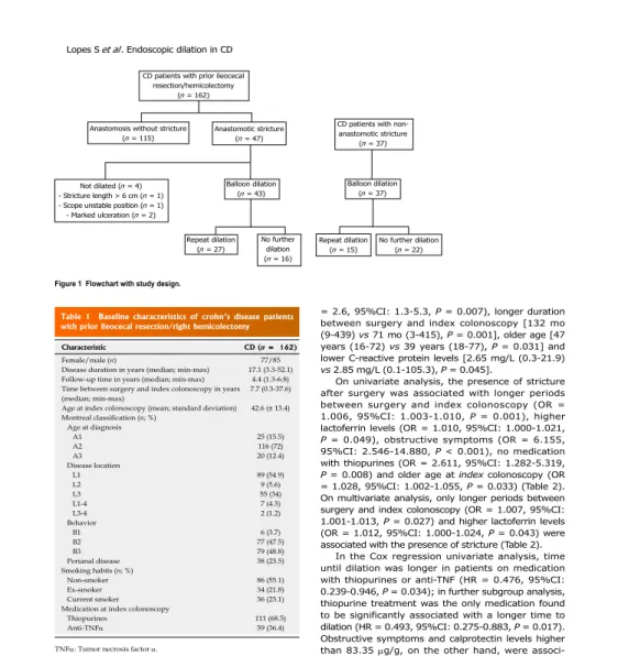
efficacy and safety of this technique. In our group of 178 patients who
underwent colonoscopy, 48 were successfully dilated and recurrence
was diagnosed only after dilation in 22 patients. Fecal markers were
good predictors of endoscopic recurrence and can be used as guidance
to EBD. EDB demonstrated to be an effective and safe alternative to
surgery, with the possibility of being repeated as needed.
In order to monitor response to therapy we evaluated the performance of
computed tomography enterography (CTE) and fecal markers compared
to endoscopy at diagnosis and in the first year after beginning therapy.
In a group of 29 consecutive patients with newly diagnosed CD we
performed endoscopy, CTE and fecal calprotectin (FC) at M0 and M12.
We found a good correlation between the 3 methods at both time
points. In patients with endoscopic remission at one year, CTE findings
of inflammatory activity significantly improved, CTE score decreased
and FC values normalized. A combination of both noninvasive markers
may be used to monitor response to therapy, with the advantage of
evaluating transmural healing.
The last aim of this project was to evaluate the impact of VCE in CD
management, since its role in treatment guidance is not completely
defined. In a group of 83 patients with long-term disease in clinical
remission, VCE identified unknown upper tract involvement in 49
patients, and its findings translated in treatment changes in 40%
of patients. These results highlight the importance of small bowel
mucosal imaging in the management of CD.
In conclusion, despite the promising results obtained with noninvasive
methods in disease monitoring, being easy to repeat and gaining
patient general acceptance, their definitive place in patients’ evaluation
26
still needs to be defined. Direct mucosal observation, either by
colonoscopy or VCE, still has a major role in IBD management. Cross
sectional imaging modalities and fecal markers may and should be
incorporated into patients’ algorithm management, selecting patients to
endoscopy. Validated scores are needed in CTE, with special attention
to mesenteric findings of inflammation, and in endoscopy, dissemination
of the Modified Rutgeerts score should be implemented. EBD should be
performed not only to induce symptoms resolution in stenotic patients
but also to gain access to otherwise inaccessible bowel segments,
allowing therapeutic adjustments as needed.
27
Resumo
A Doença Inflamatória Intestinal (DII) é uma doença crónica, inflamatória
e recidivante do tubo digestivo, englobando a Doença de Crohn (DC) e
a Colite Ulcerosa (CU). A associação entre o aumento da sua incidência
e prevalência e a industrialização e ocidentalização das populações tem
despertado muito interesse para um potencial papel de agentes
ambi-entais na sua etiopatogénese. Assume-se que a DII se desenvolve num
indivíduo geneticamente predisposto que reage de forma inadequada
ao seu microbioma após exposição a um estímulo externo. Neste
contexto, nos últimos anos o viroma humano tem despertado muita
atenção da comunidade científica. Alguns estudos demonstraram uma
interação entre vírus eucarióticos e genes de susceptibilidade para a DII,
sugerindo um papel para o viroma na patogénese da DII. O
reconhe-cimento de que a terapêutica imunossupressora influencia a população
vírica entérica, coloca a hipótese de alguns vírus específicos poderem
aproveitar o estado imunossuprimido do hospedeiro para promoverem
o desenvolvimento de doença. Apesar de evidência cientifica
implicando a disbiose viral na patogénese da DII, ainda não foi possível
confirmar estes resultados em coortes de validação. Os vírus da família
Herpesviridae humana são vírus de ADN englobando o CMV, EBV e
HSSV- 6. Estes vírus têm em comum a elevada prevalência na idade
adulta (> 95%) e persistirem num estado de latência ao longo da vida
do hospedeiro. Durante períodos de imunossupressão a sua reativação
pode ocorrer, com consequente exacerbação de doenças crónicas. Os
dados existentes da transplantação demonstram que em estados de
imunossupressão intensa se verifica a reativação vírica com implicações
clínicas, sugerindo que estes efeitos também possam ocorrer em
doentes com DII. Até ao momento, continua por inequivocamente
demonstrado, se as alterações de número e variabilidade de vírus na
DII são biológica e clinicamente relevantes em termos de fenótipo,
comportamento e resposta da doença ao tratamento.
As várias modalidades endoscópicas disponíveis garantem o acesso á
mucosa doente e saudável, e permitem a observação directa e
histológica das manifestações inflamatórias no tubo digestivo. A
endos-copia para além de contribuir para o esclarecimento da patogénese da
28
doença tem também um papel na sua monitorização e na avaliação da
resposta ao tratamento.
Os conceitos de cicatrização da mucosa, cicatrização transmural e dano
intestinal (“bowel damage”), introduzidos recentemente na abordagem
da DII, modificaram os objetivos terapêuticos. Na actualidade o seguimento
ideal destes doentes implica a monitorização regular da carga
inflamatória de forma a evitar a irreversibilidade das lesões. Como os
sintomas e os biomarcadores séricos demonstraram ser imprecisos na
avaliação da atividade da doença, e pela invasividade que a endoscopia
representa, foi explorada a utilização de outros métodos não-invasivos
alternativos. Os marcadores fecais, os métodos de imagem radiológicos
e a vídeo cápsula endoscópica (VCE) têm vindo a ganhar interesse na
monitorização da doença.
Apesar dos resultados encorajadores da acuidade destes métodos
na avaliação da actividade da doença, a colonoscopia continua a ser
a referência para comparação e validação dos restantes métodos. A
endoscopia, além da determinação da gravidade e extensão das
lesões, permite a realização de técnicas terapêuticas, como a dilatação
endoscópica com balão (DEB). Esta técnica para além da resolução
sintomática, permitindo a preservação do segmento intestinal, possibilita
o diagnóstico da recorrência da doença no pós-operatório.
Com esta Tese, procuramos estudar como alguns métodos endoscópicos,
os marcadores fecais e a imagiologia podem contribuir para o
acompanhamento clínico dos doentes com DII em contextos clínicos
específicos.
No primeiro estudo, investigamos a prevalência e o papel de alguns
membros da família Herpesviridae na patogénese e na evolução da
doença. Realizamos colonoscopia a 95 doentes com DII (DC e CU) e 50
indivíduos saudáveis (HC). Os vírus EBV e CMV foram mais prevalentes
na população com DII e em áreas de mucosa com atividade endoscópica.
A prevalência do HHV6 foi semelhante entre doentes e HC. A carga vírica
média do CMV foi mais elevada na mucosa ulcerada dos doentes com
29
CU, enquanto a carga vírica média do EBV foi maior na mucosa ulcerada
dos doentes com DC. Não encontramos qualquer correlação entres os
regimes terapêuticos imunossupressores e a prevalência vírica sérica.
O segundo objetivo desta tese foi avaliar a acuidade dos marcadores
fecais como predictores de recorrência após a cirurgia e comparar dois
scores endoscópicos - o score de Rutgeerts e o score de Rutgeerts
modificado. Neste estudo com 99 doentes, os níveis de calprotectina
fecal e lactoferrina fecal demonstraram ser mais elevados em doentes
com recorrência endoscópica em comparação com doentes em
remissão endoscópica. O score de Rutgeerts modificado demonstrou
ser superior ao score de Rutgeerts na predição de recorrência, com
maior sensibilidade, valor preditivo negativo e acuidade. Foram
estabelecidos valores de cut-off para os dois marcadores fecais.
O terceiro e o quarto objetivos foram avaliar a acuidade dos marcadores
fecais na predição de recorrência em doentes com estenose assintomática
da anastomose ileocólica selecionando os doentes para DEB, e avaliar
a eficácia e segurança desta técnica. No nosso grupo de 178 doentes
submetidos a colonoscopia, 48 foram dilatados com sucesso e foi feito o
diagnóstico de recorrência apenas após a dilatação em 22 pacientes. Os
marcadores fecais revelaram ser preditores de recorrência endoscópica
podendo ser utilizados como indicadores da necessidade de DEB. A
DEB demonstrou ser uma alternativa eficaz e segura à cirurgia, com a
possibilidade de poder ser repetida se necessário.
A fim de monitorizar a resposta à terapêutica, avaliamos o desempenho
da enterografia por tomografia axial computorizada (TAC) e dos
marcadores fecais em comparação com a endoscopia, á data do
diagnóstico e no primeiro ano após o início de tratamento. Num grupo
de 29 doentes consecutivos com DC recém-diagnosticada, foi realizada
endoscopia, enterografia por TAC e doseamento de calprotectina fecal
ao mês 0 e ao mês 12. Os resultados demonstraram existir uma boa
correlação entre os 3 métodos ao diagnóstico e ao primeiro ano de
seguimento após o início do tratamento. Nos doentes em remissão
endoscópica ao ano, os sinais de atividade inflamatória na enterografia
30
por TAC melhoraram significativamente, o score da enterografia por
TAC diminuiu e os valores de calprotectina fecal normalizaram. A
combinação dos dois marcadores não invasivos poderá ser utilizada
para monitorizar a resposta á terapêutica, com a vantagem de avaliar
a cicatrização transmural.
O último objetivo deste projeto foi avaliar o impacto da VCE no
seguimento da DC, uma vez que o seu papel no estabelecimento da
estratégia terapêutica não está totalmente definido. Em 83 doentes com
doença de longa duração em remissão clínica, a VCE diagnosticou o
envolvimento do trato digestivo superior em 49 doentes, condicionando
alterações terapêuticas em 40% dos doentes. Estes resultados salientam
a importância da observação da mucosa do intestino delgado na
abordagem da DC.
Em conclusão, apesar dos resultados promissores dos métodos não
invasivos na abordagem da DII, com facilidade de repetição e maior
aceitação pelos doentes, o seu posicionamento no algoritmo de
avaliação precisa ser definido. A observação direta da mucosa, seja
por colonoscopia ou VCE, ainda possui um papel fundamental na
abordagem da DII. Os métodos de imagem radiológicos e os
marcadores fecais poderão e deverão ser incorporados no algoritmo de
seguimento, selecionando os doentes para endoscopia. É necessário
o desenvolvimento e validação de scores na enterografia por TAC,
incorporando os achados mesentéricos de inflamação. Na endoscopia,
a disseminação do score de Rutgeerts modificado deve ser fomentada.
A DEB deve ser realizada não só para resolução sintomática em
doentes com estenoses, mas também para permitir a avaliação de
segmentos intestinais de outra forma inacessíveis, possibilitando
modificações terapêuticas se necessário.
33
37
45
45
52
57
59
63
65
69
71
79
89
Balloon Dilation in Crohn´S Disease Patients with Anastomotic
Strictures.
99
109
137
139
147
159
163
31
Table of Contents
Rational and Introduction
Metabolomics
Imaging Modalities
Endoscopy
Enterography
The Lémann Score
(The Crohn´s Disease Digestive Damage Score)
Fecal Biomarkers
Fecal Biomarkers in the Postoperative Setting
Aims
Results - Publications
Looking into Enteric Virome in Patients with IBD: Defining Guilty
or Innocence?
Correlation Between Calprotectin and Modified Rutgeerts Score.
Fecal Markers Levels as Predictors of the Need for Endoscopic
Balloon Dilation in Crohn´S Disease Patients with Anastomotic
Strictures.
Endoscopic Balloon Dilation of Crohn’s Disease Strictures: Safety,
Efficacy and Clinical Impact.
Monitoring Crohn´s Disease Activity: Endoscopy, Fecal Markers
and CT Enterography.
Transmural Healing in Crohn´s Disease: Beyond Mural Findings.
Capsule Enteroscopy is Useful for the Therapeutic Management
of Crohn´s Disease.
Discussion
Conclusions and Future Research
References
33
37
45
45
52
57
59
63
65
69
71
79
89
Balloon Dilation in Crohn´S Disease Patients with Anastomotic
Strictures.
99
109
137
139
147
159
163
33
Rational
34
35
(1)
Crohn BB et al. The Mount Sinai
Journal of Medicine. 2000.
Inflammatory bowel disease (IBD) is a chronic, relapsing, inflammatory
condition of the gastrointestinal tract, encompassing two different
diseases: Crohn´s disease (CD) and Ulcerative colitis (UC). CD was
described for the first time in 1932 at the American Medical Association
Meeting. In this session, a paper by Crohn, Ginzburg and Oppenheimer
was presented, entitled “Regional Enteritis, a Pathological and Clinical
Entity”
(1). A disease description was made, based on a study of 14 cases
up to 1932. The first report of UC goes back to the second half of
nineteen century when Samuel Wilks reported a case of a young woman
who died of severe bloody diarrhea. Initially reported as sporadic
diseases, since the middle of the 20
thcentury, the incidence of both
diseases has increased in the western world. Currently it is estimated
that the prevalence of IBD in western countries is up to 0,5% of the
general population. Defined as a chronic, incurable disease with a low
mortality rate diagnosed predominantly at a young age, the pool of
newly diagnosed patients every year exponentially increases the number
of prevalent cases.
Since its first report, much as evolved in terms of diagnosis and treatment
in IBD.
The evidence that incidence and prevalence of IBD correlates with
industrialization and westernalization of populations has driven much
attention to the environmental triggers of disease. IBD is believed to
manifest in a genetically predisposed individual who reacts
inappro-priately to the gut microbiome after exposure to an external trigger. A
great deal of investment and research has focused on how immune
dysfunction induces and maintains a chronic inflammatory state in IBD,
resulting in an improved understanding of its immunopathogenesis.
Despite that, a full comprehension of the cause and mechanisms of
IBD is still lacking. The immune pathogenic mechanisms are only one
of the arms of the equation, interacting with genetic, microbiome and
environmental factors.
At the present time, recognition of immunological mediators of the
disease has led to the development of more directed therapies but data
are still missing in terms of unravel the causal agent(s). It is widely
accepted that no single component of this equation can alone trigger
Rational and Introduction
36
the disease and a combined disruption of all the elements controlling
intestinal homeostasis is necessary to disease initiation and mediation.
The follow-up of IBD patients is challenging, since it implies an
individualized strategy, which needs to be adjusted to the disease and
the patient. In the last decades, the way we approach and manage
this condition has evolved tremendously. The development of new
and more powerful drugs, capable of reverting structural damage, the
increasing use of more accurate biological markers of inflammation to
early detect and predict disease activity and the availability of more
accurate imaging technics, have changed the goal of the follow-up.
Actually, the standard of care implies a close monitoring of the
inflammatory burden of the disease in order to avoid disability. This
includes taking into account different parameters such as symptoms,
biological markers, endoscopy and other imaging modalities, and
adjust therapy according.
37
Metabolomics
The increasing progress in recent years in metabolomics as shifted
again the attention to disease pathogenesis, mainly to the importance of
the microbiome. There are some pathological features of CD suggestive
of an infectious etiology, such as aphthous ulcers of the mucosa,
mural abscesses, suppurative fistulas, and macrophage and epithelioid
cell granulomas. The possible implication of an infectious agent in CD
was first postulated by Dalziel in 1913, before the classic description
of CD, when he noted the similarities with Johne’s disease (a diarrheal
disease in animals, caused by Mycobacterium avium paratuberculosis,
with epithelial granulomas). However, no infectious agent has yet
been unequivocally identified as inducer of CD.
It has been well demonstrated that the composition of the luminal and
mucosal bacteria differs in patients with IBD compared to the general
population, with a decreased prevalence of anti-inflammatory bacteria.
The increased incidence of IBD worldwide and especially in countries
with previous low incidences, and the increased risk for developing IBD
among people who migrate from low to high incidence zones appear
to support the notion of environment-driven epigenetic modifications.
It is known that geography and ethnicity, as well as diet, antibiotic
consumption early in life, and certain lifestyle (like smoking and oral
contraceptive use) influences microbiota composition. This may explain
the recent changes in the incidence of microbiota-related disorders, in
which IBD is included. The identification of several gene loci associated
with the development of IBD has highlighted the existence of a
gene-microbe-environment interaction in IBD pathogenesis. Key
IBD risk genes include genes involved in the pathway of sensing and
response to microbiota. Whether tissue damage results from an abnormal
immune response to a normal microbiota or from a normal immune
response to an abnormal microbiota remains to be definitely and
unequivocally answered. Possibly more important than the type and
quantity of bacteria present in the gut and altered in IBD, is the functional
consequence of that alteration, interfering with pathways involved in
38
Much less investigated has been the role of virome in IBD. In fact,
virus comprise the most abundant biological entities within the gut,
greatly outnumbering bacteria. It is known that the viral component
of the microbiome has the potential to influence host physiology
and homeostasis. Enteric virome consists of bacteriophages and
eukaryotic viruses
(2-7). Like the bacterial microbiome, the human
gut virome is characterized by significative changes during the first
2 years of life. In adult life bacteriophages decrease in richness and
diversity, and their composition shifts significantly; these changes
are associated with increased richness of eukaryotic viruses, which is
believed to depend strictly on environmental influences
(8).
Animal studies have proved an interaction between eukaryotic
viruses and IBD risk genes, indicating that members of the virome
may contribute to IBD
(9-12). In a study of germ-free or antibiotic treated
mice infected with murine norovirus (MNV), Kernbauer et al
(13)demonstrated that the beneficial function of commensal bacteria
in the gut may be replaced by virus. This study supports the
hypothesis that similarly to bacteria, eukaryotic viruses have the
capacity to support intestinal homeostasis and shape mucosal
immunity. Most of the research on the potential role of virus in IBD
has focused on bacteriohages due to their influence on bacterial
populations. The interaction of persistent virus infection with commensal
microbiome and immune system is believed to be responsible for a
particular immunophenotype. Virus-susceptibility gene interaction
could explain the clinical heterogeneity and the great variability of
response to specific treatments options between different patients.
The role of gut virome is IBD pathogenesis is just beginning to be
understood, but there is evidence of being altered in patients with
IBD with specific changes assessed between UC and CD. Normal et
al demonstrated that patients with IBD have an increased number
and richness of phage virus, with specific populations identified
in each disease type, associated with bacterial dysbiosis
(14). It has
also been demonstrated that viral population may be influenced by
immunosuppressive therapy, with some specific viruses taking
advantage of the immunosuppressive status of the host
(15-18).
Despite the amount of evidence trying to implicate bacterial and viral
dysbiosis in the pathogenesis of IBD it has been difficult to confirm
(2)
Breitbart M et al. Journal of
Bacterio-logy. 2003;185(20):6220-3.
(3)
Finkbeiner SR et al. Virology Journal.
2008;5:117.
(4)
Minot S et al. Proceedings of the
National Academy of Sciences of the
United States of America. 2013.
(5)
Minot S et al. Proceedings of the
National Academy of Sciences of the
United States of America. 2012.
(6)
Minot S et al. Genome Research.
2011.
(7)
Reyes A et al. Nature. 2010.
(8)Lim ES et al. Nature Medicine. 2015.
(9)Basic M et al. Inflammatory Bowel
Diseases. 2014.
(10)
Cadwell K et al. Cell. 2010.
(11)Irving PM et al. Nature Clinical
Practice Gastroenterology &
Hepato-logy. 2008.
(12)
Sun L et al. Current Opinion in
Gastroenterology. 2011.
(13)
Kernbauer E et al. Nature. 2014.
(14)Norman JM et al. Cell. 2015.
(15)Perez-Brocal V et al. Inflammatory
Bowel Diseases. 2015.
(16)
Madsen CD et al. HIV Clinical Trials.
2002.
(17)
Thom K et al. Journal of Medical
Virology. 2007.
(18)
McElvania TeKippe E et al. PloS
One. 2012.
39
results across validation cohorts. Other potential interest of virus in
IBD would be their role as biomarkers of disease, as several works have
demonstrated an increased richness and biodiversity of phage population
compared to controls
(14,19,20).
It has been shown that latent viral infections, like herpesvirus
infec-tions, possess a role in exacerbating chronic inflammatory diseases
(21-24).
The Human Herpesviridae family is a DNA virus family encompassing
cytomegalovirus (CMV), Epstein-Barr virus (EBV) and Human Herpes
simplex virus (HSSV)-6. They have in common being highly prevalent in
adulthood (>95%) and inducing lifelong latency in the host, with
reac-tivation during periods of immunosuppression. There is a large number
of studies trying to implicate CMV and EBV in IBD pathogenesis and/
or disease course. Although CMV colitis in immunocompetent patients
is extremely rare, its association with IBD has been reported for more
than half a century
(25). The exact prevalence of CMV in IBD population
is not entirely known. Several studies have shown a higher prevalence
of CMV in IBD patients, that have not been replicated by others. This
may be explained by selection bias and the methods used to diagnose
the infection. While latent or subclinical infection is diagnosed by the
presence of a positive CMV IgG, the diagnosis of CMV colitis should not
be based on the presence of CMV IgM, but rather on viremia detection
and presence of the virus in the colon.
There are 3 diagnostic methods to detect CMV in colonic mucosa:
haematoxylin and eosin staining, immunohistochemistry and polymerase
chain reaction (PCR). PCR is the most sensitive and specific of the 3
methods in diagnosing CMV disease, being able to determine viral load.
Qualitative PCR can detect the presence of CMV DNA in colonic mucosa,
but that does not discriminate between infection and disease. On the other
hand, the quantitative method seems more attractive, allowing for the
quantification of viral DNA. That has proved to be useful on the context
of EBV and CMV infection in the post-transplant population. Nevertheless,
in IBD no cut-off has until now been defined, although some papers
state that a CMV viral load > 250 copies/mg of tissue in patients with
active UC is predictive of nonresponse to steroids, infliximab and
cyclosporine
(26).
(19)
Wagner J et al. Inflammatory Bowel
Diseases. 2013.
(20)
Perez-Brocal V et al. Clinical and
Translational Gastroenterology. 2013.
(21)
Barton ES et al. Nature. 2007.
(22)White DW et al. Blood. 2010.
(23)Yager EJ et al. Viral Immunology.
2009.
(24)
Canny SP et al. Journal of Virology.
2014.
(25)
Powell RD et al. The American
Journal of Medicine. 1961.
(26)
Roblin X et al. The American Journal
of Gastroenterology. 2011.
40
It is also assumed that the difference in the immunological milieu
between UC and CD is responsible for the lower rate of reactivation of
CMV in CD compared to UC
(27-29).
Another issue that remains to be clarified is the role of CMV in disease
severity. Some reports and studies suggest that colonic superimposed
CMV infection is associated with an increased rate of complications,
namely toxic megacolon and need for surgery
(30-32). On the other hand,
there are more recent papers suggesting that the viral load does not
impact on clinical outcome
(26, 33).
Although the appealing nature of this association, it has been
difficult to prove if CMV is really a causative factor in the pathogenesis
of severe colitis or whether it simply represents a surrogate marker of
a severe or steroid-refractory disease.
At the present time there are several questions concerning CMV without
a definitive answer: whether it is more prevalent in IBD population
than in general population; if there is a significative difference in
prevalence between CD and UC; whether viral load is higher in ulcerated
compared to normal mucosa; if the presence of CMV DNA in the colonic
mucosa determines the severity of the disease and has therapeutical
implications.
Regarding EBV, the major concern is its association in immunocompromised
patients with several malignancies namely Hodgkin´s disease, T/NK-cell
lymphoma, Burkitt’s lymphoma and gastric carcinoma. The implication
of EBV on this malignancy risk translates from the transplant setting and
its association with post-transplant lymphoproliferative disease (PTLD)
(34).
In IBD, information regarding the role of EBV in disease course and
prognosis is scant, due to the lack of a consensual and proved more
specific method to define disease vs infection, and a greater focus
on CMV infection. Increasing interest in EBV has derived from the
published evidence of the increased risk of lymphoma in IBD, especially
among young male patients under thiopurines. In immunosuppressed
patients, this increased risk seems to be related to reactivation of a
latent or a primary EBV infection
(35). There is the notion that EBV-positive
cells can be found in the colonic mucosa of more than half of IBD
patients, predominantly in the inflamed areas
(36-38). The reason for this
high percentage is the increased number of infiltrating B-lymphocytes
(27)
Nakase H et al. Digestive Diseases
and Sciences. 2010.
(28)
Knosel T et al. Pathology, Research
and Practice. 2009.
(29)
Takahashi Y et al. Diseases of the
Colon and Rectum. 2004.
(30)
Domenech E et al. Inflammatory
Bowel Diseases. 2008.
(31)
Kojima T et al. Scandinavian Journal
of Gastroenterology. 2006.
(32)
Kambham N et al. The American
Journal of Surgical Pathology. 2004.
(33)
Leveque N et al. Journal of Medical
Virology. 2010.
(34)
Green M et al. Am J Transplant.
2013.
(35)
Beaugerie L et al. Dig Dis. 2009.
(36)
Wakefield AJ et al. Journal of Medical
Virology. 1992.
(37)
Ryan JL et al. Digestive Diseases
and Sciences. 2012.
(38)
Spieker T et al. The American Journal
of Pathology. 2000.
41
in the mucosa due to inflammation, and the increased EBV replication
rate as a result of immunosuppression
(39).
Several authors have studied the prevalence of EBV in IBD patients. In
a work by Knosel T et al
(28), EBV DNA detected by PCR was the second
most prevalent agent in patients with CD, and not detected in the
control group. This higher prevalence in CD patients compared to
healthy individuals in a German population, was also reported by
Spieker et al
(38)and Ruther et al
(40). This evidence was not reproduced
in a Belgian and French population
(41)and the clinical relevance of
EBV-positivity in colon cells remains unclear, either relatively to
disease course and malignant complications.
A recent prospective study by Ciccocioppo et al
(42)evaluated the
prevalence of CMV and EBV in both blood and colonic mucosa, by
quantitative PCR and immunohistochemistry. The authors concluded
that quantitative real-time PCR was the best method to define
disease. They found a higher prevalence of both CMV and EBV in
refractory patients compared to non-refractory and the control group,
a higher median mucosa viral load in refractory IBD, and a significantly
higher viral DNA load in disease vs non-disease mucosa. Mucosal
viral load positively correlated to the degree of endoscopic activity,
both for EBV and CMV. No difference was found between median
viral DNA levels of non-diseased mucosa and those of non-refractory
IBD patients and controls.
A relevant finding was the determination of a cut-off value, above
which viral related disease was considered, suggesting a closer
follow-up and early recognition of patients at risk. Regarding the
impact of therapy on viral prevalence, the authors found systemic steroid
use a significant risk factor for EBV and CMV colitis, and no correlation
with de use of immunosupressants. By contrast, our group found a
higher prevalence of EBV in patients under infliximab, irrespective of
associated use of other immunosupressors
(43). On the other hand we
could not find any correlation between the number of copies in serum
and therapeutic regimen or C-reactive protein (CRP) level. Despite this
evidence, it remains to be established if and which IBD patients should
be tested and followed in the long-term. In the post transplant context
serial monitoring of EBV viral load is advocated, and preventive
treatment of the infection can reduce the morbidity and mortality of
(39)
Kumar S et al. The American Journal
of Surgical Pathology. 2000.
(40)
Ruther U et al.
Hepato-Gastroen-terology. 1998.
(41)
Van Kruiningen HJ et al. APMIS:
Acta Pathologica, Microbiologica, et
Immunologica Scandinavica. 2007.
(42)
Ciccocioppo R et al. World Journal
of Gastroenterology. 2015.
(43)
Magro F et al. Inflammatory Bowel
Diseases. 2013.
42
EBV-related lymphoproliferative disorders in PTLD
(44). If this approach
is to be implemented in IBD needs further evidence.
Another HHV related to gastrointestinal symptoms in immunocompromised
patients is Human Herpes Virus-6. The seroprevalence in adulthood is
estimated to exceed 95%. In IBD, data on HHV-6 is scarse. The
importance of HHV-6 reactivation in immunocompromised patients is
being recognized in the post-transplant population, being responsible
for severe morbidity and sometimes graft lost. It has been identified as
a possible trigger to other herpesvirus infections, especially CMV
(45-48),
being suggested that immunosuppressed patients harboring HHV-6
should be more closed monitored. As HHV-6 DNA has been identified
in the mucosa of post-transplant patients with gastrointestinal
symptoms, it was hypothesized that in IBD immunosuppressed
patients it could be a trigger of relapse. Some studies have shown a high
prevalence of HHV-6 DNA in the colon of both UC and CD patients
(36, 49). By contrast, archival tissue examination of CD patients using PCR
technique found HHV-6 positivity in only 3.6% (2/56) of samples
(28).
A prospective study by Sipponem T et al
(50)evaluated the prevalence
of CMV and HHV-6 antigens in the mucosa in endoscopically active
and inactive CD and UC patients and in a non-IBD control population.
CMV and HHV-6 antigenemia were also evaluated in IBD patients. In
this study, the authors found a higher positivity for HHV-6 in both UC
(45%) and CD (44%) patients. This number was comparable with that
seen in solid organ transplant recipients
(51). Both virus were shown
to be more prevalent in endoscopically active mucosa compared to
endoscopically inactive segments, with a significant difference in
HHV-6 antigen expression in more severe endoscopic disease (p=.042).
One interesting finding was the simultaneous expression of both virus
in patients under more than one immunosupressive medication and
with endoscopically more severe disease (at least moderate disease).
In IBD patients, clinical significance of this coexistence is unknown,
but in transplant recipients the presence of HHV-6 in tissue has the
potential to trigger other herpesvirus infections, especially CMV
(45-48, 52).
Another feature that remains to be elucidated is the role of anti-TNF
alpha agents in patients testing positive for these viruses, with some
conflicting results. In this series, the CMV expression in the mucosa
was more intense in patients under biologicals or cyclosporine, but the
same was not proved for HHV-6.
(44)
Comoli P et al. American Journal
of Transplantation: Official Journal of
the American Society of
Transplan-tation and the American Society of
Transplant Surgeons. 2007.
(45)
Halme L et al. Clinical Infectious
Diseases: an Official Publication of
the Infectious Diseases Society of
America. 2008.
(46)
Halme L, et al. APMIS: Acta
Patho-logica, MicrobioPatho-logica, et
Immuno-logica Scandinavica. 2008.
(47)
Mendez JC et al. The Journal of
In-fectious Diseases. 2001.
(48)
DesJardin JA et al. The Journal of
Infectious Diseases. 1998.
(49)
Sura R et al. APMIS: Acta Pathologica,
Microbiologica, et Immunologica
Scan-dinavica. 2010.
(50)
Sipponen et al. Scandinavian Journal
of Gastroenterology. 2011.
(51)
Razonable RR et al. American
Jour-nal of Transplantation: Official JourJour-nal
of the American Society of
Transplan-tation and the American Society of
Transplant Surgeons. 2009.
(52)
Lautenschlager I et al. Clinical
In-fectious Diseases: an Official
Publica-tion of the Infectious Diseases Society
of America. 1998.
43
Until the present time, it remains to be proved with no margin of doubt
if the changes observed in number and richness of virus in IBD is
biologically and clinical relevant in terms of disease phenotype, behavior
and response to therapy.
45
Imaging Modalities
Endoscopy
One of major difficulties in IBD is establishing the correct diagnosis.
Being a chronic, lifelong condition, needing in the majority of patients
of prolonged and intensive immunosuppression, an unequivocal
diagnosis is mandatory. Despite typical, symptoms are not
pathognomonic of either disease (CD or UC), and objective data are
required to make the diagnosis. Ileocolonoscopy is the gold standard,
allowing to rule out other conditions that may mimic IBD symptoms
and to obtain tissue samples for histological assessment. It is accurate
in differentiating CD from UC and has a role in evaluating disease
extent and severity, serving as a prognostic tool.
Endoscopic scores developed to standardize reports of mucosal
lesions in IBD, both in clinical trials and clinical practice. In CD, the first
endoscopic score developed and validated was de Crohn´s disease
endoscopic index of severity (CDEIS)
(53). This score proved to be
complex and cumbersome, with limited application in clinical practice.
In 2004 a simplified version of CDEIS, the simple endoscopic score for
Crohn´s disease (SES-CD)
(54)was proposed and validated. This score
ranges from 0 to 56, and mucosal healing is defined as a SES-CD<3.
In UC, the most commonly used endoscopic scores are the Mayo
Endoscopic subscore
(55)and the Ulcerative Colitis Endoscopic Index
of Severity (UCEIS)
(56), a more recent and validated score. Endoscopic
remission is defined as a Mayo Endoscopic subscore of 0 or 1 but for
UCEIS no cut-off has yet been defined. UCEIS has proved to be useful
in predicting medium and long term outcomes in UC patients.
If initially mainly limited to establishing the diagnosis, nowadays the
role of endoscopy has evolved tremendously, not only due to technical
developments but also to a change in the treatment paradigm of IBD.
The severity of endoscopic findings and the extent of the disease are
known prognostic factors predicting response to therapy and need for
(53)Mary JY et al. Gut. 1989.
(54)
Daperno M et al. Gastrointestinal
Endoscopy. 2004.
(55)
Schroeder KW et al. The New
England Journal of Medicine. 1987.
46
surgery. During disease flares, endoscopic evaluation of the mucosa
allows to exclude other causes of exacerbation, namely infections.
If traditionally the clinical effectiveness of therapy was evaluated by
clinical indices, nowadays it is known that these indices have clear
limitations and do not correlate with mucosal findings or clinical
outcomes in the long time. In recent years, with the advent of new
molecules capable of inducing mucosal healing (MH) (assessed by
endoscopy), this became the new treatment goal both for CD and UC.
In several clinical trials, the achievement of mucosal healing has been
associated with improved outcomes, such as lower rate of relapse,
steroid free remission, fewer hospitalizations and surgeries. There is
also some evidence in UC that healed mucosa may be associated with
a decrease rate of dysplasia and colon adenocarcinoma. Endoscopy
has become an important evaluation tool of response to therapy and
before any treatment adjustment. The Selecting Therapeutics Targets
in Inflammatory Bowel Disease (STRIDE) program
(57)developed
recommendations for potential treatment targets to be used in clinical
practice. Both in CD and UC, endoscopic remission was agreed as a target,
and therapy adjusted if this goal was not achieved in the reevaluation
endoscopy.
The performance of endoscopy in IBD goes beyond being merely a
diagnostic or prognostic tool because it allows therapeutic procedures.
One major complication in CD is stricture formation, still the main
reason for surgery (either strictureplasty or bowel resection). Small
bowel strictures occur in approximately 25% of Crohn’s disease
patients, and colonic strictures in about 10%. Up to 50% of CD patients
undergo surgical resection within the first 10 years of diagnosis
(58).
After surgery, symptomatic recurrence is as high as 38% at one year,
with need to repeat surgery that can lead to short bowel syndrome.
Endoscopic balloon dilation (EBD) has become an accepted alternative
to surgery, with overall favorable results in terms of safety, efficacy, and
patient satisfaction. Being a bowel conserving procedure with high
technical (73%-100%) and clinical success rate (64%-70%), and a low
rate of adverse events (2%-6.4%), its major drawback is the high rate
of recurrence. Re-dilation may be required in up to 20% and 50% by
1 and 5 years, respectively(59-61) with the same high success rate,
supporting the evidence that repeated dilations do not reduce the
procedural efficacy
(62, 63).
(57)
Peyrin-Biroulet L et al. The
Ameri-can Journal of Gastroenterology. 2015.
(58)
Bernell O et al. Annals of Surgery.
2000.
(59)
Morar PS et al. Alimentary
Pharma-cology & Therapeutics. 2015.
(60)
Morini S et al. Digestive and Liver
Disease: Official Journal of the Italian
Society of Gastroenterology and the
Italian Association for the Study of
the Liver. 2003.
(61)
Thomas-Gibson S et al. European
Journal of Gastroenterology &
Hepa-tology. 2003.
(62)
Chen M et al. Inflammatory Bowel
Diseases. 2014.
(63)