UNIVERSIDADE DE LISBOA FACULDADE DE CIÊNCIAS
DEPARTAMENTO DE BIOLOGIA VEGETAL
Role of IL-4 in the lineage plasticity of human
CD4 single positive thymocytes
Andreia Sofia Ramalho dos Santos
Mestrado em Biologia Molecular e Genética
Dissertação orientada por:
Dra Helena Nunes-Cabaço (PhD, IMM)
Dra Margarida Telhada (PhD, FCUL)
CONTENTS ACKNOWLEDGEMENTS ... i ABSTRACT ...ii RESUMO ... iv ABBREVIATIONS ... vii 1 INTRODUCTION ... 1
1.1 Thymus – the primary organ for T cell generation ... 1
1.2 Thymic structure ... 1
1.3 T cell differentiation ... 2
1.3.1 Early T cell development ... 2
1.3.2 Positive and Negative selection... 3
1.3.3 CD4/CD8 lineage specification ... 4
1.4 Human T cell differentiation ... 5
1.5 Transcription factors regulating lineage choice ... 6
1.5.1 Maintenance of lineage commitment ... 7
1.6 CD4-helper T cells ... 9
1.7 CD8-cytotoxic T cells ... 9
1.8 Role of IL-4 in immunity ... 10
1.8.1 Role of IL-4 in T cell development... 11
2 OBJECTIVES ... 12
3 METHODOLOGY ... 13
4 RESULTS AND DISCUSSION ... 17
4.1 IL-4 induces upregulation of CD8αβ in CD4SP thymocytes ... 17
4.2 IL-13 is not capable of upregulating CD8 ... 19
4.3 Role of proliferation in IL-4-induced CD8 upregulation on CD4SP thymocytes ... 19
4.4 CD8 upregulation potential decreases with CD4 T cell maturation ... 20
4.5 Co-receptor reversal capacity is lost in the periphery... 21
4.6 CD8 expression is stable upon IL-4 removal ... 22
4.8 IL-4 is mainly present in the medulla of the human thymus ... 24
4.9 IL-4 did not produce an effect on T cell differenctiation in Thymic Organ Cultures (TOCs)... 27
4.10 Future perspectives ... 28
5. CONCLUSION ... 29
6 REFERENCES ... 31
i ACKNOWLEDGEMENTS
Dizem que estamos sozinhos na vida e que só temos nós próprios com quem contar. Eu tenho a sorte de ter comigo pessoas muito importantes que tiveram igual parte em tornar possível a conclusão desta etapa da minha vida e a quem não posso deixar de, pelo menos, tentar agradecer.
Em primeiro lugar gostaria de deixar o meu apreço à Dra. Ana Espada de Sousa pela oportunidade de trabalhar no seu laboratório, por me desafiar a pensar cientificamente e por apoiar este projeto.
Agradeço à Professora Doutora Margarida Telhada por coordenar a tese ao nível da FCUL, pelos conselhos e pela disponibilidade.
À Helena, não há palavras para descrever a imensa admiração não só pela profissional mas também pela pessoa maravilhosa que és. Obrigada por guiares este projeto ensinando-me tanto com uma paciência inquebrável, por me dares espaço para cometer os meus próprios erros e aprender com eles, por confiares no meu trabalho, por seres uma grande mentora e amiga.
Ana Rita, obrigada por me ensinares tudo sobre histologia e por fazeres qualquer pessoa que trabalhe contigo apaixonar-se por ela. Foste não só uma grande professora mas também uma boa amiga que me fez ver sempre que o caminho é para a frente.
Ana Antão e Vânia Passos, vocês foram realmente as minhas companheiras de guerra com quem no terreno pude partilhar momentos bons e maus e acabando por ganhar duas amigas fantásticas que me deram energia e confiança todos os dias. Não posso agradecer o suficiente por isso.
Catarina Chinita, Ana Luísa e Cheila Rocha, os vossos sorrisos e simpatia encheram os dias no laboratório de luz e boa disposição que não deixam ninguém indiferente.
A todos no laboratório sem exceção: Paula Matoso, Adriana Albuquerque, Yumi Tokunaga, Alcinda Melo, Professora Conceição, Russell Foxall, Francisca Matos, Henrique Machado, Robert e Susana Lopes. Fizeram-me sentir verdadeiramente parte desta família. Obrigada a cada um por me ensinarem algo todos os dias e por ajudarem o meu projeto a crescer.
Aos meus amigos, obrigada por estarem sempre presentes esteja a montanha russa a descer ou a subir. As minhas conquistas serão sempre também vossas.
Nada seria possível sem o melhor sistema de apoio incondicional, a minha família. Sei que nunca poderei agradecer-vos o suficiente por acreditarem em mim e por apoiarem as minhas decisões nunca me deixando cair sem me ajudarem a levantar logo de seguida. Vocês são o meu bem mais valioso, a minha maior sorte. Obrigada por me ajudarem a atingir mais um objetivo.
ii ABSTRACT
The thymus is the primary organ for the production of T cells, critical for the generation of adaptive immune responses. T cell progenitors proceed through a series of developmental stages, including selection, lineage commitment and maturation in the thymus, in order to differentiate into functional CD4 helper or CD8 cytotoxic T cells. The decision to commit into either lineage is known to involve T-cell receptor (TCR) binding, cytokine signaling and lineage-specific transcriptions factors, and is believed to be virtually irreversible.
Here, we report a role for IL-4 in the plasticity of CD4 single positive (CD4SP) thymocytes. We showed that IL-4 has the capacity to induce CD8α and CD8β upregulation in CD4SP thymocytes, generating mature double positive (DP) and CD8SP thymocytes, and further investigate the mechanism underlying this co-receptor reversal.
CD8 upregulation in purified CD4SP thymocytes was not dependent on cell proliferation, as a significant proportion of the CD8+ cells generated had not divided in culture. CD8 upregulation was very low in the presence of IL-2, while IL-7 generated some DP but few CD8SP cells. IL-13, a cytokine that shares IL4Rα signaling with IL-4, did not increase CD8 expression in CD4SP thymocytes. Of note, IL-4 did not have an impact on CD4 expression when added to CD8SP thymocytes.
IL-4-induced co-receptor reversal was observed even when CD4SP cells expressed the human maturation and egress markers CD27 and CD45RA, respectively, though to a lesser extent. In agreement, IL-4 induced lower upregulation of CD8 expression in CD4 T cells from cord blood. Notably, this ability was lost in CD4 T cells from adult blood, thus confirming a gradual loss of upregulation capacity from the human thymus towards the periphery.
CD8SP cells generated from CD4SP thymocytes upon IL-4 exposure appear to be bona fide CD8 T cells, as CD8 expression was stable upon IL-4 removal from culture. This was not observed for DP cells, which depended on the presence of IL-4 to maintain CD8 expression. Furthermore, the transcriptional pattern of IL-4-induced CD8SP cells in culture supported the modulation of lineage-specific genes towards CD8 commitment, as revealed by the decrease in the transcriptional levels of CD4 and ThPOK and the increase of CD8.
Moreover, we found that IL-4 was expressed in the medulla of the human thymus, in agreement with a possible physiological role for IL-4 in T cell development.
Collectively our results support a role for IL-4 in human T cell development and differentiation, specifically in driving co-receptor reversal in CD4SP thymocytes, a mechanism unknown until now. Additionally, this system provides a valuable model for the study of the regulation of T cell lineage commitment in the human thymus, and merits further study.
iii Keywords: Human T cell development; Lineage commitment; IL-4; Plasticity of CD4 single positive thymocytes; CD8α and CD8β upregulation.
iv RESUMO
Os progenitores de células T sofrem uma série de transformações durante o seu desenvolvimento incluindo seleção, comprometimento para uma determinada linhagem (CD8 ou CD4) e maturação no timo de modo a poderem diferenciar-se em células T funcionais CD4 auxiliares ou CD8 citotóxicas. A decisão de se diferenciar numa das duas linhagens envolve o recetor de células T (TCR), sinalização por parte de interleucinas (IL) e fatores de transcrição (TF) específicos para cada linhagem, crendo-se, atualmente, ser um processo vitualmente irreversível.
Compreender as bases que fundamentam a decisão de uma célula se diferenciar numa determinada linhagem é de extrema importância para a compreensão dos mecanismos envolvidos no desenvolvimento dos timócitos humanos. Deste modo, este modelo é dos mais debatidos em imunologia apesar de se recorrer maioritariamente ao modelo animal do ratinho que, apesar de ter proporcionado um vasto leque de conhecimentos, nem sempre se aplicam ao modelo humano, raramente estudado.
O modelo de sinalização cinética definido em ratinho para explicar o desenvolvimento dos timócitos aponta para a IL-7 como força motriz na especificação e diferenciação em células CD8, sendo a sua ausência durante o desenvolvimento no timo responsável pela manutenção do fenótipo CD4 definido nos estadios mais precoces de maturação. A IL-4 tinha já sido testada no modelo do ratinho tendo tido fraco desempenho no sentido de conduzir à diferenciação dos timócitos em células T CD8 quando comparada com a IL-7.
A IL-4, membro da família de recetores com cadeia γ, foi originalmente descrita em 1982 como sendo uma citocina pleiotrópica de tipo I produzida maioritariamente por linfócitos T auxiliares do tipo 2 (Th2), basófilos, eosinófilos, mastócitos, células natural killer (NK), células T foliculares (Tfh) e células T γ/δ. Esta citocina tem a função de induzir a polarização do fenótipo Th2 e suprimir a diferenciação das células Th1. Adicionalmente, a IL-4 promove o crescimento e diferenciação dos linfócitos B, controlando a especificidade das alterações de classe de imunoglobulinas tal como o desenvolvimento de células B de memória.
As funções fisiológicas desta citocina são mediadas pelo recetor de IL-4, composto por uma cadeia α de 140kDa e uma cadeia γ partilhada com os recetores das citocinas IL-2, IL-7, IL-9 e IL-15. Este dímero pode ser encontrado em células hematopoiéticas ativadas exclusivamente após ligação da IL-4. Após ligação da IL-4 ao seu recetor é promovida ativação do ativador de transcrição e transdutor de sinal 6 (STAT6), que por sua vez promove a regulação de genes responsáveis pela diferenciação de células T CD4 ativadas em células Th2 e Th9, expressão de GATA binding protein 3 (GATA3), produção de quimiocinas e pelo
v aumento da expressão da imunoglubina IgE responsável pelo desenvolvimento de uma resposta imunológica alérgica.
Neste projeto sugerimos um papel para a IL-4 na manutenção da plasticidade dos timócitos CD4+CD8- (single positive, CD4SP) em humano. Mostramos que esta citocina apresenta a capacidade de induzir a expressão dos co-recetores CD8α e CD8β em timócitos CD4SP, gerando timócitos duplos positivos (DP) e CD8SP, e aprofundamos os mecanismos na base desta reversão de co-receptores.
A indução da expressão de CD8 em timócitos CD4SP purificados não mostrou ser dependente de proliferação das células uma vez que uma significativa porção de células CD8 positivas geradas não se dividiu em cultura. A indução da expressão de CD8 foi muito baixa na presença de IL-2, enquanto que na presença de IL-7 surgiram DP mas muito poucas CD8SP. A IL-13, uma citocina que partilha da sinalização através do recetor α da IL-4, não aumentou a expressão de CD8 em timócitos CD4SP. É de notar que a IL-4 não teve qualquer impacto na expressão de CD4 quando adicionada a culturas de timócitos CD8SP.
A reversão de co-receptor induzida pela IL-4 observou-se mesmo no caso de timócitos CD4SP já com expressão dos marcadores de maturação e de saída do timo CD27 e CD45RA, respetivamente, apesar de apresentarem níveis mais baixos de indução de expressão de CD8 quando comparados com timócitos CD4SP CD27-.
Na periferia, a IL-4 foi capaz de induzir expressão de CD8 em células T CD4 provenientes do cordão umbilical. É de notar que esta capacidade se perde com células T CD4 provenientes de sangue adulto, confirmando assim uma perda gradual desta capacidade de induzir a expressão de CD8 desde o timo para a periferia.
As células CD8SP geradas a partir de timócitos CD4SP parecem ser verdadeiras CD8, uma vez que a expressão deste co-receptor se manteve estável mesmo na ausência de estímulo da IL-4 em cultura. O mesmo não se observou no caso das DPs geradas, as quais parecem depender do estímulo de IL-4 para manter a expressão de CD8.
O padrão transcripcional das células CD8SP induzidas pela IL-4 em cultura apoiam uma modulação dos genes responsáveis pela especificação da linhagem e comprometimento das células T CD8, revelado pela diminuição dos níveis transcripcionais de CD4 e ThPOK e pelo aumento dos níveis de CD8.
Por fim, encontrámos expressão de IL-4 na medula do timo humano, apontando, assim, para um possível papel fisiológico desta citocina no desenvolvimento das células T humanas.
Em estudos futuros pretendemos descobrir qual a via envolvida na sinalização de IL-4 responsável pela indução da expressão de CD8 em timócitos CD4SP usando inibidores das várias vias usadas pela IL-4. Deste modo poderemos identificar a via exata usada pela IL-4 para induzir a reversão de co-recetor de CD4 para CD8 no decurso do desenvolvimento das células T no timo humano.
vi De igual modo, em estudos adicionais pretendemos estudar a capacidade funcional das células T CD8 geradas no sentido de determinar se possuem ou não a característica função citotóxica, possivelmente através da medição de fatores citolíticos tais como Granzima-B, interferão γ (IFN-γ) e Perforina. Outros estudos funcionais podem ser realizados usando linhas de células alvo para testar o poder citotóxico das células T CD8 geradas.
Uma vez que um circuito epigenético estabelece a identidade das linhagens auxiliar vs citotóxica, será importante compreender quais os mecanismos epigenéticos a atuar durante a diferenciação das células T no timo, tal como quais os mecanismos mediados pela cromatina envolvidos no comprometimento CD4/CD8. Determinação da acetilação/metilação do DNA e das histonas apresenta-se como um possível método a usar neste sentido.
O estudo de culturas com pedaços de timo (TOCs) permitirá revelar um possível papel fisiológico da IL-4 durante a diferenciação dos timócitos, visto que os pedaços de tecido tímico mantêm as células o mais próximo possível do seu ambiente natural com todos os mecanismos envolventes intrínsecos, difícil de replicar usando células purificadas.
Dada a importância da utilização do modelo animal, assumimos a relevância de testar se uma semelhante indução de CD8 poderá ocorrer em timócitos de murganho.
Em suma os nossos resultados revelam um papel da IL-4 no desenvolvimento e diferenciação das células T humanas, em especial na condução da reversão de co-recetor e manutenção de um certo nível de plasticidade das células T CD4 do timo humano, algo desconhecido até ao momento. Adicionalmente, este sistema providencia um valioso modelo de regulação do comprometimento das células T para uma determinada linhagem no timo humano, merecendo mais estudo.
Palavras-chave: Desenvolvimento e diferenciação das celulas T humanas;
Comprometimento das células T para uma determinada linhagem; IL-4; Plasticidade dos timócitos CD4SP; Expressão dos co-recetores CD8α e CD8β.
vii ABBREVIATIONS
Abbreviation Description
ACTB β-actin
AHR Airway hyperresponsiveness
APC Antigen-presenting cell
BM Bone marrow
CCR7 C-C chemokine receptor type 7
CD Cluster of differentiation
c-Kit Mast/stem cell growth factor receptor
CLP Common lymphoid progenitor
CMP Common myeloid progenitor
cTEC Cortical thymic epithelial cell
DC Dendritic cell
DN Double negative
DP Double positive
EDP Early double-positive
ERC Epithelial reticular cell
FACS Fluorescence-activated cell sorting
FCS Fetal bovine serum
Flt3 FMS-like tyrosine kinase-3 FTOC Fetal thymic organ culture
FVD Fixable viability dye
GATA3 GATA Binding Protein 3
Gfp Green fluorescent protein
GM Granulocyte/monocyte
GzmB Granzyme B
HIV Human Immunodeficiency Virus
HSC Hematopoietic stem cell
IEL Intraepitlhelial Lymphocyte
IFN Interferon
Ig Immunoglobulin
IL Interleukin
IL-7R Interleukin 7 receptor
IONO Ionomycin
ISP Immature single positive
KO Knock out
viii MHC Complex of major histocompatibility
Mke Megakaryocyte/erythrocyte
MPP Multipotent progenitor
mTEC Medullary thymic epithelial cell
NK Natural killer
PBMC Peripheral blood mononuclear cell
PBS Phosphate-buffered saline
RAG Recombination activating gene
RT Reverse transcriptase
RT-PCR Reverse Transcriptase polymerase chain reaction Runx Runt related transcription factor
SEM Standard error of the means
SOCS1 Suppressor of cytokine signaling 1 STAT Signal transducer and activator of transcription
TCR T cell receptor
TEC Thymic epithelial cell
Tfh Follicular helper T cell
Th Helper T cell
Th-POK T-helper inducing POZ-Kruppel like factor
TN Triple negative
TNF Tumor necrosis factor
1 1 INTRODUCTION
1.1 Thymus – the primary organ for T cell generation
T cells are key mediators of cellular immunity. They are highly specialized in the defense against bacterial and viral infections, mediate immune surveillance against tumor cells and react to foreign tissues. (1)
The thymus is the organ that supports the differentiation and selection of T cells. Thymic T cell development consists of several processes that require the dynamic relocation of developing lymphocytes into, within and out of the multiple environments of the thymus (2-4). The importance of the thymus is revealed, for example, by the rescue of T-cell deficiency in some patients with DiGeorge syndrome (a rare congenital disorder that in some cases is characterized by the inexistence of a thymus and parathyroid glands, resulting in complete T-cell deficiency and severe immunodeficiency) that received transplantation of allogeneic thymus tissue (5).
1.2 Thymic structure
The human thymus is composed of 2 lobes and many lobules. Histologically, it can be subdivided into four major compartments with distinct functions that are specific to the microenvironment and that guide different stages of T cell development (6). The four compartments include the subcapsular zone, the cortex, the medulla, and the cortico medullary junction (Figure 1). The subcapsular zone is comprised mainly of cortical thymic epithelial cells (cTECs), whereas the cortex contains an abundant mix of cTECs, fibroblasts, and macrophages. In the medulla, a stromal network of dendritic cells (DCs) and medullary TECs (mTECs) is present, as well as Hassal Corpuscles (HC), which are concentric structures of degenerating epithelial reticular cells (ERC). The cortico-medullary junction contains a dense network of endothelial cells that allows the entry and exit of thymocytes to and from the blood (6).
2
Figure 1. Histology of the human thymus. The thymus is a primary lymphoid organ composed of two lobes
divided into many lobules. The outer region is the cortex (C), full of immature lymphocytes undergoing differentiation and surrounded by the subcapsular region (SR). The inner lighter region is the medulla (M) where Hassal’s corpuscles (HC), which are keratinized degenerated epithelial cells arranged in a concentric layers, are located. The cortico medullary junction (CMJ), between the Medulla and the Cortex, allows entry and exit of thymocytes to and from the blood.
1.3 T cell differentiation
Throughout development, bi-potential cells use environmental cues to determine cell fate. The mechanisms of lineage decision are currently under intense investigation (7).
The thymus provides the microenvironment essential for the development of T cells from hematopoietic stem cells (HSCs). As the cells residing in the thymus have little self-renewing potential, thymopoiesis temporarily depends on continued recruitment of hematopoietic precursors from the bone marrow (8).
T cell progenitors originated in the bone marrow (BM) enter the thymus and undergo a series of defined and coordinated developmental stages, including differentiation, selection and maturation, in order to become functional T cells. This development through specific stages takes place in discrete areas of the thymus and can be identified by tracing the gradual alterations in cell-surface marker expression (9).
1.3.1 Early T cell development
HSCs leave the BM as MPPs (Multipotent progenitors) and undergo the first lineage commitment step in the thymus which results in a strict bifurcation of myeloid and lymphoid driven lineages (10) originating common myeloid progenitors (CMPs) and common lymphoid progenitors (CLPs) respectively (Figure 2) (11). CLPs can originate B, T and natural killer (NK) cells, but not myeloid cells. In parallel, CMPs differentiate into megakaryocyte/erythrocyte (MkE) and granulocyte/monocyte (GM) progenitors. Notch ligands Delta-like 1 and 4, members of a highly conserved family of transmembrane receptors, in combination with IL-7 and Flt3 ligand, are determining factors in the choice between T- and B-cell fate (12-14).
Bone marrow-derived thymic precursors expressing CD34 (marker of hematopoietic progenitors) enter the thymus through the cortico-medullary junction and migrate to the cortical region, where interactions with the thymic stroma induce their proliferation and differentiation (15, 16).
Human
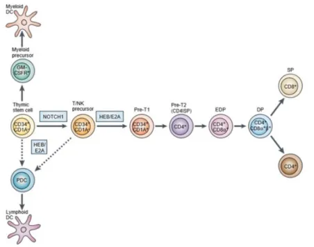
CD34+ cells committed to the T cell lineage express CD1a but still lack CD4 and CD8 coreceptors, and are thus double-negative (DN) cells (Figure 2) (15-17). CD34+CD1a+ cells start expressing CD4, but not yet CD8, being therefore referred to as CD4 immature single-positive (CD4ISP) cells (or pre T1) (18-20). This population contains precursors of both αβ and γδ T cells and is, therefore, upstream of the β-selection checkpoint (21-23). β-selection3 is the process by which precursor T cells (pre-T1) with a productive rearrangement of the TCRβ locus are selected for further differentiation.
1.3.2 Positive and Negative selection
After β-selection, rearrangements at the TCRα locus are initiated in cells expressing CD4 but only one of the two CD8 chains, CD8α giving rise to early double positive (EDP) cells (17). The CD8 molecule is composed of two chains, α and β, and is expressed as an αβ (in conventional CD8 T cells) or as an αα (such as in CD8αα intra-epithelial lymphocytes - IELs ) dimer. The CD8β extracellular domain is important to increase the avidity of CD8 binding to the complex of major histocompatibility I (MHC I) enabling pathogen recognition (18).
Upon CD8αβ expression DP thymocytes undergo a strict selection of functional TCRs that can engage self-MHC molecules. Only these cells are signaled to survive and differentiate into functionally mature T cells (8). Too little TCR signaling results in apoptosis/death by neglect and in return, too much signaling can also promote acute apoptosis (negative selection). The proper, intermediate level of TCR signaling will initiate effective maturation also called positive selection. Thymocytes that express TCRs that bind self-peptide–MHC class I complexes become CD8SP T cells, whereas those that express TCRs that bind self-peptide–MHC class II become CD4SP T cells, reallocating in the thymic medulla (24).
Once thymocytes have completed their development, they exit the thymus into the blood. On reaching a peripheral lymphoid organ they leave the blood to migrate through the lymphoid tissue, returning to the bloodstream to recirculate between blood and peripheral lymphoid tissue until they encounter their specific antigen (17).
Figure 2. T cell development in the thymus. CD34+ precursors originated in the bone marrow populate the
4
CD4, but not yet CD8, and are referred to as CD4 immature single-positive (CD4ISP) cells. At this stage some cells begin rearrangement of their TCR β (pre-T1 cells) and undergo β selection. This stage is followed by cells that express CD4 and only one of the two CD8 chains, CD8α, and are referred to as early double-positive (EDP) cells. Subsequently, acquisition of a CD8αβ dimer generates DP cells that, in the thymic cortex, interact with cells that express MHC molecules. Only a small portion of DP thymocytes survive positive/negative selection and differentiate into SP CD4 or CD8 T cells (adapted from Spits et al, 2002)
1.3.3 CD4/CD8 lineage specification
Thymocytes positively selected differentiate into either CD4 helper T cells or CD8 cytotoxic T cells, a critical decision known as CD4/CD8‑lineage choice (25).
In the mouse model, two classical models of lineage choice were suggested which fall into two main categories: ‘stochastic’ - The stochastic selection model; or ‘instructive’ - Strength-of-signal instructional model and Duration-Strength-of-signal instructional model, assuming, respectively, a random or an instructed termination of co-receptor transcription. Both models agree on some fundamental principles such as: positive selection and lineage commitment are simultaneous events that are induced by the same TCR signals, which can selectively terminate either Cd4 or Cd8 gene expression, a process known as lineage commitment (25).
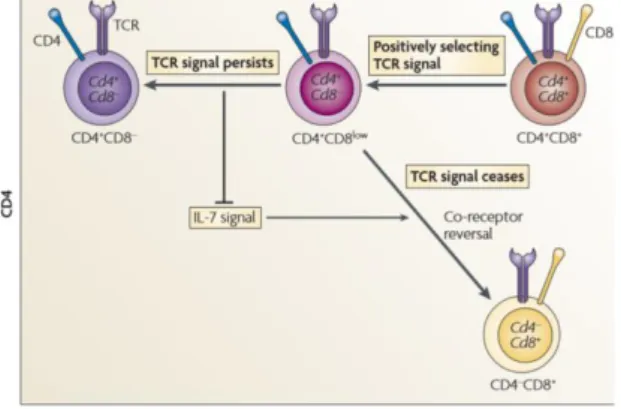
The currently accepted, Kinetic signaling model, is a non-classical model best explaining the CD4/CD8-lineage choice (Figure 3) (25, 26, 27). This model proposes that CD4/CD8-lineage choice is defined by TCR-signal duration, in contradiction with the first models, and that cytokines of the common cytokine receptor γ-chain (γc) family, such as IL7, serve as sensors that detect the duration of the TCR signal (26, 28, 29). This model suggests that cytokine-unresponsive DP thymocytes become cytokine-responsive intermediate thymocytes that are transcriptionally Cd4+Cd8−, since Cd8 gene transcription is terminated, upon TCR-mediated positive selection (26, 28, 29). In the case of persistent TCR-mediated signals, and in the absence of Cd8 gene transcription, thymocytes differentiate into CD4 T cells. If, in the absence of Cd8 transcription, TCR-mediated signalling ceases promoting signaling by intrathymic γc cytokines such as IL-7, the cells differentiate into CD8 T cells.
In this sense, assessment of TCR-signalled thymocytes can identify cell fate, once MHC class II-restricted TCR signaling does not depend on Cd8 gene transcription. In contrast, signaling by MHC class I-restricted TCRs is dependent on CD8 and so would cease in the absence of Cd8 gene transcription (25).
For uncommitted Cd4+Cd8– intermediate thymocytes to differentiate into CD8+ T cells, they must terminate Cd4 gene transcription and reinitiate Cd8 gene transcription, a process designated ‘coreceptor reversal’. This process is, in fact, the foundation of the kinetic signalling model uncouvering the dependence of signalling by IL-7 and possibly other intrathymic γc cytokines during T cell development and lineage choice (28-31).
5 In respect to previous studies, DP thymocytes are in truth unresponsive to intrathymic cytokines such as IL- 7 and IL-4 (31, 32), but solely before receiving a TCR signal. Moreover, although DP thymocytes are located in the thymic cortex, which lacks IL-7-producing cells, induction of Il7r and Ccr7 expression allow TCR-signaled DP thymocytes to migrate into cytokine-rich areas of the thymus (such as the cortico medullary junction and thymic medulla) and to bind IL-7 allowing differentiation of DP thymocytes into mature T cells (33, 34).
Overall, in the mice model, TCR-mediated positive selection converts DP cells into cytokine-responsive thymocytes, though, differentiation into cytotoxic-lineage T cells relies on the following signaling by intrathymic cytokines.
Figure 3. The kinetic signaling model of CD4/CD8 lineage specification. Positively selecting TCR signals
induce DP thymocytes to terminate Cd8 gene expression and to convert into CD4+CD8– intermediate thymocytes. Persistence of TCR signaling blocks IL‑7‑mediated signaling and induces differentiation into mature CD4 T cells. Cessation or disruption of TCR signaling allows IL‑7‑mediated signaling, which induces CD4+CD8– intermediate thymocytes to undergo co‑receptor reversal, gain a CD4–CD8+ phenotype and differentiate into CD8 T cells (from
Singer et al, 2008).
1.4 Human T cell differentiation
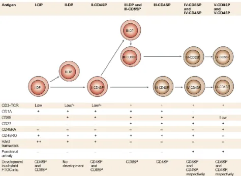
Regarding human T cell differentiation, phenotypic analysis using hybrid fetal thymic organ cultures (FTOC) revealed the markers specifying each phase of late T cell differentiation (17). Upon acquirement of a rearranged αβ TCR, immature double-positive (I-DP) cells arise lacking the early activation antigen CD69 marker (Figure 4). After positive selection, expression of CD69 is upregulated. Two populations CD69+CD27− arise: the CD4+CD8+ (II-DP) cells that fail to develop further, and the CD69+CD4+ cells (II-CD4SP), with low levels of CD8, which are the precursors for both CD4 and CD8 SP T cells. Next, the surface CD3-TCR and CD27 (marker of commitment to the helper lineage) markers are upregulated and the CD69+CD27+CD4+ (III-CD4SP) cells differentiate into CD4SP T cells. Both CD4SP and CD8 SP cells then loose CD1a expression, related with the acquisition of in vitro expansion capacity and the production of high levels of cytokines, determining functional maturation. SP cells
6 finally acquire the egress marker CD45RA (V-SP stage) and still express low levels of CD69, expression that will be lost when the cells migrate out of the thymus (17).
Figure 4. Model of late differentiation/maturation of T cells in the human thymus. In this model the sequential
steps of T cell differentiation and maturation of single-positive cells are defined by cell-surface markers. Immature DP T cells start by acquiring the early activation antigen CD69 marker of positive selection. Acquisition of CD27 in CD4SP thymocytes marks the commitment toward the helper-lineage. Upon completion of the T cell development both CD4SP and CD8SP thymocytes express the naïve T cell marker CD45RA enabling the migration of thymocytes into the blood (from Spits et al, 2002).
1.5 Transcription factors regulating lineage choice
In humans, the signals involved in lineage decision are poorly defined but, in mice, the transcription factors that specify CD4 or CD8 lineage choice in positively selected thymocytes have largely been identified: CD4 lineage choice is specified by the zinc-finger transcription factor ThPOK (35-37), whereas CD8 lineage choice is specified by the Runt-family transcription factor Runx3 (38-40). ThPOK and Runx3 negatively regulate each other’s expression, thus reinforcing lineage choices (35-39).
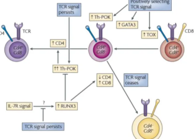
In agreement with predictions of the kinetic signaling model, persistent TCR MHC class II-restricted signaling in DP thymocytes induce ThPOK expression, upregulating the GATA binding protein 3 (GATA3) and preventing CD4 silencing, thus specifying CD4 lineage choice (Figure 5) (41-43). Alternatively, if positively selecting MHC class I restricted TCR signals are disrupted or cease, there is subsequential loss of GATA3 and ThPOK expression. Also, IL-7R signaling and Runx3 expression are upregulated (25).
ThPOK, a zinc-finger protein encoded by the zbtb7b gene, appears to be required and sufficient in determining CD4-lineage choice. Consistent with this perspective, retroviral
7 transduction of mature CD8 T cells with ThPOK led to decreased T-cell cytotoxicity and induced CD4 T-cell characteristics, indicating that ThPOK can induce the helper-lineage even in mature CD8 T cells (35-39). When the developmental potential of CD4 thymocytes to express ThPOK at intermediate or high levels was tested in vitro, CD4-CD8+ T cells emerged specifically from the cells expressing intermediate amounts of ThPOK. Thus, simple induction of ThPOK expression is insufficient for full commitment to the helper lineage, relying on the escalation of ThPOK levels (44). ThPOK as also an effect in both Cd4 and Cd8 gene transcription. It maintains Cd4 gene transcription by preventing factors, such as Runx3, from silencing it, while it decreases Cd8 gene expression by downregulating the enhancer activity of e8I (43).
It was revealed that Runx proteins also bind to a sequence in the ThPOK gene inhibiting its expression. Runx3 is able to bind to the Cd4 silencer element and silences Cd4 gene transcription (40, 45). Indeed, Runx protein deficiency results in Cd4 gene de-repression (45, 46), whereas overexpression of a Runx3 transgene downregulates Cd4 gene transcription (47, 48). Current evidence demonstrates that Runx3 also associates with genes encoding Granzyme B, Perforin and interferon-γ (IFNγ) characteristic of cytolytic CD8 effector cells (49).
The factors that promote Runx3 activation are poorly known, though, STAT5, which acts downstream of IL-7 signaling, is thought to promote Runx3 expression after cessation of TCR signaling in post-selected thymocytes (50).
GATA3, an enhancer binding zinc finger protein, functions as a lineage specific transcription factor in T cells at various stages of development (51, 52). GATA3 is expressed in the earliest progenitor T cells and is required for thymocytes to differentiate beyond the DN stage of development (52). The GATA3 role in CD4 lineage choice is revealed by its upregulation by TCR signaling in DP thymocytes and its sustained expression blocks the generation of CD8+ T cells (53). However, unlike ThPOK, GATA3 does not seem to be a CD4 lineage specifying factor because forced expression of GATA3 does not redirect MHC class I-restricted thymocytes to differentiate into CD4+ T cells. In fact, as it is expressed in positively selected thymocytes earlier than either ThPOK or Runx3, GATA3 might function upstream of these other factors. Indeed, the lack of ThPOK expression in post-selection GATA3-deficient thymocytes and the presence of GATA3 binding at two regions in the ThPOK gene provides further evidence of its upstream role (51-52).
1.5.1 Maintenance of lineage commitment
Regarding the role of ThPOK in maintaining commitment of peripheral CD4 cells to the helper T lineage, other studies revealed, upon transfer into immunodeficient recipients, CD4 T cells from which Thpok had been conditionally excised, upregulated genes characteristic of the cytotoxic T lineage, including Cd8 and Granzyme B (GzmB). This finding revealed that
8 continuous extrathymic expression of ThPOK is necessary to maintain helper-lineage gene signatures, which are established during differentiation in the thymus (37).
Also, although the intronic Cd4 silencer is essential to establish the initial repressive state at Cd4 during CD8 lineage commitment in the thymus, this repression is maintained after removal of the silencer from mature CD8 T cells in the periphery by epigenetic mechanisms in place in peripheral cells that maintain Cd4 silencing (54). In opposition, the epigenetic mechanisms regulating repression of Cd8 during helper-lineage specification remain unclear.
A recent study suggested a potential form of lineage plasticity in the mice gut. Upon transfer of naïve CD4 T cells containing the Thpok-gfp reporter allele into lympho-deficient RAG KO mice, some cells down-regulated Thpok-gfp re-expressing CD8α in the small and large intestine, but not in other peripheral lymphoid tissues. This recovery of Cd8 co-receptor was independent of MHC-restriction and confirmed by in vivo fate mapping showing that CD4+CD8αα+ IEL differentiate from CD4 lineage cells under normal conditions (55). This experiment also confirms the importance of lineage decision regulating genes such as ThPOK and its capacity of reverting lineage decision even in the periphery.
Another experiment tried, in conventional CD4 T cells, overexpression/upregulation of Runx3 and endogenous RUNX3, but failed to downregulate ThPOK (56).
Overall, current evidence support an antagonistic interplay between ThPOK and Runx3 as the master regulators of lineage decision. This negative feedback acts as the crucial mechanism controlling helper versus cytotoxic fate decision and seems to be an evolutionary strategy for the emergence of new developmental branch points from ancestral progenitors.
Figure 5. Signaling and transcriptional regulation of CD4/CD8 lineage fate choice. During positive selection,
TCR signals upregulate GATA3 and ThPOK expression. GATA3 upregulation is important for the differentiation of CD4+CD8low thymocytes into CD4 T cells. ThPOK expression in TCR‑signaled thymocytes, upregulated by persistent TCR signaling, is required for CD4‑lineage commitment and for preventing Cd4 gene silencing by Runx proteins. Runx expression may be upregulated by interleukin‑7 receptor (IL‑7R) signaling (from Singer et al, 2008).
9 1.6 CD4-helper T cells
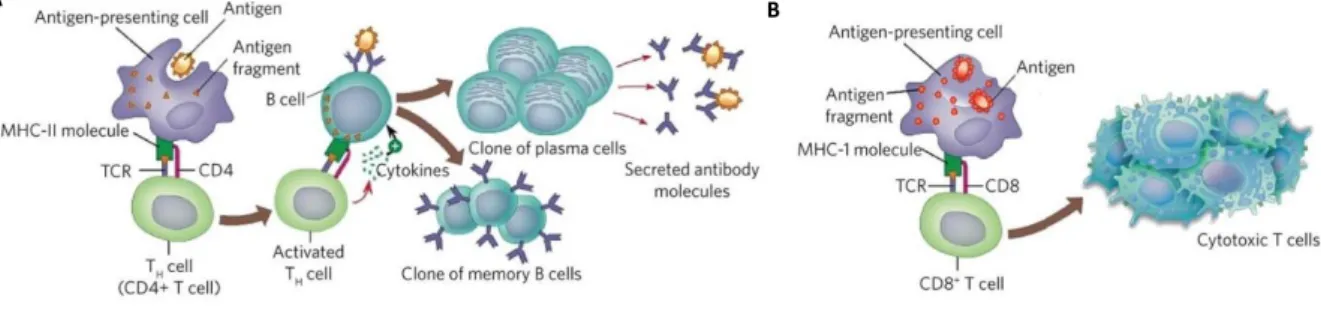
CD4 T cells play central roles in mediating adaptive immunity to a variety of pathogens. These cells help B cells generating antibodies (Figure 6A), enhance and maintain responses of CD8 T cells and regulate macrophage function. Helper cells control immune responses against a wide range of pathogenic microorganisms, and regulate/suppress immune responses both to control autoimmunity and to adjust the magnitude and persistence of responses (57).
Additionally, CD4 T cells are central mediators of immunologic memory, meaning that decreased numbers of CD4 T cells induce function loss rendering the individual susceptible to an extensive range of infectious diseases. For example, in HIV infection, as CD4 T cell numbers in blood fall, opportunistic infections are most likely to occur.
1.7 CD8-cytotoxic T cells
CD8 T cells are also key mediators of adaptive immunity. These cytotoxic T cells, which are important for directly killing cancerous or virally infected cells (Figure 6B), are of particular interest for vaccine development to induce potent CD8 T cell-mediated cytotoxic responses to viruses and tumor cells (58).
Activation and differentiation of naïve CD8 T cells into cytotoxic effector cells requires help from CD4 T-helper cells. There is a first interaction of the TCR complex with antigenic peptide and MHC-I on antigen-presenting cells (APCs) followed by a second costimulatory signal that promotes CD8 responses, including secretion of cytokines such as tumor necrosis factor-α (TNF-α) and IFN-γ, which have anti-tumor and anti-viral microbial effects.
Figure 6. CD4 vs CD8 T cell response. A. CD4-helper responses. APCs present antigens to CD4 T cells through
MHC Class II molecules inducing cell activation. Activated Th cells promote B cell priming and antibody production. Also, memory B cells arise from this interaction B. CD8-cytotoxic response. CD8 T cells express a dimeric co-receptor, CD8, composed of one CD8α and one CD8β chain. These cells recognize peptides presented by MHC Class I molecules binding to this molecule and become activated, responding with secretion of cytokines, primarily TNF-α and IFN-γ, which have anti-tumor and anti-viral microbial effects (from Wolfert, M.A. et al, 2013).
10 1.8 Role of IL-4 in immunity
Interleukin 4 (IL-4), a short four-helix bundle peptide member of the γ-chain receptor cytokine family, originally described in 1982, is a pleiotropic type I cytokine produced mainly by Th2 lymphocytes, basophils, and mast cells in response to receptor-mediated activation (59-60). Other cell types that can produce IL-4 include NKT cells, a specialized subset of T cells that express NK1.1 and CD1, eosinophils, follicular T helper cells (Tfh) and γ/δ T cells (61).
Classically, IL-4 drives CD4 T cell polarization in the Th2 phenotype together with suppression of Th1 cells (62). IL-4 also supports the growth and differentiation of B lymphocytes, controlling the specificity of the immunoglobulin G (IgG) class switching and the development of memory B cells.
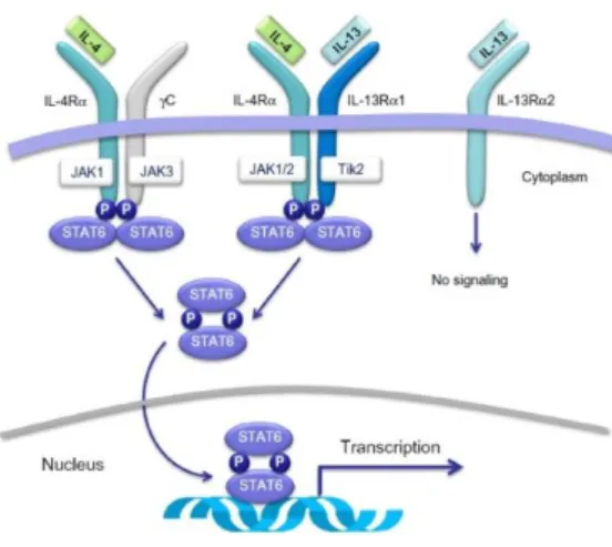
The physiologic functions of IL-4 are mediated by the IL-4 receptor (IL-4R) complex (Figure 7). Several studies have demonstrated the existence of two types of IL-4 receptors. The type I receptor complex comprises a 140-kDa chain (the IL-4Rα chain) and the γ-common chain, which is also shared by IL-2, IL-7, IL-9, and IL-15 receptors (63). This is the prevailing heterodimer in hematopoietic cells and is activated exclusively upon IL-4 binding. The IL-4Rα chain may also heterodimerize with the IL13Rα1 chain to form the type II receptor, which is able to transduce both IL-4 and IL-13 signals and is often the exclusive IL-4 receptor found in non-hematopoietic cells (63). The heterodimerization step is always necessary for the transduction of IL-4 signals (64-65).
IL4-induced activation of STAT6 promotes gene regulation, differentiation of activated CD4 T cells to the Th2 phenotype, development of the Th9 phenotype (66), IL-4–induced GATA-3 expression, production of chemokines, and production of IgE and IL-4–inducible allergic responses (67) .
Another cytokine, IL-13 also activates STAT6 after binding onto the IL-4Rα chain (Figure 6) (68). Due to its ability to induce signaling pathways through a similar receptor complex, some effects in vivo are shared by both cytokines, for example, induction of Th2 responses against parasites and control of T cell function in allergic diseases (69). Though, these two cytokines present some differences such as the restricted expression of IL-4 to Th2 lymphocytes and the inability of IL-13 to regulate T cell differentiation deriving from the lack of IL-13 receptors in T lymphocytes (70).
Regulation of CD8 T cell development, frequency and function seems to be promoted by IL-4 as endogenously it stimulates CD8 T cell proliferation and regulates Eomes (Eomesodermin) expression levels (71). In addition, exogenous IL-4 induces memory-like phenotype in peripheral CD8 T cells (72). Thus, IL-4 negatively regulates the quality of the CD8 T cell response during viral infection, impairing the protective response these cells have diminishing perforin/granzyme-mediated cytotoxicity (73-75). However, in models of tumor immunity, it was previously demonstrated that higher IL-4Rα expression reduced the functional capacity of
anti-11 viral CD8 T cells. IL-4 upregulates its own expression and in parallel downregulates cytotoxic capacity, IFN-γ production and CD8 expression during the primary activation of CD8 T cells (69). On the other hand, all these effects of IL-4 described above have been brought in question by several studies showing that IL-4 could potentiate the cytotoxic activity of CD8 T cells during tumor clearance, increasing tumor rejection in vivo (76). Moreover, in some cases IL-4 producing CD8 T cell populations displayed strong cytotoxic-T-lymphocyte activity and CD8 expression (77). Overall, the effect of IL-4 in CD8 T cells is not aggreed.
1.8.1 Role of IL-4 in T cell development
Regarding the role of IL-4 in thymocyte differentiation, studies in murine systems revealed that IL-4 blocks the differentiation of early thymocyte precursors, whereas precursor cells that are already committed to the differentiation process are no longer affected (78).
In humans, IL-4 has an impact in pro-T cells by inducing proliferation and differentiation into functionally mature γδT cells which present a TCR repertoire similar to that of gamma delta T cells that can be found in post-natal thymus (79). Thus, IL-4 effect on human T cell development is poorly described. Nonetheless, it is known that its presence is required during the initial activation of naïve T cells, promoting a specific differentiation program. Acting through STAT6 and GATA3, IL-4 can determine Th2 cell fate originating helper cells capable of producing IL-4 (80).
Figure 7. Membrane receptors and intracellular signaling pathways activated by IL-4 and/or IL-13. IL-4
binds to the IL-4Rα subunit of the type II receptor complex, whereas IL-13 binds to the IL-13Rα1 subunit of the same receptor complex. Both IL-4 and IL-13, which share the IL-4Rα subunit in their cognate receptors, activate STAT6 which dimerises, migrate to the nucleus, and bind to the promoters of the IL-4 and IL-13 responsive genes, such as those associated with T-helper type 2 (Th2) cell differentiation, airway inflammation, airway hyperresponsiveness (AHR) and mucus production (from Vatrella, A. et al, 2014).
12 2 OBJECTIVES
T cell development taking place in the human thymus is a process of extreme importance allowing differentiation and maturation of immune cells responsible for surveillance and response to pathogens that attack the organism. Though, the mechanisms underlying cell’s fate determination are still poorly described in humans.
The main aim of this project was to elucidate the role of IL-4 in the plasticity of the CD4SP thymocytes subset and in CD4 T-cell lineage fate.
The specific objectives were to:
o Characterize the modulation of CD8 expression in human CD4SP thymocytes in response to IL-4 and its kinetics;
o Compare the modulation of CD8 expression in human CD4SP thymocytes with that of cord blood and adult naïve CD4 T cells;
o Study the expression of key transcription factors in human CD4SP thymocytes upon culture with IL-4, and the relationship with the degree of CD8 upregulation;
o Determine the landscape of IL-4 and IL-4Rα expression in the human thymus; o Understand if there is a physiological role for IL-4 in CD4SP T cell differentiation.
13 3 METHODOLOGY
3.1 Samples
Thymic tissue was obtained from thymectomy during pediatric corrective cardiac surgery (newborns to 4-year old) at Santa Cruz Hospital, after parent’s informed consent. Study was approved by the Ethical Boards of the Faculty of Medicine of Lisbon, Santa Maria and Santa Cruz Hospitals.
Blood was collected from volunteer healthy donors at IMM (Instituto de Medicina Molecular), Lisbon, Portugal.
Cord Blood was collected in the obstetric department of the Hospital de Santa Maria, Lisbon, Portugal, after parent’s written informed consent. The study was approved by the Ethical Board of the Faculty of Medicine of the University of Lisbon.
3.2 Tissue and whole blood processing
Total thymocytes were recovered through tissue dispersion and separation on a Ficoll-Paque Plus (GE Healthcare) density gradient.
T cells from peripheral and cord blood were recovered after Ficoll separation from diluted whole blood in PBS.
Cell number was estimated using a Neubauer chamber and trypan blue for dead cell exclusion.
3.3 Cell Sorting/Isolation
From total thymocytes the cells were sorted (Table 1 and Supplementary figure 1) with purity >98% using FACSAria high-speed cell sorter (BD Biosciences).
From peripheral blood, CD4 naïve and memory T cells were also FACS-sorted using FACSAria high-speed cell sorter (BD Biosciences) (Table 1 and Supplementary figure 2).
From cord blood, cells were separated using the EasySep Human naïve CD4+ T cell isolation kit (Stemcell).
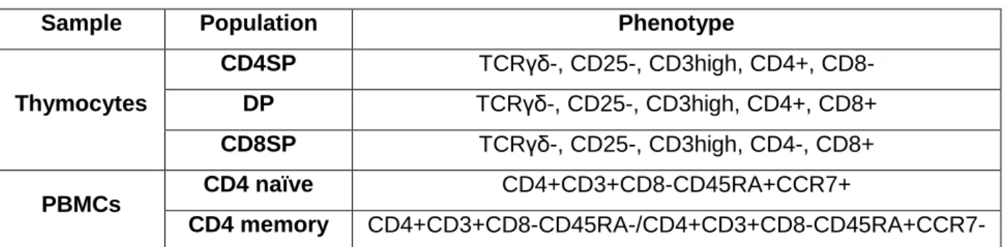
Table 1. Definition of FACS-sorted subsets
Sample Population Phenotype
Thymocytes CD4SP TCRγδ-, CD25-, CD3high, CD4+, CD8- DP TCRγδ-, CD25-, CD3high, CD4+, CD8+ CD8SP TCRγδ-, CD25-, CD3high, CD4-, CD8+ PBMCs CD4 naïve CD4+CD3+CD8-CD45RA+CCR7+ CD4 memory CD4+CD3+CD8-CD45RA-/CD4+CD3+CD8-CD45RA+CCR7-
14 3.4 Cell cultures
Sorted/isolated cells were ressuspended in complete medium (RPMI 1640 with 10% of FCS (thymocytes and PBMCs) or human AB serum (cord blood cells), 2 mM L-glutamine and 50 U/ml penicillin-streptomycin).
Thymocytes or T cells were then cultured for 7 to 14 days in the absence or presence of CM, IL-2 (20 U/ml), IL-4 (1, 10, 20, 50 ng/ml; R&D), IL-7 (1, 10, 50 ng/ml; R&D) or IL-13 (10, 50, 100ng/mL) at 37°C/5% CO2.
Some cells were re-sorted as CD4+CD8-, CD4+CD8+, CD4-CD8+ at day 7 of culture with IL-4 and re-cultured for 7 more days in the absence or presence of IL-2 (20 U/ml) and IL-IL-4 (20ng/ml; R&D).
3.4.1 Thymic Organ Cultures (TOCs)
Thymic tissue blocks (diameter 1 to 2 mm) were placed on 0,8 uM isopore membrane filters (Millipore) in a 6-well plate containing 2 ml of TOC medium. TOCs were cultured at 37°C in 5% CO2 for 7 days in the presence of CM (RPMI1640, 15% FCS, 1% L-Glutamine, 1% Non-essential aminoacids, 1% HEPES, 1% NaPyr, 0,5% Pen/Strep, 1% Gentamycin), IL-4 (20 ng/ml; R&D), anti-IL-4 blocking antibody (1, 5, 10 ug/mL, eBioscience) and IgG1 isotype control for the IL-4 blocking antibody. On days 0 or 7 TOCs were mashed and stained, or fixed in formol for further immunohistochemistry analysis.
3.5 Flow cytometry staining
Surface staining was performed for 20 minutes at room temperature and always included fixable viability dye (FVD; eBioscience) for dead cell exclusion. Thymocytes were further fixed and permeabilized using an intracellular staining kit (eBioscience) and stained with intracellular antibodies. The antibodies used and conjugated fluorophores are specified in Table 2. At days 0, 7 and 14 the thymocyte number per well was determined using 10-μm latex beads (Coulter), and cells were also analyzed by flow cytometry. Cells were acquired using LSRFortessa cell analyzer (BD Biosciences), and data was analyzed with FlowJo software (TreeStar).
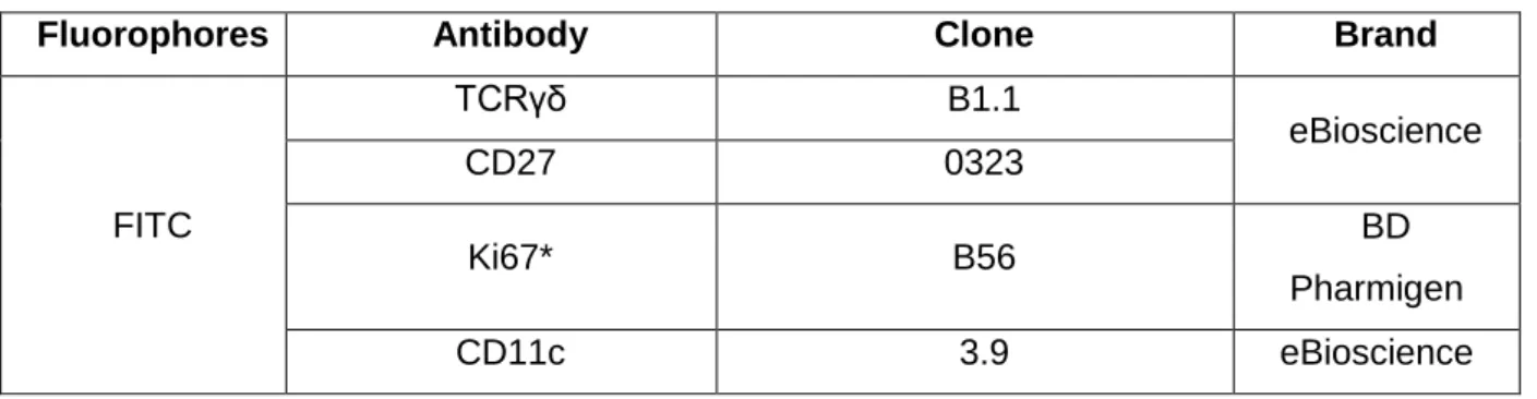
Table 2. Antibodies used and respective fluorophores
Fluorophores Antibody Clone Brand
FITC TCRγδ B1.1 eBioscience CD27 0323 Ki67* B56 BD Pharmigen CD11c 3.9 eBioscience
15 CD14 61D3 CD16 CB16 CD19 HIB19 CD123 6H6 PE CD4 RPA-T4
PerCp-Cy5.5 CD8a RPA-T8
PeCy7 CD45RA HI100
eF660 CD8β SIDI8BEF eF450 CD45RA HI100 CD127 eBioRDR5 FoxP3* PCH101 V500 CD3 UCHT1 BD Horizon - α-IL-4 MP4-25D2 eBioscience
*Antibodies used intracellularly
3.6 Immunohistochemistry staining
Pieces of human thymic tissue were preserved in 4% formaldehyde, embedded in paraffin, deparaffinised and cut into 3-μm sections (Minot microtome Leica RM2145). Samples underwent antigen retrieval (Leica Buffer Ph9) by heat for 15 min. Samples were stained with the appropriate primary antibodies anti-IL-4 (single and double staining), IL-4Rα (single staining), Pan-cytokeratin (single and double staining) and c-kit (single staining).
Single or double immunohistochemistry stainings were revealed by enzymatic substrate with horseradish peroxidase (Dako EnVision Detection Systems Peroxidase/DAB, Rabbit/Mouse kit (Agilent Technologies, Santa Clara, CA)) and alkaline phosphatase (EnVision G|2 Doublestain System). Rabbit/Mouse (DAB+/Permanent Red (Agilent Technologies, Santa Clara, CA)) stain in brown and red, respectively.
All slides were counterstained with Hematoxylin and mounted with Quick-D mounting medium (single staining) or Glycergel (double staining) from DakoPower. Bright-field images were acquired using a Leica DM 2500 Microscope or a NanoZoomer (Hamamatsu’s).
3.7 RNA extraction, cDNA synthesis and quantification by RT-qPCR
Transcriptional expression of lineage specific genes was analyzed by reverse transcription– polymerase chain reaction (RT-PCR) of total RNA extracted at day 0 or day 7/9 after cytokine culture.
RNA purification was performed using a ZR-Duet DNA/RNA Miniprep isolation kit (Zymo Research). The purified RNA was then used to synthesize cDNA using random primers and
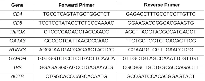
16 SuperScript III RT (Invitrogen), according to the manufacturer's instructions. Real-time PCR was performed using Power SYBR green PCR master mix (Applied Biosystems) with adequate primers (Table 3), using GAPDH, β-actin (ACTB) and 18S as reference genes. Quantification was performed using an Applied Biosystems ViiA7 real-time PCR system. Results were expressed using the ΔCt method.
Table 3. Primers used in the study
Gene Forward Primer Reverse Primer
CD4 TGCCTCAGTATGCTGGCTCT GAGACCTTTGCCTCCTTGTTC
CD8 TCCTCCTATACCTCTCCCAAAAC GGAAGACCGGCACGAAGTG
ThPOK GTCCCCAGAGCTACGAACC AGCTTAGGTAGGCCATCAGGT
GATA3 GCCCCTCATTAAGCCCAAG TTGTGGTGGTCTGACACTTCG
RUNX3 AGGCAATGACGAGAACTACTCC CGAAGGTCGTTGAACCTGG
GAPDH GGTGGTCTCCTCTGACTTCAACA GTTGCTGTAGCCAAATTCGTTGT
18S GGAGAGGGAGCCTGAGAAACG CGCGGCTGCTGGCACCAGACTT
ACTB CTGGCACCCAGCACAATG GCCGATCCACACGGAGTACT
3.8 Graphic analysis
Graphic analysis were performed using GraphPad Prism (v5.01) software (GraphPad Software Inc.) and are presented as mean±SEM (standard error of the means).
17 4 RESULTS AND DISCUSSION
4.1 IL-4 induces upregulation of CD8αβ in CD4SP thymocytes
Previous observations from our lab suggested that IL-4 induced the upregulation of CD8 in CD4SP thymocytes. We thus cultured CD4SP thymocytes with two other c cytokines, IL-2 or IL-7, in order to understand whether that mechanism was particular of IL-4. IL-7 was already described to play a role in T cell survival and development, particularly in the co-receptor reversal that promotes differentiation of CD8 T cells in mice (kinetic signaling model) (28-29), and IL-2 represents a primary T-cell growth factor (81) and was chosen as a control.
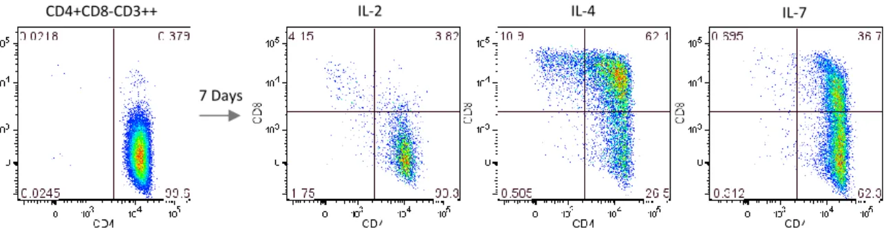
Our results showed that CD8 upregulation was much higher in CD4SP thymocytes treated with IL-4 for 7 days than in those treated with IL-2 or IL-7 (Figure 1). In contrast to what was expected, IL-7 had lower capacity than IL-4 to upregule CD8 in CD4SP thymocytes, and generated mostly DP cells. IL-2 had no specific pattern of CD8 upregulation in CD4SP thymocytes, indeed representing a control. Of note, the same effect of IL-4 was found when CD4SP cells were cultured with two other recombinant IL-4s (Supplementary figure 4).
Our results show that, in humans, co-receptor reversal may be induced by IL-4, and indicate a possible role for IL-4 in T cell development.
Figure 1. IL-4-induced upregulation of CD8 in CD4 thymocytes. FACS-sorted CD4SP thymocytes were
cultured for 7 days with IL-2 (20U/mL), IL-4 (20ng/mL) or IL-7 (10ng/mL). CD8α upregulation and generation of DP and CD8SP cells from CD4SP thymocytes was increased upon IL-4 exposure as compared to the other cytokines tested.
We further tested several concentrations of IL-4 or IL-7 in order to understand their capacity of upregulating CD8 on CD4SP T cells, as the dose of IL-7 could be insufficient to induce a complete co-receptor reversal. CD4SP cells were cultured for 7 days in the absence or presence of IL-2 or of different concentrations of IL-4 or IL-7. Analyses at day 7 revealed that CD8 upregulation on CD4SP thymocytes was lower for IL-7 than for IL-4 independently of the concentration tested, indicating that the effect of IL-7 was indeed less marked (Figure 2). In
7 Days
18 addition, we observed no difference in the frequency of DP T cells generated with the three highest concentrations of IL-4 tested (Figure 2A). On the other hand, an increasing number of CD8SP T cells were generated in parallel with increasing concentrations of IL-4, with exception of the highest concentration (50ng/mL) (Figure 2B). We thus chose IL-4 at 20ng/mL as the ideal concentration for future experiments, since it represented the concentration that yielded higher levels of CD8SP cells with less variation.
Figure 2. Dose dependent response to IL-4. FACS-sorted CD4SP thymocytes were cultured for 7 days with
Media (CM), IL-2 (20U/mL), IL-4 (1, 10, 20 and 50ng/mL) or IL-7 (1, 10 and 50ng/mL) in order to determine the dose-dependent response to each cytokine. Graphs represent the frequency of DP (A) or CD8SP (B) thymocytes at day 7. IL-7 was not able to induce the same extent of CD8 upregulation as IL-4, independently of the concentration used. 20ng/mL was the ideal concentration of IL-4 to induce CD8SP T cells from CD4SP T cells, and was the concentration chosen for future experiments. Values are represented as mean±SEM (n=3).
Mature functional CD8 T cells express on the surface a complex of two chains, α and β which are the products of two closely linked but separate genes (82-84). As data so far had been obtained using the CD8α antibody, which is the molecule routinely identified in CD8 T cell analyses, we further wanted to determine whether the CD8β chain was also being expressed in the surface of IL-4-treated CD4SP T cells. Our results confirmed that the heterodimer CD8αβ was expressed on the surface of the newly generated CD8 T cells (Figure 3) thus indicating that IL-4 is abble not only of inducing activation of thymocytes but also induces a co-receptor reversal from CD4+CD8- into CD4-CD8+ T cells.
A B C D A B 7 Days IL-4 CD4+CD8-CD3++ Within DPs Within CD8SP
19
Figure 3. IL-4 induces upregulation of the CD8αβ heterodimer. FACS-sorted CD4SP thymocytes were
cultured for 7 days with Media, IL-2 (20U/mL), IL-4 (20ng/mL) or IL-7 (10ng/mL) and by flow cytometry expression of both CD8α and β chains was assessed. Results showed that both CD8 chains were upregulated in CD4SP thymocytes cultured with IL-4. Values are represented as mean (n=4).
4.2 IL-13 is not capable of upregulating CD8
Since Interleukin-13 (IL-13) binds to the IL-13Rα1, promoting dimerization with IL-4Rα (85), it was plausible to think that IL-13 could induce similar CD8 upregulation in CD4SP T cells. However, we saw no capacity of upregulation of CD8α on CD4SP thymocytes by IL-13 (Supplementary figure 5). This can be explained by the reported absence of IL-13R in human T cells (86).
4.3 Role of proliferation in IL-4-induced CD8 upregulation on CD4SP thymocytes
To gain further insight on the mechanisms behind IL-4-induced CD8 upregulation we asked whether it was driven by proliferation. Using the proliferation marker Ki67 we found that, although there are some cells proliferating, CD8 upregulation can be observed in IL-4-treated CD4SP thymocytes in the absence of proliferation (Figure 4A). Of note, IL-2 did not induce proliferation, while IL-4 and IL-7 mostly induced the proliferation of CD8- thymocytes (Figure 4B).
Overall, we can attest that proliferation does not seem to be necessary for IL-4-promoted co-receptor reversal. 7 Days IL-4 IL-7 IL-2 Total Thymocytes A B
20
Figure 4. CD8 upregulation is observed in the absence of proliferation. FACS-sorted CD4SP thymocytes
were cultured for 7 days with IL-2 (20U/mL), IL-4 (20ng/mL) or IL-7 (10ng/mL) in order to evaluate cell proliferation using the Ki67 marker. A. CD8 upregulation appears to be independent of proliferation, as most of CD8SP T cells generated do not express the proliferation marker Ki67. B. IL-4 and IL-7 induced, majorly, upregulation of CD8- thymocytes. Values are represented as mean±SEM (n=3).
4.4 CD8 upregulation potential decreases with CD4 T cell maturation
In order to evaluate the ability of CD4SP thymocytes at different stages of maturation/commitment to perform co-receptor reversal we analyzed the various stages of CD4 development (as described by Spits, 2002) and stimulated these subsets with IL-4 for 7 days. We sorted CD4+CD3hi CD27-CD45RA- (lineage-uncommitted cells), CD4+CD3hiCD27+CD45RA- (CD4-helper lineage-committed cells) and CD4+CD3hiCD27+CD45RA+ (CD4-helper lineage-committed cells ready to egress the human thymus) thymocytes and cultured these three subsets with IL-4, IL-2 or IL-7 for 7 days.
As shown on Figure 5, the potential to generate CD8SP thymocytes was highest in the more immature subset, and this ability was gradually lost with increasing CD4 commitment. Thus, CD27-CD45RA- CD4SP thymocytes were the more plastic cells, followed by CD27+CD45RA- and by CD27+CD45RA+, which still retained some capacity to undergo co-receptor reversal, although very low. Importantly, co-receptor reversal was observed even in CD27+ CD4SP thymocytes, indicating that CD27 may not be such a definitive marker for human CD4 T cell commitment.
Figure 5. The potential to induce co-receptor reversal is gradually lost with increasing CD4SP maturation.
FACS-sorted CD4+CD3hiCD27-CD45RA- (A), CD4+CD3hiCD27+CD45RA- (B) or CD4+CD3hiCD27+CD45RA+ (C) thymocytes were cultured for 7 days with Media, IL-2 (20U/mL), IL-4 (20ng/mL) or IL-7 (10ng/mL). A. Sorted thymocyte subsets at day 0 according to human the CD4-lineage commitment developmental stages. B. We observed a decreasing capacity of inducing the CD8SP phenotype in parallel with an increase in the CD4SP maturation. Values are represented as mean±SEM (n=3).
B C TCRγδ-/CD25-/CD3hi/CD4+/CD8- A B A
21 4.5 Co-receptor reversal capacity is lost in the periphery
T cells leaving the thymus incorporate the peripheral naïve T cell pool. We asked whether CD4 T cells in the periphery retain the ability to upregulate CD8 in the presence of IL-4, and whether this ability was age-dependent. With this purpose, isolated naïve CD4 T cells from cord blood or naïve and memory CD4 T cells from adult blood were cultured with IL-4 for 7 days, and CD8 expression was assessed by flow cytometry. Newborn cord blood naïve CD4 T cells, sorted as untouched CD4 naïve T cells, were still able to upregulate CD8 upon IL-4 exposure but did not complete co-receptor reversal (Figure 6A). In contrast, naïve and memory CD4 T cells from adult blood showed a complete loss of CD8 upregulation capacity (Figure 6B). It is possible that the kinetics of CD8 upregulation is slower in the periphery, and this could be addressed by prolonging the cultures. Nonetheless, our results support a continuous loss of plasticity of CD4 T cells from the thymus to the periphery, both with differentiation and with age.
Figure 6. The capacity to undergo co-receptor reversal is gradually lost in the periphery. A. CD4 T cells
from cord blood were separated using the EasySep Human naïve CD4+ T cell isolation kit and cultured for 7 days with IL-4 (20ng/mL). CD4 T cells retain some CD8 upregulation capacity, but generate only DP T cells; B. Naïve
7 Days
7 Days IL-4
IL-4 CD4 T cells from adult blood
7 Days IL-4 CD4 T cells from Cord Blood A
Naïve
Memory B
22
and memory CD4 T cells from adult blood were FACS-sorted as CD4+CD3+CD8-CD45RA+CCR7+ (naïve) or CD4+CD3+CD8-CD45RA-/CD4+CD3+CD8-CD45RA+CCR7- (memory) and were cultured with IL-4 (20ng/mL) for 7 days. CD4 T cells from adult blood lost the capacity to undergo co-receptor reversal, irrespective of the maturation stage.
4.6 CD8 expression is stable upon IL-4 removal
We next investigated the stability of the CD8 upregulation and of the co-receptor reversal induced by IL-4 in CD4SP thymocytes.
In order to do so, we cultured CD4SP thymocytes with IL-4 for 7 days, and then sorted the CD4+CD8-, CD4+CD8+ and CD4-CD8+ populations generated and cultured them separately for 7 more days with IL-4 or IL-2, which we have confirmed does not significantly induce CD8 expression.
Our results showed that sorted CD8SP cells maintained their phenotype after 7 days of culture in the absence of IL-4 (IL-2 condition), including CD8α and β chain expression, supporting the stability of the co-receptor reversal (Figure 7).
Interestingly, DPs with IL-4 continued to proceed to the CD8SP phenotype, as did to some extent CD4SP cells, which means that the capacity of CD8 upregulation in the presence of IL-4 was not lost. Thus, as the co-receptor reversal was sparse in time, it may require more or less stimulation possibly depending on the degree of CD4SP commitment, in accordance with our previous results.
Surprisingly, when analyzing the DP T cells cultured for 7 days without IL-4 stimuli (but with IL-2), we observed a loss of CD8 expression. This suggests that co-receptor reversal is a two directional process and that the DP stage constitutes a transitory phase of activation only.
Figure 7. IL-4-induced CD8SP cells maintain the acquired phenotype without continuous IL-4 stimuli.
FACS-sorted CD4 T cells were cultured in IL-4 (20ng/mL) for 7 days. At day 7 CD4, DP and CD8 T cells were again FACS-sorted and cultured for 7 more days in IL-2 (20U/mL) or IL-4 (20ng/mL). CD8SP cells maintained the acquired phenotype without IL-4 stimuli. DPs and CD4SP cells cultured in IL-4 for 7 more days maintained the ability to upregulate CD8. However, DPs cultured didn’t seem fully committed to their phenotype, as they revert to CD4SP cells upon IL-2 culture.
23 4.7 IL-4 induces modulation of lineage-specific genes
Lineage commitment is highly regulated at the transcriptional level (34-56). To gain further insight into the phenotypically characterization of generated CD8 T cells we assessed their expression of CD4 and CD8, as well as the expression of key transcription factors driving lineage specification and commitment: ThPOK and GATA3 (commitment to CD4) and RUNX3 (commitment to CD8). To do so we sorted CD4SP thymocytes and cultured them with IL-4 for 7 days. At days 7/9 CD4SP, DP and CD8SP cells were once again sorted and cell pellets were stored for further RNA purification. Gene expression was assessed by real time RT-PCR.
In agreement with the upregulation of CD8 protein assessed by flow cytometry, we found an increase in CD8 gene expression in DP and CD8SP cells generated in culture (Figure 8). In accordance with the literature (26), and with the loss of commitment towards the CD4 lineage,
ThPOK levels of expression were decreased in newly generated CD8 T cells, as well as CD4
gene expression. Indeed, CD4, CD8 and ThPOK levels of CD8 T cells at day 7/9 were not very different from day 0 levels of these genes, in ex vivo CD8 T cells (Figure 8). Of note, ThPOK levels in DP were comparable to those found in CD4SP thymocytes after IL-4 culture, supporting the uncommitment of newly-generated DP cells to the CD8SP lineage, as indicated by the loss of the CD8 expression in DP upon IL-4 removal (Figure 8).
7 Days 7 Days IL-2 IL-4 CD4SP (Day14) DP (Day14) CD8SP (Day14) CD4+CD8-CD3++ (Day0) Thy (Day7) IL-4
24 Surprisingly, GATA3 and RUNX3 expression wasn’t altered with IL-4 culture, indicating that these transcription factors may not be playing an essential role in IL-4 induced co-receptor reversal.
Overall, the modulation of lineage-related genes found suggests that IL-4 induced real commitment toward the CD8 lineage fate.
In relation to the predicted kinetic signaling model in mice (50), being this mechanism a possible co-receptor reversal in humans it should be highly transcriptionally regulated and IL-4 may be the force inducing this mechanism.
Figure 8. IL-4 induces modulation of lineage-specific genes. FACS-sorted CD4SP cells were cultured for 7 or
9 days with IL-4 (20ng/mL). CD4, DP and CD8 T cells were then sorted at day 7/9, the RNA was extracted and cDNA was synthesized by retro transcription. Gene expression of day 0 or day 7/9 cells was assessed by real time PCR. Generated CD8SP T cells present an increased expression of CD8, and decreased expression of CD4 and
ThPOK. Values are represented as mean±SEM (n=3).
4.8 IL-4 is mainly present in the medulla of the human thymus
In order to determine whether there might be a physiological role for IL-4 in CD4SP T cell differentiation we assessed the expression of IL-4 and of its receptor in the human thymus by immunohistochemistry.