www.elsevier.com/locate/bjid
The Brazilian Journal of
INFECTIOUS DISEASES
1413-8670/© 2012 Elsevier Editora Ltda. All rights reserved. A R T I C L E I N F O
Article history:
Received 15 April 2011 Accepted 29 May 2011
Keywords:
Dendritic cells Apoptosis
Epstein-Barr virus infections
* Corresponding author at: Department of Clinical Immunology/Department of Nephrology and Rheumatology, Children’s Hospital of Shanghai, Shanghai Jiao Tong University, 1400 Beijing Western Road, Shanghai 200040, China
E-mail address: [email protected] (Tong-Xin Chen) A B S T R A C T
Objective: Epstein-Barr virus (EBV) is a ubiquitous human γ-herpes virus, which can adapt and evade host immune defense. Dendritic cells (DCs) play a pivotal role in the initiation and maintenance of immune responses. This study investigated the effects of EBV on cord blood monocytes derived DCs (CBDC).
Methods: Monocytes were isolated from cord blood and cultured in medium containing recombinant IL-4 and GM-CSF to induce DCs development. B95-8 supernatant was added in monocytes culture medium for EBV infection at day 0. Phenotypic characterization of DCs, apoptotic cells, and mitochondrial membrane potential (MMP) were detected by flow cytometry. The morphology was observed by Hoechst 33258 staining and TUNEL staining, the expression of X-linked inhibitor of apoptosis protein (XIAP) was detected by Western blotting assay and caspase 3, 8 and 9 activity was measured.
Results: Phenotypic characterization of DCs was changed in EBV-treated group. Chromatin condensation and DNA fragmentation were observed in EBV induced CBDC apoptosis. In addition, caspase 3, caspase 8, and caspase 9 activation were enhanced in the EBV-treated group. This was accompanied by the loss of MMP. Furthermore, XIAP expression was down-regulated in the EBV-treated group and compared to mock-infected group.
Conclusion: These results suggested that EBV could inhibit CBDC phenotypic differentiation, and induce CBDC apoptosis in caspase-dependent manner with involvement of the mitochondrial pathway. This might help EBV to evade host immune responses to establish persistent infection.
© 2012 Elsevier Editora Ltda. All rights reserved.
Introduction
Epstein-Barr virus (EBV) is a ubiquitous human γ-herpes virus present in 90% of the world population. Primary EBV infection is usually asymptomatic at an early age. The infected individuals harbor the virus without further expression of
clinical symptoms. Under certain conditions, EBV infections may also be associated with the development of malignancies, such as nasopharyngeal carcinomas, lymphoproliferative syndrome of immunocompromised patients and with some lymphomas (Burkitt’s lymphoma, Hodgkin’s lymphoma or T cell lymphoma).1,2
Original Article
Effects of Epstein-Barr virus on the development of dendritic
cells derived from cord blood monocytes: an essential role for
apoptosis
Juan-Juan Wang
a,b, Yu-Feng Li
b, Ying-Ying Jin
b, Xi Wang
b, Tong-Xin Chen
a*
aDepartment of Clinical Immunology/Department of Nephrology and Rheumatology, Children’s Hospital of Shanghai, Shanghai Jiao Tong
University, Shanghai, China
Dendritic cells (DCs) are antigen-presenting cells that play key roles in linking innate and adaptive immunity. DCs play a crucial role in anti-viral immune responses. DCs might also be involved in the immune escape mechanisms of EBV. The relationship between EBV and DCs remains unclear, although more and more evidence has accumulated. CD14+ monocytes can be induced to differentiate into DCs both in vitro and in vivo, indicating divergent pathways for monocytes differentiation.
The outcome of EBV infection is dependent on the monocyte maturation state. Recently, Li et al.3 have demonstrated that
EBV infection inhibits the development of DCs by promoting apoptosis of their monocyte precursors cultured with granulocyte-macrophage colony-stimulating factor (GM-CSF) and IL-4. Thus, modulation of monocytes survival and maturation may represent an important strategy used by EBV to interfere with virus-specific immune responses.
Apoptosis plays an important role in host immune response to viral infection and is an efficient mechanism for killing infected cells containing virion spillage. Apoptosis will be favored by the virus to enhance virus fitness. Several experiments have shown that EBV is associated with altered regulation of cell differentiation or apoptosis.4 However, there
are few reports concerning the activity of human cord blood dendritic cells (CBDC).
In this study, the potential of human CBDC, generated from umbilical cord blood monocytes in the presence of GM-CSF and IL-4, was analyzed. The results demonstrated that EBV could infect monocytes and inhibit their differentiation into DCs. EBV induced cell death in vitro, and the cell death was associated with nucleosomal cleavage of cellular DNA and expression of phosphatidylserines on cell surface, an early event of apoptosis occurring before membrane disruption. We demonstrated that EBV-induced apoptosis was dependent on activation of caspases. These findings might help us well understand the immunoregulation and pathogenesis of EBV infection.
Materials and methods
Isolation of monocytes
Human umbilical cord blood samples were collected from normal, full-term infants. Informed consent was obtained from the mothers prior to delivery. The protocol was approved by Institutional Review Board of Xinhua Hospital. All cord blood samples were collected in heparinized flasks. Cord blood mononuclear cells were isolated from whole blood using Ficoll-Hypaque gradients (Hengxin Chemical Reagent Co., Ltd, Shanghai, China). Mononuclear cells at the interface were collected, washed twice with PBS, then cells were plated at a density of 1×10⁶ cells/mL in complete
culture medium (RPMI 1640 supplemented with 10% heat-inactivated fetal calf serum, 2 mM L-glutamine, 1 mM sodium pyruvate, 10 mM Hepes, 100 U/mL penicillin and 100 U/mL streptomycin) on plastic plates and incubated at 37°C, 5% CO2 for 2 hours. The adherent cells were detached with cell scraper. Cell viability, was measured using trypan blue exclusion. If viability was more than 95%, cells could
be harvested. The purity of monocytes (CD14+ cells) was
measured by flow cytometry. If purity was more than 90%, cells could be harvested.
Generation of immature DCs in vitro
Detached monocytes (1×10⁶ cells/mL) were maintained
in complete culture medium containing IL-4 (10 ng/mL, PeproTech, USA) and GM-CSF (50 ng/mL, PeproTech, USA) and incubated in a humidified incubator with 5% CO2 at 37°C. The cultures were fed with fresh medium and cytokines every 3 days. Cell differentiation was monitored by light microscopy.
Virus preparation, titration and infection
EBV was obtained from culture supernatants of B95-8 cell line. Briefly, B95-8 cells were incubated in RPMI-1640 medium supplemented with 10% heat-inactivated fetal bovine serum. The viability of cells was determined by trypan blue-dye exclusion. When the viability reached 20% or less, the cell culture supernatant was harvested, and filtered through a 0.22 μm pore size filter. The virus was purified by ultracentrifugation at 16000xg for 90 min at 4°C. Virus stocks were resuspended in RPMI-1640, aliquoted and frozen at -70°C until use. EBV infection was performed by resuspending EBV preparation in 1×10⁶ monocytes (1:2v/v). This was set as
the 0 day point of infection (POI) for the experiments described below. After incubation for 1 h at 37°C, cells were washed with PBS and cultured in medium contained IL-4 and GM-CSF to induce DCs development. Mock infected cells were treated in parallel, without virus.
Immunofluorescence staining
After a 7-day culture as described above, cells were mixed with fluorochrome-conjugated antibodies (CD14-FITC, CD40–FITC, CD80-FITC, CD83-FITC, CD86-FITC, MHC class I-FITC, MHC class II-FITC, CD11c-PE, MR-PE, and CD1a–PC5, HLA-DR-PC5) at a concentration of 4 μg/106 cells and incubated
in dark for 15 min. After incubation, cells were resuspended in 300 μL PBS and analyzed by flow cytometry (FACSCalibur, Becton Dickinson, USA). Control cells were stained with isotype-matched antibodies. The proportions of positive cells were calculated using CellQuest software. FITC, PE and PC5 conjugated antibodies were all from BD PharMingen (San Diego, CA, USA).
Apoptosis assay by flow cytometry
Cells were gently scraped and collected from the wells at the defined time. The percentage of cells undergoing apoptosis was detected by using the Annexin V (AV)-FITC Apoptosis Detection Kit (BioVision, USA). Cells were resuspended in 1×binding buffer at a concentration of 1×10⁶ cells/mL. A volume of
100 μL of solution containing 1×10⁵ cells was incubated with
Measurement of nuclear morphology by Hoechst 33258 staining
Cells were harvested on day 7. Briefly, cells were fixed with 4% paraformaldehyde for 30 min and washed three times with PBS. Then cells were exposed to 10 mg/L Hoechst 33258 (Beyotime, China) in the dark at room temperature for 10 min. Morphologic changes in apoptotic nuclei were observed under fluorescence microscope with excitation at 350 nm and emission at 460 nm. The cells with condensed chromatin and shrunken nuclei were classic as apoptotic. The apoptotic percentage was calculated by counting the number of apoptotic cells in three different fields per slide. At least three slides were counted.
Measurement of DNA fragmentation using the TUNEL assay
To detect cells containing DNA fragments, the TUNEL (TdT-mediated dUTP nick-end labeling) method was employed. EBV-infected or mock-infected DCs were harvested on day 7 post infection. The FITC-labeled TUNEL-positive cells were stained with the One Step TUNEL Apoptotic Assay Kit (Beyotime, China) according to the manufacturer’s instruction. All samples were detected by fluorescence microscope. Three randomly areas per slide were used to calculate the rate of apoptotic cells. At least three slides were counted.
Caspase 3, 8 and 9 activity assay
Caspase activity was determined on the basis of transformed ability of caspase 3, 8 and 9 (Beyotime, China). Cells were lysed in a lysis-buffer (100 μL per 2×10⁶ cells ) for 15 min
on ice. The cell lysate was centrifuged at 12000xg for 15 min at 4°C and the precipitates were discarded. Protein concentrations were measured by the Bradford Protein Assay Kit (Beyotime, China). The mixture composed of 10 μL of cell lysate, 80 μL of reaction buffer and 10 μL of 2 mM caspase substrate (for -8,-9, and -3) was incubated in 96-well plate at 37°C for 2 hours. Caspase activity was quantified by a microplate spectrophotometer (Biotek, USA) at 405 nm. The specific caspase activity, normalized for total proteins of cell lysate, was then expressed as fold of the baseline caspase activity of control cells.
Detection of XIAP by Western blot analysis
X-linked inhibitor of apoptosis protein (XIAP) was quantified by Western blotting analysis. Briefly, total proteins were prepared by standard procedures and quantified by Bradford Protein Assay Kit (Beyotime, China). Equal amounts of protein (10 μg) from each sample were loaded to electrophoresis on a 10% SDS/PAGE gel. Separated protein was transported onto nitrocellulose membrane. The membrane was incubated with antibody against XIAP (1:1000, MBL, Japan) overnight at 4°C. After washing, the membrane incubated with horseradish peroxidase-conjugated goat anti-mouse IgG before detection using the ECL chemoluminescence system. Membranes were probed for β-actin to normalize for equal protein loading and transfer.
Measurement of mitochondrial membrane potential
Mitochondrial membrane potential (MMP) was measured by the retention of Rhodamine 123 (Rh123), a membrane-permeable fluorescent cationic dye. The uptake of RH123 by mitochondria is proportional to the MMP. Briefly, cells were adjusted to a density of 106/mL in PBS. 150 μL
Rh123 (1 μg/mL, Sigma, USA) was added to 150 μL of the cell suspension while 150 μL PBS was added to another 150 μL cell suspension as a negative control. The cell suspensions were incubated at 37°C for 30 min, and then cells were washed with PBS and analyzed by flow cytometry. The relative fluorescence intensity (RFI) was determined by: [MFI (mean fluorescence intensity) of the stained group] / [MFI of the unstained group]
Statistical analysis
All data were expressed as mean ± SD. Statistical analyses were processed with SPSS 11.5 statistical software program. Student’s t test was performed between EBV-infected DCs and mock-infected DCs. Significant differences were indicated as *p < 0.05 and **p < 0.01.
Results
Surface molecule expressions on CBDC stimulated with EBV
Immunophenotypic studies were performed on CBDC generated from purified monocytes cultured with IL-4 and GM-CSF. After 7 days of culture, the cells exhibited characteristic DCs morphology and surface molecule expressions. Flow cytometric analysis identified DCs as CD11c+, CD1a+, CD14- cells (Fig. 1). They did not express monocyte surface maker, CD14, indicating that monocytes were induced to DCs. After EBV treatment, CD14 expression did not decrease completely. A lower percentage of CD1a+ cells, a marker of immature monocyte-derived DCs, was generated from the EBV-treated group. There was no difference in the percentages of CD11c+ cells generated from control and EBV-treated groups. The percentage of mannose receptor (MR)-positive cells in EBV-treated DCs was also significantly lower than that in cord blood DCs. However, CD40, CD83 and CD86 on the EBV-treated group could be up-regulated markedly (Fig. 2).
EBV induced CBDC apoptosis
Fig. 1 - Mock-infected immature DCs and EBV-infected immature DCs were cultured for 7 days. Expression levels of CD14, CD11c and CD1a in mock-infected immature DCs and EBV-infected immature DCs were examined by three-color flow cytometry. (A) Phenotypic characterization of the cord blood monocyte-derived dendritic cells in a representative case. (B) Comparison of percentages of CD14-, CD1a- and CD11c- positive cells between control and EBV-treated cord blood monocytes after a 7-day culture with GM-CSF and IL-4. Significantly lower CD1a+ and higher CD14+ cells
were found in EBV-treated group than in the control group. Results are expressed as mean ± SD, n = 8. *p < 0.05.
Fig. 2 - Expression levels of CD40, CD80, CD83, CD86, MHC class I, MHC class II and MR molecules on control DCs and EBV-treated cord blood DCs. CD40, CD83 and CD86 were higher in EBV-treated cord blood DCs compared to control DCs, while MR was lower in EBV-treated cord blood DCs compared to control DCs. Statistical significance was confirmed in 8 experiments. Results are expressed as mean ± SD, n = 8. Compared with control, *p < 0.05.
Fig. 3 - DCs viability and apoptosis percentage were analyzed by flow cytometry on three different days after infection (days 5, 6, and 7). Only the AV+/PI- cells were gated as apoptotic cells. * mean p < 0.05 compared with control group (mean ± SD, n = 8).
EBV-induced nuclear condensation and DNA fragmentation
Cells undergoing apoptosis show a sequence of morphological features, such as chromatin condensation and DNA fragmentation. Chromatin condensation of apoptotic cells was detected by Hoechst 33258 staining. After 7 days of culture, the two groups had a homogeneous pattern of staining for Hoechst 33258. The nuclei of control group appeared with regular contours and were round or ellipse in shape. In contrast, many cells with smaller nuclei and condensed chromatin were observed in EBV-treated group (Fig. 4). DNA fragmentation was detected by TUNEL staining. Many yellowish green TUNEL-positive cells were observed in EBV-treated group (Fig. 4), while very few TUNEL-positive cells were observed in the control group. These data show that EBV induced monocytes derived DCs death in apoptotic way.
Caspases activation in EBV-mediated apoptosis
Caspases, a family of cysteine proteases, play an important role in the execution phase of apoptosis. To assess the activation of caspase 3, 8, 9 in EBV-mediated apoptosis, cells were treated with EBV for up to 7 days. Whole cell extracts were obtained on days 5, 6, and 7. The activation of caspase
Control EBV 120 100 80 60 40 20 0 120 100 80 60 40 20 0
CD40 CD80 CD83 CD86 MHC I MHC II MR CD14 CD1a CD11c
* % % * CD1a CD14/CD11c/CD1a.002 CD14/CD11c/CD1a.002 13.10 88.00 5.81 6.06 17.01 2.69 66.13 14.17 0.12
1.22 0.89 1.37
80.15 17.60 4.28 81.40 10 010 1 10 2 10 3 10 4 10 0 10 1 10 2 10 3 10 4 10 0 10 1 10 2 10 3 10 4 10 0 10 1 10 2 10 3 10 4 10 0 10 1 10 2 10 3 10 4 10
0 10
1 10 2 10 3 10 4 10
0 10
1 10 2 10 3 10 4 10
0 10
1 10 2 10 3 10 4 PI
PI PI PI
PI PI 10 0 10 1 10 2 10 3 10 4 10 0 10 1 10 2 10 3 10 4
100 101 102 103 104
100 101 102 103 104 100 101 102 103 104 100 101 102 103 104
100 101 102 103 104 100 101 102 103 104 100 101 102 103 104
100 101 102 103 104 100 101 102 103 104
100 101 102 103 104
CD1a.PCS CD1c.PE CD1c.PE CD1a.PCS CD14FITC CD14FITC Control EBV CD14 CD14FITC Control
5d 6d 7d
EBV CD14FITC (A) (B) ** * CD14/CD11c/CD1a.002 Apoptosis .002
Apoptosis .002 Apoptosis .002 Apoptosis .002 Annexin V FITC Annexin V FITC Annexin V FITC
Annexin V FITC Annexin V FITC
Annexin V FITC
Control EBV
Apoptotic incidence (%)
Apoptosis .002 Apoptosis .002
0.02 0.03 0.95 0.01 0.18
6.48 92.51 93.02 0.01 89.12 9.64 0.02 80.35 15.41
4.22 0.02 2.77
78.31 18.91 1.23 6.78 90.92 1.31 7.75 A D B E C F CD14/CD11c/CD1a.002 CD11c 25 20 15 10 5 0
5d 6d 7d AV+/PI-
Fig. 4 - Morphological changes of DCs on day 7 culture (×400). (A) DCs were stained with Hoechst 33258, and nuclei were imaged. There are visible condensed nuclei (red arrows) (B) TUNEL assay showed that EBV-induced DNA fragmentation (red arrows). (C) Analysis of Hoechst 33258 staining and TUNEL assay. Apoptotic rate was determined by comparing the number of apoptotic cells to the total cells. Compared with control * p < 0.05.
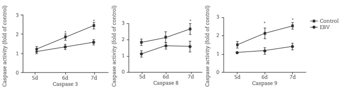
Fig. 5 - Activity of caspase 3, 8, and 9 is assessed on three different days after EBV infection (days 5, 6, and 7). *p < 0.05, (mean ± SD, n = 8).
Caspase acti
vity (fold of contr
ol)
Caspase acti
vity (fold of contr
ol)
Caspase acti
vity (fold of contr
ol)
3
2
1
0
3
2
1
0
3
2
1
0 5d 6d 7d
Caspase 3
5d 6d 7d Caspase 8
5d 6d 7d Caspase 9 *
* * * * Control
EBV Control
*
* (A)
Hoechest 33258
(B)
(C) TUNEL
Control EBV
Hoechst 33258 staining Tunel assay
Control EBV
Hoec
hest 33258 positi
v
e
n
uclein/total n
umber cells
T
unel positi
v
e n
uclein/total
n
umber cells
25
20
15
10
5
0
30
20
10
3, 8, 9 in EBV-treated group were similar to control group at day 5. Whereas, caspase 3, 8, 9 activity was increased progressively during the following days. No such cleavage was observed in the control cells (Fig. 5). These data indicate that activation of caspase family of proteases was, at least in part, associated with EBV-induced apoptosis.
Down–regulation of XIAP expression
To further confirm that caspases were activated in EBV-induced apoptosis, we assessed XIAP expression. DCs were treated with EBV for up to 7 days. XIAP level was analyzed by Western blotting. As shown in Fig. 6, XIAP was markedly decreased in EBV treated group compared to control cells.
Fig. 6 - The expression of XIAP was detected by Western blot on day 7. Mock-infected immature DCs and EBV- infected immature DCs were cultured with GM-CSF and IL-4 for 7 days. Then cells were harvested, and total proteins were extracted. XIAP were analyzed by Western blot. β-actin were normalized for equal protein loading.
Fig. 7 - Mitochondrial membrane potential was detected by flow cytometry on three different days after infection (days 5, 6, and 7).
Mitochondrial alterations in EBV-induced apoptosis
Changes in the membrane potential of mitochondria occur in the early phase of apoptosis. To examine the role of mitochondria in EBV-mediated apoptosis, MMP was monitored by mitochondria sensitive dye, Rh123, and analyzed by flow cytometry. As shown in Fig. 7, mitochondria of untreated control cells retained the ability to take up Rh123. However, the MMP of DCs derived from EBV-treated monocytes significantly decreased from day 5 to day 7 in a time-dependent manner.
Discussion
Despite the fact that monocytes/DCs constitute the key elements in nonspecific and specific immune defenses against viral infection, very little is known about the interactions of EBV with these cell types. Few studies of human neonatal DCs in umbilical cord blood have shown them to be immunocompetent as they can stimulate a mixed leukocyte reaction, although these responses tend to be reduced when compared with adult peripheral blood DCs.5
We have previously demonstrated that CB monocytes can be infected by EBV as shown by the expression of EA and VCA and BcLF-1, BALF-2, and LMP1 mRNA expression (data not shown). Our results suggested that EBV infection could alter biological functions of cord blood monocytes and affect development to CBDC, which might be a new mechanism to disrupt the immune response and promote viral propagation during the early stages of infection.
It is different in susceptibility to EBV treatment between monocytes and DCs, indicating phenotypical alterations during monocyte differentiation into DCs. Immature DCs can be identified by their expression of HLA class I and II molecules, CD1a, CD40, CD80 and CD86, but not CD83. Mature DCs express CD83 and have enhanced HLA class I and II, CD80 and CD86. Some researchers have introduced semi-mature DCs and pathogen-driven regulatory mature DCs. The characteristics of semi-mature DCs are high expression of co-stimulatory molecules and CCR7, low excretion of cytokines, which have been related to immune tolerance and homeostasis.6 The characteristics of
pathogen-driven regulatory mature DCs are high expression of co-stimulatory molecules and CCR7, down-regulation of IL-12 secretion and up-regulation of IL-10 secretion, which have been related to protection of host and immune evasion of pathogens.7-12 According to the phenotype, EBV-infected
DCs in our culture system were similar to the two kinds of DCs mentioned above, which showed mature phenotype and down-regulation of MR expression, with inhibition of differentiation and without enhancement of antigen presenting capacity.
DCs are critical for efficient induction of primary immune responses and inhibition of their function is likely to delay the development of both cellular and humoral specific immunity in EBV-infected individuals. EBV latent membrane protein 1 (LMP-1) protects infected B cells from apoptosis by up regulation of the bcl-2. EBNA-2, another EBV latent protein, can increase the effect of LMP-1 on bcl-2 expression. Bcl-2 is anti-apoptosis
Control EBV
XIAP
β-Actin
Control 5d 6d 7d
EBV
M1
528.29 427.47.3 419.30
380.53 270.90 208.76
M1 M1
M1 M1
M1
0
40
80
120
160
200
0
40
80
120
160
200
0
40
80
120
160
200
0
40
80
120
160
200
0
40
80
120
160
200
0
40
80
120
160
200
Counts
RH123 RH123
RH123 RH123
RH123 RH123
100 101 102 103 104 100 101 102 103 104
100 101 102 103 104 100 101 102 103 104
100 101 102 103 104
protein.13,14 This mechanism does not seem to be used by
EBV with regard to DCs, because EBNA-2 protein was not detected in DCs; moreover, expression bcl-2 and bcl-x are likely to be absent in this type. EBV-induced apoptosis of DCs is a complex phenomenon necessitating multiple events. Although enormous strides made in our understanding of cell death, the mechanism by which EBV cause apoptosis is still unknown.
Viral counterattack could lead to immunosuppression. It could be particularly efficient if it targets DCs, which play a pivotal role in both innate and adaptive immune responses. In this study, we investigated whether EBV might induce apoptosis in DCs derived from cord blood monocytes. We dectected apoptotic DCs by staining with Annexin V and PI. Our results demonstrated that EBV could induce CBDC apoptosis. EBV induced DCs apoptosis which was dependent on caspase activation. We also found that caspase 3, 8, and 9, which belong to the class of proteases involved in apoptosis, were activated. Mitochondrial contribution was validated by MMP loss during apoptosis. We also assessed MMP in apoptotic DCs. The loss of MMP in EBV-treated group suggested that mitochondrion might modulate at an early stage the EBV-mediated death signal. Our findings thus suggested that, in addition to the inhibition of DCs maturation, the immunosuppressive effects of DCs are mediated, at least in part, by inducting DCs apoptosis.
There are two distinct pathways of cell death leading to caspase activation, as already reported. Mitochondrial proteins that cause caspase-dependent cell death include cytochrome C (Cyt C), which triggers caspase-9 activation by binding and activating the apoptotic protease activating factor-1 (Apaf-1), and Smac/Diablo, which potentiates caspase activation by binding inhibitor of apoptosis proteins (IAP) and blocking their caspase-inhibitory activity.
Caspase-3 is activated during most apoptotic processes and is believed to be the main executioner caspase.15 Its activation
has been shown to be essential for DNA fragmentation as well as chromatin condensation and plasma membrane blebbing. Biochemically, these alterations are associated with the translocation of phosphatidylserine to the outer leaflet of the plasma membrane and the activation of an e n d o n u c l e a s e w h i c h c l e a v e s g e n o m i c D N A i n t o internucleosomal fragments. The conserved IAP family of proteins can potently inhibit the enzymatic activity of active caspases. In mammals, caspase-3, -7 and -9 are subject to inhibition by IAPs. Interestingly, although caspase-9 binds to several IAPs, it is primarily inhibited by XIAP. By contrast, caspase-3 and -7 are inhibited by XIAP and to a lesser extent, by c-IAP1, c-IAP2 and NAIP.16,17 XIAP almost disappeared in
EBV-treated group compared to the control group.
Our results suggested the involvement of different pathways for the differential regulation of co-stimulatory molecule expression and apoptosis. DCs maturation and survival are regulated by different signaling pathways, as previously described.18
In this study, we have compared the outcomes of EBV infected and mock-infected monocytes on the development of primary human dendritic cells, important antigen-presenting cells for initiating adaptive immune responses.
EBV not only inhibited the maturation of cord blood monocyte-derived DCs but also promoted the apoptosis of CBDC. It might be one strategy that EBV evades host immune responses to establish persistent infection.
Financial support
This research was supported by grants from National Natural Science Foundation of China (30471620, 81001314); Science and Technology Commission of Shanghai Municipality, China (044119628); PhD Programs Foundation of Ministry of Educa-tion of China (20090073110085); Shanghai Municipal Education Commission, China ( 0 8 Z Z 6 1 ) ; S h a n g h a i M u n i c i p a l H e a l t h B u r e a u (2007Y76); Pudong New District Social Development Bureau (PW 2008D-6); PhD Programs Foundation of Shanghai Jiao Tong University School of Medicine (BXJ0919).
Conflict of interest
All authors declare to have no conflict of interest.
R E F E R E N C E S
1. Kawa K. Epstein-Barr virus-associated diseases in humans. Int J Hematol. 2000;71:108-16.
2. Khanna R, Burrows SR, Moss DJ. Immune regulation in Epstein-Barr virus-associated diseases. Microbiol Rev. 1995;59:387-95.
3. Li L, Liu D, Hutt-Fletcher L, et al. Epstein-Barr virus inhibits the development of dendritic cells by promoting apoptosis of their monocyte precursors in the presence of granulocyte macrophage-colony-stimulating factor and interleukin-4. Blood. 2002;99:3725-34.
4. Salek-Ardakani S, Lyons SA, Arrand JR. Epstein-Barr virus promotes human monocyte survival and maturation through a paracrine induction of IFN-γ. J Immunol. 2004;173:321-31.
5. Liu E, Tu W, Law HK, et al. Decreased yield, phenotypic expression and function of immature monocyte-derived dendritic cells in cord blood. Br J Haematol. 2001;113:240-6. 6. Lutz MB, Schuler G. Immature, semi-mature and fully
mature dendritic cells: which signals induce tolerance or immunity? Trends Immunol. 2002;23:445-9.
7. McGuirk P, Johnson PA, Ryan EJ, et al. Filamentous hemagglutinin and pertussis toxin from Bordetella pertussis modulate immune responses to unrelated antigens. J Infect Dis. 2000;182:1286-8.
8. Van der Kleij D, Latz E, Brouwers JFHM, et al. A novel host-parasite lipid cross-talk. Schistosomal lyso-phosphatidylserine activates toll-like receptor 2 and affects immune polarization.
J Biol Chem. 2002;277:48122-29.
10. Servet-Delprat C, Vidalain P-O, Bausinger H, et al. Measles virus induces abnormal differentiation of CD40 ligand-activated human dendritic cells. J Immunol. 2000;164:1753-60.
11. Schnorr J-Jr, Xanthakos S, Keikavoussi P, et al. Induction of maturation of human blood dendritic cell precursors by measles virus is associated with immunosuppression. Proc Natl Acad Sci USA. 1997;94:5326-31.
12. Marie JC, Kehren J, Trescol-Biémont M-C, et al. Mechanism of measles virus-induced suppression of inflammatory immune responses. Immunity. 2001;14:69-79.
13. Kieff E. Epstein-Barr virus and its replication. In: Fields BN, Knipe DM, Howley PM, eds. Fields Virology. 3rd ed. Philadelphia: Lippincott-Raven, 1996:2343-96
14. Gregory CD, Dive C, Henderson S. Activation of Epstein-Barr virus latent genes protects human B cells from death by apoptosis. Nature. 1991;349:612-14.
15. Shi Y. Mechanisms of caspase activation and inhibition during apoptosis. Mol Cell. 2002;9:459-70.
16. Salvesen GS, Duckett CS. IAP proteins: blocking the road to death’s door. Nature Rev Mol Cell Biol. 2002;3:401-10. 17. Maier JK, Lahoua Z, Gendron NH, et al. The neuronal
apoptosis inhibitory protein is a direct inhibitor of caspases 3 and 7.
J Neurosci. 2002;22:2035-43.