Universidade de Lisboa
Faculdade de Medicina de Lisboa
Study the function of the newly discovered
TrkB receptor fragment (TrkB-ICD) formed by
calpain cleavage
João Filipe Fonseca Gomes
Orientadores:
Professora Doutora Maria José de Oliveira Diógenes Nogueira
Doutor André Jerónimo Santos
Dissertação especialmente elaborada para obtenção
do grau de Mestre em Neurociências
2016
Todas as afirmações efectuadas no presente documento são da
exclusiva responsabilidade do seu autor, não cabendo qualquer
responsabilidade à Faculdade de Medicina de Lisboa pelos conteúdos
nele apresentados.
A impressão desta dissertação foi aprovada pelo Conselho Científico da Faculdade de Medicina de Lisboa em reunião de19 de janeiro de 2016.Publications
In parallel with my Master project, I collaborated in other project whose results are now published (1). Moreover, I was invited to participate in the preparation of a review article already published (2):
1. Jerónimo-Santos A, Fonseca-Gomes J, Guimarães DA, Tanqueiro SR, Ramalho RM, Ribeiro A, Sebastião AM, Diógenes MJ (2015). Brain-Derived Neurotrophic Factor mediates neuroprotection against Aβ-induced toxicity through a mechanism independent on adenosine 2A receptor activation.
Growth Factors. 33(4):298-308.
2. Ribeiro FF, Xapelli S, Miranda-Lourenço C, Tanqueiro SR, Fonseca-Gomes J, Diógenes MJ, Ribeiro JA, Sebastião AM (2015). Nucleosides in neuroregeneration and neuroprotection. Neuropharmacology. S0028-3908(15)30170-2.
Index
Figure Index _________________________________________________________ v Tables Index _________________________________________________________ ix Abbreviations and symbol list __________________________________________ xi Acknowledgments ___________________________________________________ xv Resumo __________________________________________________________ xvii Abstract ___________________________________________________________ xxi 1 Introduction ______________________________________________________ 1 1.1 Neurotrophins _________________________________________________ 1 1.1.1 Brain-derived neurotrophic factor _____________________________ 3 A) BDNF: processing and release ________________________________ 3 B) BDNF receptors ____________________________________________ 5 C) BDNF/TrkB-FL system _______________________________________ 7 1.2 Neurodegeneration ___________________________________________ 11 1.2.1 Alzheimer’s disease ________________________________________ 13 A) Molecular features of AD ___________________________________ 15 B) Consequences of Aβ accumulation ____________________________ 16 C) BDNF/TrkB-FL system in AD _________________________________ 18 1.3 Calpains ____________________________________________________ 21 1.3.1 Fragments produced by calpain cleavage ______________________ 22 A) Src protein _______________________________________________ 22 B) β-catenin protein _________________________________________ 23 C) p35 protein ______________________________________________ 23 D) mGluR1α protein _________________________________________ 23 E) TrkB-FL receptor __________________________________________ 24 2 Aim ____________________________________________________________ 27 3 Methods _______________________________________________________ 29 3.1 Cells cultures _________________________________________________ 29 3.1.1 H4 Cell line – Neuroglioma cells ______________________________ 29 3.1.2 Primary neuronal cultures __________________________________ 29 3.2 Cloning _____________________________________________________ 30 3.3 Transfection _________________________________________________ 33 3.4 Drug treatments ______________________________________________ 34 3.5 Subcellular Fractionation _______________________________________ 35 3.6 Western-blot _________________________________________________ 37 3.7 Immunofluorescence __________________________________________ 38 3.8 Statistical analysis ____________________________________________ 38 3.9 Reagents, drugs and antibodies __________________________________ 39 3.9.1 Culture reagents __________________________________________ 39 3.9.2 Drugs ___________________________________________________ 403.9.3 Antibodies _______________________________________________ 40 4 Optimization and characterization of the transfection process ____________ 43 4.1 Rational ____________________________________________________ 43 4.2 Determination of optimal time transfection and moment of medium renewal 43 4.3 TrkB-ICD expression on primary neuronal cultures ___________________ 46 4.4 Discussion ___________________________________________________ 48 5 Study of TrkB-ICD stability _________________________________________ 51 5.1 Rational ____________________________________________________ 51 5.2 Determination of TrkB-ICD half-life time ___________________________ 51 5.3 Discussion ___________________________________________________ 54 6 Subcellular expression of TrkB-ICD fragment __________________________ 55 6.1 Rational ____________________________________________________ 55 6.2 Distribution of TrkB-ICD fragment on H4 cells _______________________ 55 6.3 Distribution of TrkB-ICD fragment on primary neuronal cultures ________ 58 6.4 Discussion ___________________________________________________ 60 7 Impact of TrkB-ICD fragment on cells phosphorylation pattern ___________ 65 7.1 Rational ____________________________________________________ 65 7.2 Evaluation of tyrosine kinase activity of TrkB-ICD fragment on H4 cells and primary neuronal cultures __________________________________________________ 65 7.3 Kinetics of phosphorylation induced by TrkB-ICD fragment ____________ 69 7.4 Signalling pathways contribution for protein phosphorylation induced by TrkB-ICD 74 7.5 Phopshorylation pattern induced by TrkB-ICD fragment: evaluation on subcellular fractions ______________________________________________________ 77 7.6 Relationship between phosphorylation pattern and TrkB-ICD nuclear translocation 79 7.7 Discussion ___________________________________________________ 81 8 Conclusions and Future Perspectives _________________________________ 89 9 References ______________________________________________________ 91
Figure Index
Figure 1.1 – Schematic representation of neurotrophins interactions with its receptors. ___________ 2 Figure 1.2 – Schematic representation of BDNF processing and release. ________________________ 4 Figure 1.3 – Schematic representation of TrkB receptors isoforms: TrkB-FL and truncated forms (TrkB-T1, TrkB-T2 and TrkB-T-Shc). __________________________________________________________ 6 Figure 1.4 – Schematic representation of BDNF/TrkB-FL signalling. ____________________________ 9 Figure 1.5 – Schematic representation concerning the effects of dysregulations on BDNF/TrkB-FL system. __________________________________________________________________________ 10 Figure 1.6 – Estimative of the proportion of all dementia cases subdivided by different dementia subtypes. _________________________________________________________________________ 12 Figure 1.7 – Comparison between (A) a normal aged brain and (B) an AD brain. ________________ 14 Figure 1.8 – Differential processing of APP: (A) non-amyloidogenic and (B) amyloidogenic pathways. 16 Figure 1.9 – Schematic representation of toxicity mechanisms induced by Aβ peptide. ___________ 17 Figure 1.10 – Role of BDNF/TrkB-FL system on neurodegeneration: a putative cycle with positive feedback. ________________________________________________________________________ 20 Figure 1.12 – Action of calpains on cell substrates. ________________________________________ 22 Figure 1.13 – Alterations of both TrkB-FL and TrkB-ICD levels in the brain of AD, when compared with an age-matched control. ____________________________________________________________ 24 Figure 3.1 - Representative images of (A) H4 cells and (B) primary cortical neurons. _____________ 30 Figure 3.2 – Expression vectors used in this work: (A) TrkB-ICD-V5 and (B) TrkB-ICD. _____________ 31 Figure 3.3 – Amplification of TrkB-ICD by PCR. ___________________________________________ 32 Figure 3.4 – (A) H4 cells transfected with TrkB-ICD-V5 and TrkB-ICD plasmids and (B) summary of plasmid cloning process _____________________________________________________________ 33 Figure 3.5 – Schematic representation of transfection process. ______________________________ 34 Figure 3.6 – Subcellular fractionation protocol: (A) summary of all steps and (B) preliminary results. 36 Figure 4.1 – TrkB-ICD expression on H4 cells transfected with pcDNA-TrkB-ICD-V5 plasmid. _______ 44 Figure 4.2 – Transfection optimization of H4 cells transfected with pcDNA-TrkB-ICD-V5: moment of medium renewal. __________________________________________________________________ 45 Figure 4.3 – TrkB-ICD fragment is mainly expressed in neurons. ______________________________ 46 Figure 4.4 – Percentage of transfected cells is approximately 15% of total cells. _________________ 47 Figure 5.1 – Time course analysis of TrkB-ICD stability after CHX (5 μM) treatment on (B1, B2) H4 cells and (C1, C2) primary neuronal cultures. ________________________________________________ 53 Figure 5.2 – First order decay function: an intermediate step to determine TrkB-ICD half-life time. __ 53 Figure 6.1 – Distribution of TrkB-ICD fragment on H4 cells transfected with pcDNA-TrkB-ICD: subcellular fractionation. ____________________________________________________________ 56Figure 6.2 – Distribution of TrkB-ICD fragment on H4 cells transfected with pcDNA-TrkB-ICD (24h): immunofluorescence assay. __________________________________________________________ 56 Figure 6.3 – Distribution of TrkB-ICD-V5 fragment on H4 cells transfected with pcDNA-TrkB-ICD-V5 (48h): immunofluorescence assay. _____________________________________________________ 57 Figure 6.4 – Nuclear translocation of TrkB-ICD fragment in 7 DIV neurons transfected with pcDNA-TrkB-ICD: subcellular fractionation. ____________________________________________________ 58 Figure 6.5 – Nuclear translocation of TrkB-ICD fragments in 7 DIV neurons transfected with pcDNA-TrkB-ICD: immunofluorescence assay. __________________________________________________ 59 Figure 6.6 – Schematic representation of transfection steps until protein release in cell cytoplasm. _ 60 Figure 6.7 – Schematic representation of TrkB-ICD translocation to the nucleus on (A) primary neuronal cultures and (B) H4 cells. _____________________________________________________ 61 Figure 6.8 – Results obtained from cNLS Mapper software about prediction of NLS on TrkB-ICD sequence. ________________________________________________________________________ 63 Figure 7.1 – Levels of tyrosine phosphorylated proteins on (A) H4 cells and (B) primary neuronal cultures. _________________________________________________________________________ 66 Figure 7.2 – Localization of tyrosine phosphorylated proteins on H4 cells transfected with pcDNA-TrkB-ICD plasmid. ______________________________________________________________________ 67 Figure 7.3 – Localization of tyrosine phosphorylated proteins on 7 DIV primary neuronal cultures transfected with pcDNA-TrkB-ICD. _____________________________________________________ 68 Figure 7.4 – Transient phosphorylation pattern on (A1, A2) H4 cells and (B) 7 DIV primary neuronal cultures transfected with pcDNA-TrkB-ICD-V5. ___________________________________________ 70 Figure 7.5 – CHX (5 μM) treatment effects on levels of tyrosine phosphorylated proteins on (A1, A2) H4 cells and (B) 7 DIV primary neuronal cultures transfected with pcDNA-TrkB-ICD. ________________ 72 Figure 7.6 – K252a (5 μM) treatment effects on levels of tyrosine phosphorylated proteins on (A) H4 cells and (B) 7 DIV primary neuronal cultures transfected with pcDNA-TrkB-ICD. ________________ 73 Figure 7.7 – Contribution of signalling pathways on TrkB-ICD levels and ratio between phosphorylation levels and TrkB-ICD fragment amount (7 DIV primary neuronal cultures). ______________________ 76 Figure 7.8 – Levels of tyrosine phosphorylated proteins on different fractions of (A) H4 cells and (B) 7
Figure 10.1 – TrkB-ICD fragment decreases cell viability on 7 DIV primary neuronal cultures. _____ 112 Figure 10.2 – TrkB-ICD fragment does not affect levels of nuclear GAPDH isoform. _____________ 113 Figure 10.3 – TrkB-ICD-V5 does not promote αII-spectrin breakdown. ________________________ 114 Figure 10.4 – K252a (5 μM) treatment effects on TrkB-ICD levels on different fractions of HEK293T cells transfected with pcDNA-TrkB-ICD. ____________________________________________________ 116
Tables Index
Table 3.1 – Expression vectors used in this experimental work. ______________________________ 30 Table 3.2 – Drugs/Mediums used in cell cultures. _________________________________________ 39 Table 3.3 – Drugs used for cells treatment. ______________________________________________ 40 Table 3.4 – Primary antibodies used in Western-blot and Immunofluorescence assays. ___________ 40 Table 3.5 – Secondary antibodies used in Western-blot and Immunofluorescence assays. _________ 41 Table 7.1 – Drugs used and respective signalling pathway inhibited. __________________________ 74 Table 10.1 – Approaches performed to study TrkB-ICD toxicity. _____________________________ 111
Abbreviations and symbol list
μg Microgram μL Microliter μM Micromolar aa Amino Acid Aβ Amyloid-Beta AD Alzheimer’s Disease ADP Adenosine Diphosphate ANOVA Analysis of Variation APP Amyloid Precursor Protein ATP Adenosine Triphosphate BACE1 Beta-site amyloid precursor protein cleaving enzyme 1 BDNF Brain-Derived Neurotrophic Factor bp Base Pairs Ca2+ Calcium (2+) Ion CDK5 Cyclin-dependent Kinase 5 C&M Fraction Enriched in Cytoplasmic and Membrane Proteins CHX Cyclohexamide CMV Cytomegalovirus CNS Central Nervous System CTR Control Dapi 4',6-diamidino-2-phenylindole DIV Days In Vitro DMSO Dimethyl Sulfoxide DNA Deoxyribonucleic Acid DTT 1,4-dithiothreitol EDTA Ethylenediaminetetraacetic Acid EGFR Epidermal Growth Factor Receptor EGTA Ethylene Glycol Tetraacetic Acid ER Endoplasmic Reticulum EV Empty Vector iAβ Intracellular Amyloid-Beta IF Immunofluorescence FL Full-Length GAPDH Glyceraldehyde 3-Phosphate Dehydrogenase GC Golgi Complex H Homogenate H2O WaterHBSS Hanks' Balanced Salt Solution HEPES N-2-Hydroxyethylpiperazine-N'-2-Ethanesulfonic Acid ICD Intracellular Domain KCl Potassium Chloride Lipof. Lipofectamine MAPK Mitogen-Activated Protein Kinase MG132 Z-Leu-Leu-Leu-CHO Mn2+ Manganese (2+) Ion mRNA Messenger Ribonucleic Acid MTT 3-(4,5-Dimethylthiazol-2-yl)- 2,5-Diphenyl Tetrazolium N Fraction Enriched in Nuclear Proteins Na3VO4 Sodium Orthovanadate NaCl Sodium Cloride NaF Sodium Floride ng Nanogram NLS Nuclear Localization Sequence n.s. Non Significant NT Neurotrophin p75NTR P75 Neurotrophin Receptor PBS Phosphate Buffered Saline PC12 Pheochromocytoma Cell Line PCN Primary Cortical Neurons PCR Polymerase Chain Reaction PD Parkinson’s Disease PDL Poly-D-Lysine Pi Phosphate Group PI3K Phosphatidylinositol 3-Kinase PIP2 Phosphatidylinositol-4,5-Bifosphate PIP3 Phosphatidylinositol-3,4,5-Trisphosphate PKA Protein Kinase A
Tc Truncated TBS Tris-Buffered Saline TBS-T Tris-Buffered Saline-Tween Trk Tropomyosin-related Kinase TrkB-ICD TrkB-Intracellular Domain (calpain-generated) TrkB-FL TrkB Full-Length TrkB-T’ TrkB Truncated (calpain-generated) TrkB-T1 TrkB Truncated isoform 1 TrkB-T2 TrkB Truncated isoform 2 TrkB-Tc TrkB Truncated (total pool) UK United Kingdom USA United States of America WB Western-Blot wt:vol Weight:volume
Acknowledgments
Para além do meu contributo, este projecto só foi possível com a participação e cooperação, directa ou indirecta, de algumas pessoas a quem só estar grato. Em primeiro lugar, gostaria de agradecer à Mizé e ao André pela forma como me receberam, por me terem rapidamente integrado no projecto e por terem guiado o início da minha carreira científica. A eles lhes agradeço a simpatia, a amizade, a paciência, a motivação, a confiança e por me terem dado espaço para pensar. Mizé, não há palavras para si, sempre foi insuperável e nem por uma vez me senti desapoiado, acho que tudo se resume à certeza de que não podia ter desejado melhor. André, graças ao teu espírito de Descobridor, encontraste o TrkB-ICD que, à partida, vai melhorar enormemente a qualidade de vida dos pacientes de Alzheimer, bem como das suas famílias. Para além disso, obrigado pelo companheirismo, alguma paciência e pelos ensinamentos que me transmitiste.Gostaria também de mostrar o meu sincero agradecimento à Professora Ana Sebastião que me permitiu realizar este projecto de investigação na sua unidade e sempre me fez sentir à vontade em me dirigir ao seu gabinete para qualquer que fosse o tema de conversa.
Catarina, ambos sabemos que não foi o melhor ano das nossas vidas, mas só tenho motivos para te agradecer teres estado a meu lado em todas as alegrias e frustrações. Obrigado por, já há algum tempo, fazeres parte dos meus dias e da minha vida. Um cumprimento especial e caloroso.
Gostaria agora de agradecer aos meus colegas de laboratório e pessoal da secretaria por todo o companheirismo, ajuda, empatia e tolerância para comigo. Uma palavra especial para as pessoas que, ao longo deste ano, tiveram mais paciência comigo e aligeiraram os meus dias ao ponto de os poder considerar meus amigos: Sara, Margarida, Mariana, Francisco, Rui, Afonso e Tatiana.
Gostava de dedicar esta tese a quem, desde sempre, esteve, está e estará comigo: os meus pais e o meu avô. Aos meus pais lhes agradeço todo o apoio, motivação, incondicional amizade e por serem sempre o meu porto de abrigo, quaisquer que sejam as situações e tenham mais ou menos razões para estarem do meu lado. Termino com um agradecimento especial ao meu avô que, esteja onde estiver, ficará feliz por me ver finalizar mais uma etapa. Embora não o tenha comigo, tenho a certeza que está orgulhoso de ver o seu neto conseguir mais uma conquista na vida.
Resumo
A Doença de Alzheimer (DA) é a forma mais prevalente de demência em todo o Mundo (totalizando entre 60 a 70% de todos os casos diagnosticados de demência). Esta é uma doença neurodegenerativa crónica caracterizada por diferentes estadios de progressão, culminando em atrofia cerebral globalizada e em perda neuronal substancial em áreas cerebrais particulares, com especial foco no hipocampo e no córtex entorrinal.
Com base nos inúmeros estudos desenvolvidos nas últimas décadas, actualmente, a comunidade científica considera a DA uma doença resultante da acumulação extraneuronal do péptido β-amilóide (Aβ) e acumulação intraneuronal da forma hiperfosforilada da proteína tau.
Para além da acumulação cerebral das proteínas anteriormente referidas, nesta doença, também se sabe estar presente uma desregulação da sinalização mediada pelo Factor Neurotrófico derivado do Cérebro (Brain-derived neurotrophic factor, BDNF). O BDNF, através da activaçao da isoforma completa do seu recetor (TrkB-FL), é responsável pela activação de vias moleculares essenciais à sobrevivência e diferenciação neuronais, bem como à transmissão e plasticidade sinápticas. Tanto em amostras de tecido cerebral post-mortem de pacientes com DA como em cérebros de modelos animais de DA foi possível detetar níveis diminuídos de BDNF e do receptor TrkB-FL bem como aumento significativo das isoformas truncadas do recetor TrkB (TrkB-Tc), consideradas inibidoras da sinalização do TrkB-FL. Este comprometimento da função do BDNF implica alterações profundas na homeostasia celular, uma vez que se assiste a uma perda significativa das funções celulares mediadas por esta neurotrofina. Desta maneira, actualmente considera-se que a perda de sinalização do BDNF poderá directamente contribuir para a morte neuronal associada à neurodegeneração.
Recentemente, foi descrito um novo mecanismo que está também subjacente à perda de sinalização do BDNF na DA: clivagem do recetor TrkB-FL. Na verdade, um dos efeitos neurotóxicos associados à acumulação do péptido Aβ está associado a um aumento significativo dos níveis de cálcio intracelulares, através de
inúmeros mecanismos, incluindo a interacção com receptores transmembranares (como os receptores NDMA), a disrupção membranar através da formação de poros e ainda a produção de espécies reactivas de oxigénios. Por sua vez, existe um grupo particular de enzimas dependentes dos níveis de cálcio que, neste contexto, são sobre-activados e conduzem à clivagem dos recetores TrkB-FL. Assim, como consequências desta clivagem, observa-se uma diminuição dos níveis do recetore TrkB-FL e a respectiva formação de dois fragmentos: 1) um novo receptor truncado (TrkB-T’) e 2) um fragmento intracelular (TrkB-ICD) que é libertado para o citosol. Contudo, tendo em conta que este mecanismo associado à perda de sinalização do BDNF foi recentemente descrito, não existia, até ao momento, qualquer tipo de caracterização destes fragmentos e do seu possível papel na patofisiologia da DA. Desta maneira, este trabalho foi delineado por forma a caracterizar o fragmento intracelular TrkB-ICD e estudar o seu possível papel na DA.
Para responder aos objectivos propostos, foram usados dois tipos celulares distintos: linha celular de neuroglioma humano (células H4) e culturas primárias de neurónios corticais de rato. Inicialmente, procedeu-se à optimização de diversos parâmetros do protocolo de transfecção, de modo a garantir a expressão óptima da proteína de interesse (TrkB-ICD) em ambos os tipos celulares usados. De seguida, usando Ciclohexamida (CHX, 5 μM), composto inibidor da síntese proteica de células eucarióticas, foi avaliada a estabilidade do fragmento TrkB-ICD. Os resultados obtidos indicam que este fragmento é uma proteína estável e com um tempo de semi-vida próximo das oito horas, período suficientemente longo para ser possível colocar a hipótese de que o fragmento possa interferir com a homeostase da célula.
actividade de cinase. Para além disso, ensaios com CHX demonstram que esta actividade de cinase se mantém constante ao longo do tempo, enquanto que, ensaios com K252a (5 μM), fármaco inibidor da actividade de cinase dos recetores Trk, indicam que as proteínas fosforiladas pelo TrkB-ICD são rapidamente desfosforiladas, possivelmente através de fosfatases. Por fim, através do uso de um conjunto de seis fármacos inibidores das principais vias de sinalização descritas em células eucariotas, observámos que a fosforilação induzida pelo fragmento em estudo poderá ser positivamente modulada pela via que inclui a Proteína Cinase A, uma vez que se assiste a uma diminuição drástica do padrão de fosforilação induzido pelo fragmento TrkB-ICD quando as células transfectadas são incubadas com o inibidor desta via (H-89, 25 μM).
Assim sendo, as evidências acima enumeradas suportam a hipótese de que o fragmento TrkB-ICD poderá ter um papel importante na patofisiologia da DA, podendo actuar como regulador de vias de sinalização ou regulador da transcrição genética, uma vez que se acumula no domínio nuclear. Desta maneira, os resultados deste trabalho poderão vir a tornar-se a base de trabalho futuro que permita identificar novos alvos farmacológicos para o tratamento da DA.
Palavras-Chave:
Neurodegeneração, Doença de Alzheimer, Factor Neurotrófico derivado do
Abstract
Alzheimer’s Disease (AD) is the most common form of dementia worldwide (60-70% of all cases) and the accumulation of amyloid-beta (Aβ) peptide in the brain is considered as one of the main hallmarks of this disease. Indeed, AD is a chronic neurodegenerative disease with different stages of progression, leading to general atrophy and neuronal loss of particular brain regions, such as hippocampus or entorhinal cortex, which promotes cognitive and functional impairment. Actually, accumulation of Aβ peptide contributes to neuronal death, since its accumulation on Aβ plaques induces neurotoxicity at different levels and through different mechanisms. Hence, AD is considered a protein misfolding disease and Aβ peptide is considered the main player of its etiology.
Simultaneously, Brain-derived neurotrophic factor (BDNF) signalling is seriously impaired in AD, which consequently compromises its physiological functions: neuronal survival, differentiation and plasticity. Actually, decreased levels of BDNF and its receptor (TrkB Full-Length, TrkB-FL), as well as increased levels of truncated TrkB receptors (TrkB-Tc), a dominant negative inhibitor of TrkB-FL, were already described in AD and led to the postulation that this loss of BDNF signalling could directly contribute to neurodegeneration.
Recently, it was discovered a new mechanism that also contributes to the impairment of BDNF signalling in AD: TrkB-FL cleavage. Indeed, accumulation of Aβ peptide promotes a sustained increase of intracellular Ca2+ levels, by distinct mechanisms, which in
turn leads to calpains overactivation and subsequent TrkB-FL cleavage. As consequences, TrkB-FL levels decreases and two fragments are generated: a membrane-bound truncated receptor (TrkB-T’) and an intracellular fragment (TrkB-ICD) that is released to cytosol. However, there was no further characterization about these new fragments. Thus, this work was designed to characterize TrkB-ICD fragment and to study its putative role in AD pathophysiology.
In this way, to reach the goal of this work, we used a human neuroglioma cell line (H4 cells) and primary rat cortical neurons. Initially, we optimized the transfection protocol in order to ensure the optimal TrkB-ICD expression on both cells type used. Then, we evaluated the stability of TrkB-ICD using cyclohexamide treatment (5 μM). The obtained data shows that this fragment is a stable protein with a half-life time of 8 hours, approximately, suggesting that TrkB-ICD is not immediately degraded. Immunofluorescence assays and subcellular fractionation protocols performed also indicate that TrkB-ICD accumulates within the cell nucleus over time, a translocation process dependent on its own
phosphorylation. Furthermore, we clearly show that TrkB-ICD induces a significant phosphorylation of several proteins at nucleus, soma and neuronal processes, showing that this fragment has constitutive tyrosine kinase activity. Finally, by using several inhibitors of the major signalling pathways in eukaryotic cells, we can conclude that protein phosphorylation induced by TrkB-ICD is probably modulated in a positive way by PKA pathway. The results obtained support the hypothesis that the TrkB -ICD fragment may play a role in AD pathophysiology either by acting as regulator of signalling pathways or, since it accumulates in the nucleus, as regulator of gene transcription. Thus, data obtained in this work could underlie future work that might allow the identification of new pharmacological targets for the treatment of AD and further research on this fragment could also unveil if TrkB-ICD fragment can be considered a biomarker for AD.
Keywords:
Neurodegeneration, Alzheimer’s Disease, BDNF, TrkB-FL, TrkB-ICD1 Introduction
1.1 Neurotrophins
Neurotrophins (NTs) are a family of growth factors that belong to the broad group of neurotrophic factors and have a crucial importance in a healthy nervous system. NTs can have different functions depending on the cellular developmental stage or depending on the type of external insults 1–3. In early developmental stage,
these proteins control some aspects of growth, survival and differentiation of developing neurons, but, in mature neurons, they can also act as synaptic plasticity regulators. In addition to the role of neurotrophins on central neurons, they have also a crucial role on the peripheric nervous system, including sensory and sympathetic neurons 4,5. Accordingly, NTs have crucial functions in both peripheral
and central nervous systems homeostasis.
Nerve Growth Factor (NGF) was the first NT identified by an Italian biologist Rita Levi-Montalcini, who described its role on the morphological differentiation of neural-crest-derived nerve cells 6. After this discovery, many other studies concerning NTs have started, resulting in the identification of new biological mediators involved in the promotion of cell growth, differentiation, function and survival of nerve cell populations. Thus, in addition to NGF, other three neurotrophins were found in mammals: Brain-derived neurotrophic factor (BDNF), Neurotrophin-3 (NT-3) and Neurotrophin-4/5 (NT-4/5) and another two NTs in fishes: Neurotrophin-6 (NT-6) and Neurotrophin-7 (NT-7) 7–12.
Each neurotrophin has different characteristics and properties, and they are recognized by specific receptors as shown in Figure 1.1. In general, there are two types of receptors for neurotrophins that mediate the activation of different signalling pathways with apparent opposite biological effects: the p75 neurotrophin receptor (p75NTR) and the tropomyosin-related kinase receptors (Trk) 2,13. Currently
there are three types of Trk receptors identified (TrkA, TrkB and TrkC) that are products of three different genes (NTRK1, NTRK2 and NTRK3, respectively). Trk
receptors share some main characteristics, such as its structure and functioning 14–17. Both Trk receptors and p75NTR are located at the cellular membrane, being activated by the binding of a ligand in their extracellular domain with consequences at intracellular level. This ligand-binding specificity is one of intrinsic characteristics of different Trk receptors that determine its classification, as mentioned above 2,3.
Briefly, the immature neurotrophins (pro-neurotrophins) are recognized by p75NTR, but after processing and maturation, the pro-neurotrophins are converted to neurotrophins that bind preferentially to Trk receptors, instead of p75NTR-binding (Figure 1.1) 4,18. The activation of these two different subsets of receptors
leads to distinct signalling cascades, which will be described in more detail below on Section 1.1.1.B.
1.1.1
Brain-derived neurotrophic factor
BDNF was the first NT identified after the discovery of NGF and, nowadays, it is considered the NT with most widespread expression in mammalian brain. In 1982, Barde and collegues, using 1.5 kg of pig brain, were able to purify 1 μg of BDNF. This study identified the molecular weight of BDNF (12.3 kDa), its isoelectric point (pI = 10.1) and its biological activity (0.4 ng/mL/unit), which is similar to NGF using same approaches7. This similarity can be explained due to highly homology between protein sequence of BDNF and NGF 19. It was also reported by Barde that this NT is important for survival and fibre outgrowth of cultured embryonic chick sensory neurons and for subpopulations not responsive to NGF, but the remaining characterization of its physiological function and structural properties was not much discussed on his initial research 1,7.BDNF expression pattern changes during neurodevelopment, indeed, while BDNF is abundantly expressed in hippocampus, neocortex, amygdala and cerebellum during the first months of postnatal development, in adult stages, BDNF expression is homogeneous throughout the brain 20. A) BDNF: processing and release
Similarly to other NTs, the initial pre-mRNA sequence of BDNF (pre-pro-BDNF) present in nucleus is converted to pro-BDNF (231 amino acids (aa)), after processing in endoplasmic reticulum (ER) and Golgi complex (GC). Then, this precursor form of BDNF can be processed to the mature protein (118-120 aa) by several cellular mechanisms. At intracellular level, pro-BDNF can be cleaved to homodimeric protein by two mechanisms: by furin or by proconvertase enzymes in regulated secretory vesicles. Thereby, BDNF can be released in a regulated-way dependent on neuronal activity or can be constitutively released, if occurs spontaneous fusion of vesicles with plasma membrane. On the other hand, when pro-BDNF is released as an unprocessed form, it can be directly processed by plasmin at extracellular level or can be recognized by p75NTR. Following this binding
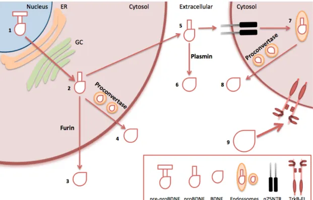
to p75NTR, BDNF is endocytosed into the cell, being then cleave to mature BDNF within endosomes by proconvertase enzymes, what leads to its release to extracellular level 21–26. In this way, mature BDNF present in extracellular side activates TrkB receptors and thereby promotes its effects, which includes neuronal survival and regulation of synaptic plasticity, as it will be described below. All these steps are schematically showed in Figure 1.2.
Figure 1.2 – Schematic representation of BDNF processing and release. Pre-mRNA sequence of BDNF present in nucleus (1) is converted to pro-BDNF, after processing in endoplasmic reticulum (ER) and Golgi complex (GC) (2). Then, this immature form can be cleaved by furin, releasing BDNF to extracellular side (3). However, BDNF can also reach
B) BDNF receptors
As mentioned above, mature BDNF binds to TrkB receptor with high-affinity and with low-affinity to p75NTR. Interestingly and on the opposite, p75NTR recognizes pro-neurotrophins with high affinity 4,18. As briefly mentioned above, the
activation of each subset of receptors leads to distinct signalling cascades, which could lead to opposite effects on cell environment. p75NTR belongs to tumour necrosis factor (TNF) receptor superfamily and the signalling mediated by this receptor is not still fully understood. However, it is already described that its activation by pro-neurotrophins triggers mainly apoptotic pathways and impairs normal synaptic function, serving Sortilin as a co-receptor. On the other hand, TrkB activation is already well characterized and is involved in main physiologic functions of neurotrophin signalling pathways, including neuronal survival, synaptic plasticity and differentiation. However, it is important to mention that in some circumstances, p75NTR can act as a co-receptor of BDNF, acting synergistically on TrkB pathways
23,27.
Regarding the structure of TrkB, this receptor has several communalities with other receptors of its family. Briefly and as shown in Figure 1.3, at extracellular level, Full-Length (FL) form of TrkB receptor (TrkB-FL) are composed by two cysteine rich domains, one domain rich in leucines and, below these domains, it has two immunoglobulin-like (IgG-like) domains, which compose the binding site responsible for selective binding site. In turn, at intracellular level, TrkB-FL receptor is mainly constituted by tyrosine kinase domain between Shc protein binding site on the top (at juxtamembrane domain) and a C-terminal tail, which includes phospholipase C gamma (PLCγ) binding site 28. Therefore, the intracellular domain of TrkB-FL is essential for signalling pathway activation (as will be detailed in Figure 1.4 and in Section 1.1.1.C).
Concerning TrkB receptors and in addition to TrkB-FL receptor, there are also truncated (Tc) isoforms (TrkB-Tc) of the receptor generated by alternative splicing of TrkB gene (NTRK2) 28–32. The main truncated TrkB isoforms characterized are the
TrkB-T1, TrkB-T2 or TrkB-T-Shc receptors. The truncated TrkB isoforms lack the tyrosine kinase domain and, thereby they are not able to trigger the canonical
signalling pathways. Moreover, these truncated receptors can act as negative modulators of TrkB-FL signalling since they can form non-functional heterodimers with TrkB-FL receptors inhibiting the downstream signalling, even in the presence of BDNF and functional TrkB-FL monomers 33. In addition, truncated isoforms of TrkB receptor also bind BDNF that is internalized with its receptor, leading to a decrease of available extracellular levels of this neurotrophin 34.
As shown in Figure 1.3, all TrkB receptors isoforms share the same extracellular domain, being only distinguished by different length of C-terminal tail.
C) BDNF/TrkB-FL system
As indicated above, the physiological consequences of BDNF signalling mediated by TrkB-FL are vital to the homeostasis maintenance of the mammalian nervous system, since it promotes neuronal growth, survival and differentiation or regulation of synaptic transmission and plasticity 2,3.
In vivo and in the presence of homodimeric BDNF, TrkB-FL receptors dimerize promoting the auto-phosphorylation of several tyrosine residues present in the catalytic domain, at intracellular level 35,36. Hence, this phosphorylation triggers
downstream signalling cascades. As represented in Figure 1.4, the TrkB-FL dimerization promotes fast phosphorylation of three tyrosine residues (Y) present on the tyrosine kinase domain (Y701, Y705 and Y706). In turn, this tyrosine kinase activity
leads to the phosphorylation of another two tyrosine residues: Y515 (in
juxta-membrane domain) and Y816 (at C-terminus), which will act as docking site for Shc
adaptor protein (directly associated with Ras/MAPK/Erk and PI3K/Akt pathways) and PLCγ, respectively 35,37–40.
Thus, the major signalling pathways mediated by the activation of TrkB-FL receptors are:
• Ras/MAPK/Erk pathway
Phosphorylated Y515 of TrkB-FL promotes the formation of a complex involving Shc protein and another proteins, such as Grb2, Gab1/2 and SOS, which leads to a transient activation of small GTPases and, in particular, Ras protein. In turn, activated Ras sequentially stimulates Raf kinase, which will activate MAPK/ERK kinase (MEK), promoting an activated cascade of events that, at the end, contributes to the activation of several transcription factors. These transcription factors activated (as it is the case of c-Fos and NF-kB) are involved in the control of expression of many proteins directly involved in neuronal survival, growth and differentiation 40–44.
• PI3K/Akt pathway
In addition to mentioned above, Ras protein is also involved in the activation of PI3K pathway, where there is a phosphorylation of PIP2, resulting in production of
PIP3 that activates serine-theronine kinase Akt. In turn, this stimulated Akt will
modulate the expression and function of a wide range of promoters directly associated with axonal growth and mainly neuronal survival, such as the inactivation of pro-apoptotic proteins. Some downstream effects of this pathway are shared with the first pathway described, since this via also promotes the activation of some transcription factors (like CREB or NF-kB) that control the expression of pro-apoptotic proteins 40,41,45.
• PLCγ pathway
Phosphorylation of Y816 promotes the binding of PLCγ1 that becomes
activated via TrkB-FL kinase domain. So, activated PLCγ1 produces IP3 and DAG from
PIP2 hydrolysis, stimulating DAG-regulated protein kinase C (PKC) and the release of
Ca2+ from intracellular sources, respectively. This increase of intracellular Ca2+ levels will activate Ca2+-regulated PKC and calmodulin-regulated kinase isoforms. Together, it contributes to the regulation of genes transcription (like CREB) with direct influence on neuronal connections, affecting synaptic plasticity and, consequently, memory and learning processes, which are dependent on long-term potentiation (LTP) 40,41,46.
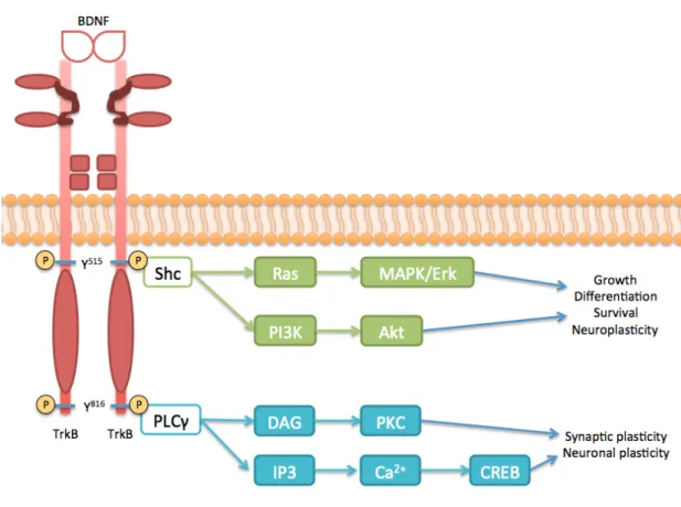
Figure 1.4 – Schematic representation of BDNF/TrkB-FL signalling. The binding of homodimeric BDNF promotes the dimerization of TrkB-FL receptors, activating the kinase domain present in intracellular side and trans-phosphorylating (P) this receptor in two tyrosine residues (Y515 and Y816). Consequently, phosphorylated Y515 recruits the binding site
of Shc adaptor protein, leading to the activation of Ras protein that in turn stimulates MAPK/Erk pathway. This phosphorylated tyrosine residue also activates and triggers PI3K/Akt pathway that promotes neuroplasticity and neuronal growth, differentiation and survival, as well as MAPK/Erk pathway. On the other hand, phosphorylated Y816 of TrkB-FL
binds to PLCγ, activating its pathway, which leads to the promotion of transcriptional factors involved in synaptic and neuronal plasticity.
In summary, it is vital that the system composed by BDNF/TrkB-FL receptor and consequent downstream pathways are tightly regulated, in order to ensure homeostasis. Therefore, dysregulations on this system, such as decrease of TrkB-FL receptor or BDNF levels and increase of TrkB-Tc levels, could result in the impairment of neurotrophic support, which ultimately can lead to neuronal death contributing to neurodegeneration 47 (Figure 1.5).
Figure 1.5 – Schematic representation concerning the effects of dysregulations on BDNF/TrkB-FL system. Considering the importance of BDNF/TrkB-FL system on neuronal functioning, dysregulation on it leads to severe consequences on cell homeostasis. Since this system provides trophic support to neurons, when this signalling is impaired, there is a marked neuronal death that ultimately promotes neurodegeneration.
1.2 Neurodegeneration
Neurodegeneration is a common event present in many brain diseases and it is characterized by the progressive and irreversible loss of neuronal function and structure, which ultimately leads to neuronal death 48,49. The absence of cell division is believed to be a core feature of neuronal identity and therefore a great loss of these cells may cause an irreversible impairment in brain functions.
Different neurodegenerative diseases, even with distinct clinical outcomes, share many characteristics, namely related to its pathogenesis. Neurodegenerative diseases can be caused by dysfunctions at molecular level, like protein misfolding, mitochondrial dysfunction, membrane damage, alterations on programmed cell death or even changes in protein degradation pathways 49,50. In this way, these
changes contribute to degeneration that supports brain and systemic dysfunction, such as movement disorders (ataxias) or dementia 49–51. For instance, Parkinson’s
disease (PD) is characterized by the presence of misfolded proteins (α-synuclein) in substantia nigra and by the degeneration of dopaminergic neurons (which promotes the depletion of dopamine in basal ganglia), leading to ataxia (tremors, stiffness and also rigidity in the major muscles of the body) 50,52. In the case of Alzheimer’s
Disease (AD), there is accumulation of β-amyloid (Aβ) peptide, hyperphosphorylation of tau protein and a marked cortical neurodegeneration contributing to dementia, which will be detailed below 53. Interestingly, some neurodegenerative diseases, such as Huntington’s disease (HD), PD or AD, have been associated with genetic mutations. However, concerning all neurodegenerative diseases, only a small part of them have genetic causes (about 5%). Therefore, intracellular dysregulations that could act simultaneously and synergistically underlying the genesis of neurodegenerative diseases have been intensively studied 48,54.
Despite all the research on this area, the current medication only alleviates the symptoms and it has no role on ethiopathology of these diseases. Hence, there is no cure for neurodegeneration. In addition, average life expectancy is being also increasing, allowing that the onset problems of this type of diseases have more time
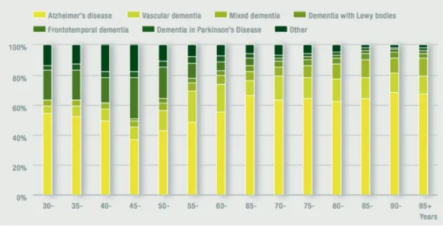
to settle. Consequently, nowadays, these factors contribute for the higher levels of AD prevalence 49,55,56. As mentioned above, there are several types of neurodegenerative diseases some of them related, in some stage, to dementia. It is estimated that there are, per year, 7.7 million new cases of dementia worldwide, implying one new case every 4 seconds, with AD representing approximately 60-70% of all cases, as demonstrated in Figure 1.6 57,58. In Portugal, the estimated number of patients with dementia
represents 6% of total population with more than 60 years and there are approximately 100 000 Portuguese people with AD, which is also a consequence of the demographic aging 59.
Figure 1.6 – Estimative of the proportion of all dementia cases subdivided by different dementia subtypes. These data (concerned women gender) show the higher proportion of
1.2.1
Alzheimer’s disease
AD is the most common form of dementia, affecting between 21 and 35 million people worldwide in accordance with an epidemiological study in 2010 58,60.
This is a number that will grow exponentially, since age is the principal risk factor for AD. In other words, age per se does not lead to AD establishment and consequent diagnosis, but the incidence of this disease doubles every 5 years after 65 years of age and, therefore, aged populations have higher prevalence of AD. This is confirmed by the estimated incidence of AD on populations older than 85 years, which one in three persons has AD 48,60,61. Besides age, in the last decades, other risk factors were also described, such as depression, female gender, rurality, head injuries, nutritional deficits and also low education or low social and professional differentiation 62–65 .
This pathology was identified in 1901 in a fifty-one old woman hospitalized on an asylum of Frankfurt, named Auguste Deter, by a German psychiatrist called Alois Alzheimer, which gave the name to this pathology. Doctor Alzheimer followed this case during five years until her death, and described other eleven cases in the following five years. In his original paper, Doctor Alzheimer refers that “clinical observation alone made the case appear so unusual that it could not be classified as one of the recognized illnesses”. Furthermore, he also described that she had a progressive presenile dementia with general cortical atrophy, which strongly compromises her orientation and memory processes, saying that “she is completely disoriented in time and space (…) her memory is seriously impaired” 66–68. These
descriptions were just the start of a new era about neurodegenerative diseases and, in particular, about AD.
AD has been extensively studied for the last decades and constitutes one of the most financially costly diseases in developed countries 69,70. Although many
aspects of AD have been unraveled, still there are no therapies that halt or reverse its progression. Nevertheless, the research done so far allowed us to have important clues about its pathogenesis (where it has been proposed several hypothesis), diagnosis, biomarkers, prognosis or even some risk factors, such as sedentary
lifestyle, smoking or diet 71–74. Nevertheless further research is needed to comprehend this complex disease and to give responses to all issues still unanswered.
As it is shown in Figure 1.7, AD is characterized by a significant reduction in the brain volume and weight, affecting differentially each brain region or neuronal population. Indeed, this decrease of brain size are directly associated with loss of neurons (mainly pyramidal cells of the entorhinal cortex and in the CA1 region of the hippocampus) and also the shrinkage and loss of neuronal processes, contributing to a dramatic reduction in neuronal branching (Figure 1.7) 75–77.
Besides general atrophy and neuronal loss, AD is also characterized by a progressive cognitive and functional impairment. Initially, the main symptoms of AD are memory impairments (directly associated with hippocampal dysfunction), but later on, this impairment converts into disorientation, judgement dysfunction, apraxia and also speech problems. The late stage of AD is characterized by a prolonged and tragic illness, where the patient loses its individuality, leading to death that normally involves pneumonia 61,65. B A
A) Molecular features of AD
In some families with early-onset familial AD, it was described an autosomal dominant mutation in the amyloid precursor protein (APP) gene and also mutations in PSEN1/2, which is a gene involved in γ-secretase complex. Both mutations are directly associated with increased Aβ production, facilitating its accumulation and the pathogenesis of AD 79–82. However these mutations are present only in 5% of all AD cases. In the remaining cases (that constitutes sporadic AD) there are no known associated mutations that could explain its pathogenesis 83. Thus, AD can be considered a major example of multifactorial diseases, since several molecular events contribute simultaneously to its pathology, including accumulation of misfolded proteins, oxidative and inflammatory agents, disruptions of Ca2+ homeostasis and its consequences on cell environment, as well as mitochondrial and synaptic dysfunction 58,61.
Currently, it is proposed that AD is predominantly a protein misfolding disease. This idea is supported by the presence of senile plaques in the AD brain, composed by aggregated Aβ peptides, and also by the accumulation of intraneuronal neurofibrillary tangles, composed by hyperphosphorylated tau protein (Figure 1.7)
84–86.
Concerning Aβ peptide, studies of genetic forms of AD, including Down’s syndrome (where patients often develop AD) and familial AD, support the “amyloid theory” 79–81. In vivo, Aβ peptides are derived from APP proteolytic cleavage by the
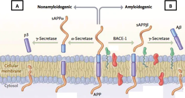
sequential enzymatic actions of beta-site amyloid precursor protein–cleaving enzyme 1 (BACE-1), also known as β-secretase, and γ-secretase, constituting the amyloidogenic pathway. On the other hand, and as shown in Figure 1.8, there is a non-amyloidogenic pathway involving sequential APP cleavage by α-secretase and γ-secretase 87–90. Therefore, an imbalance between those pathways, mitochondrial dysfunction or impairment on clearance of Aβ peptides, promote the release of several amyloid fragments (being Aβ1-42 the most neurotoxic form) and consequent
aggregation into senile plaques, which stay outside of the cells 53,91,92.
Figure 1.8 – Differential processing of APP: (A) non-amyloidogenic and (B) amyloidogenic pathways. APP can be processed by different sets of enzymes, leading to the establishment of two processing pathways: amyloidogenic pathway and non-amyloidogenic pathway. This last pathway mentioned (A) involves the action of α-secretase (cleaving the interior of Aβ peptide) and γ-secretase, releasing a soluble peptide (p3). However, when APP processing is initiated by β-secretase beta-site amyloid precursor protein–cleaving enzyme 1 (BACE-1) and then cleaved by γ-secretase, it is generated Aβ peptide that is released and can aggregated into amyloid plaques (B) (image adapted from LaFerla and Querfurth 58). B) Consequences of Aβ accumulation
Several works with distinctive approaches conclude that Aβ peptide has different neurotoxic effects, sustaining the hypothesis that it is the primary trigger of
AD. These effects can act at different levels and through different mechanisms, but always contributing to neuronal death and other hallmarks of AD 61,93,94.
homeostasis, mitochondrial dysfunction or even interactions with different transmembranar receptors, such as NMDAR or RAGE 58,76,95.
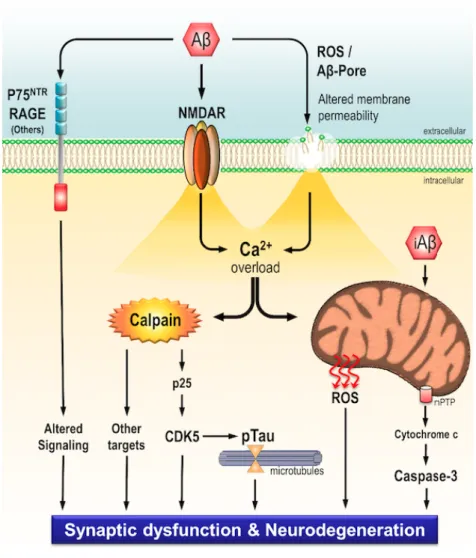
Figure 1.9 – Schematic representation of toxicity mechanisms induced by Aβ peptide. Deposition of extracellular Aβ peptides leads to neurodegeneration and synaptic dysfunction through several mechanisms. Aβ peptides can interact with multiple transmembranar receptors (such as: p75NTR, RAGE, NMDAR), leading to dysregulation of signalling pathways and also a sustained Ca2+ influx. Aβ peptide also promotes cell membrane leakage through a
pore formation or reactive oxygen species (ROS) production, leading again to a sustained Ca2+ influx that induces calpains overactivation and also mitochondrial Ca2+ overload.
Concerning overactivation of calpains, these enzymes cleave several substrates (such as p25 or mGluR1α), changing its function and leading, for instance, to tau hyperphosphorylation, an event mediated by CDK5 activity (as described below). On the other hand, mitochondrial dysfunction is also mediated by intracellular Aβ peptides, increasing ROS formation and caspase-3 activation. Taken together all these events, Aβ peptides strongly induces neurodegeneration and synaptic dysfunction (image from Jerónimo-Santos PhD thesis 96).
In the recent years, it has been shown that Aβ peptide disrupts Ca2+ homeostasis, promoting a sustained increase of intracellular Ca2+ levels. This increase is a result of: i) the interaction between Aβ peptide with neurotransmitters receptors and ion channels (including AMPA and NMDA glutamate receptors, nicotinic receptor α7-nAChr-Ca2+ dependent), ii) the formation of new Ca2+
-permeable membrane pores that allow the entry of Ca2+ into intracellular space and iii) the promotion of an uncontrolled release of Ca2+ from ER 97–102. In turn, this
increase of Ca2+ intracellular levels leads to excitotoxicity, oxidative stress,
mitochondrial and synaptic dysfunction and also overactivation of calpains, resulting in several damages that compromise cell viability 103–105.
Interestingly, recent data demonstrate that calpains overactivation induces the cleavage of TrkB-FL receptors, impairing dramatically the BDNF function, which ultimately may contribute to neuronal dysfunction and death (as detailed in next Section) 106.
C) BDNF/TrkB-FL system in AD
As mentioned before, the signalling system composed by TrkB-FL receptor and its ligand, BDNF, has a crucial role on mammalian nervous system, especially because its downstream signalling cascades are directly involved in neuronal survival
36,41. However, in vitro assays and even analysis of post-mortem human brain
samples have been describing several dysregulations affecting this system on neurodegenerative diseases, with special attention to AD. Actually, existing data
correlates in a positive way the cognitive decline of patients, as what was observed for BDNF expression 118. Moreover, the increase of TrkB-FL and BDNF levels in AD mice models reduces cognitive impairment and ameliorates spatial memory deficits
93. Thus, it is consensual that an impaired BDNF/TrkB-FL system plays a central role in
the pathogenesis of AD 93,94,113–115. There are still some aspects that are not fully
understood, such as if the impairment on BDNF/TrkB-FL system is a cause or a consequence of neurodegeneration. It is proposed that there is a cycle, with positive feedback loop, involving the role of BDNF/TrkB-FL system and neurodegeneration, however it is hard to disclose where is the starting point or end point (Figure 1.10).
Figure 1.10 – Role of BDNF/TrkB-FL system on neurodegeneration: a putative cycle with positive feedback. As mentioned above, dysregulations on BDNF/TrkB-FL system leads to neurodegeneration, where it is described that there is a notorious decrease of BDNF and TrkB-FL levels and also upregulation of TrkB-T1 mRNA levels. Consequently, these indicated changes will then promote again dysregulations of signalling pathways mediated by BDNF/TrkB-FL, leading to the establishment of a closed cycle, where causes or consequences are not fully understood yet.
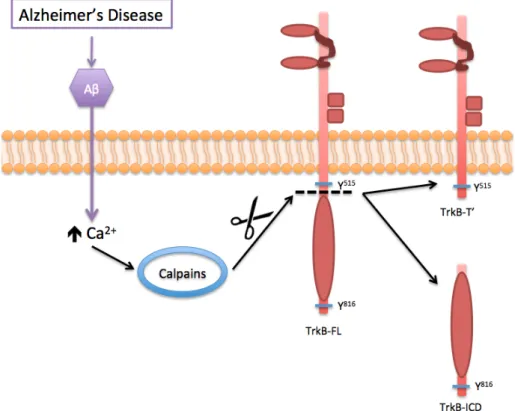
Concerning calpain-cleavage of TrkB-FL receptor, it is important to describe that are generated two new fragments: a new truncated TrkB receptor (TrkB-T’) containing transmembranar domain and an intracellular fragment that is released to
cytosol composed by the intracellular kinase domain of TrkB-FL receptor (TrkB-ICD) 106. So, we can hypothesize that these fragments can play a role on cell functioning. Indeed, being TrkB-T’ a truncated receptor one could speculate whether it could act as negative regulator of TrkB-FL signalling, as other truncated isoforms. Moreover, being equided with tyrosine kinase domain, TrkB-ICD, could also have a role.
Figure 1.11 – Schematic representation of Aβ-induced overactivation of calpains and consequent cleavage of TrkB-FL receptor, generating TrkB-ICD and TrkB-T’. The accumulation of extracellular Aβ peptide has several consequences and different neurotoxic effects, being one of them associated with the dysregulation of Ca2+ homeostasis that will
overactivate the calpains. In turn, this overactivation cleaves several substrates, including TrkB-FL receptor, promoting the release of TrkB-ICD to the cytosol and forms a truncated receptor TrkB-T’.
1.3 Calpains
Calpains are Ca2+-activated proteases with biological and physiological importance, contributing to the regulation of cell homeostasis 116,117. There are
several isoforms of calpains, being the most expressed calpain-1 (μ-calpain) and calpain-2 (m-calpain). These isoforms have a key role on the regulation of many cellular processes, by cleavage of several substrates in interdomain regions 118,119. In particular, these enzymes have a crucial role in several physiological processes, such as proliferation, motility, differentiation, apoptosis and memory 120. Calpains are also involved in cytoskeletal turnover and remodelling, since these enzymes cleave some cytoskeletal proteins, including tau protein, microtubule-associated proteins (MAP), neurofilaments, spectrin and also actin 121–125.
Although calpains play an important role in physiology, they can also contribute to degeneration when persistently overactivated 118,126. Multiple
sclerosis, cancer or even allergic encephalomyelitis are examples of diseases where calpains may have a role 119,127,128. And indeed, the sustained activation of calpains is
considered as a pathogenic mechanism of AD. Actually, there are several reports showing calpains overactivation on AD post-mortem human brain samples. Moreover, in vitro studies demonstrated that calpain-1 overactivation promotes amyloid plaque formation and correlates calpains location with neurofibrillary tangles 118,129–133. Importantly, studies with AD brains after short post-mortem
intervals demonstrated that calpains overactivation really contributes to neuronal death, discarding the hypothesis that the activation of calpain-1 isoform could be a consequence of post-mortem autolytic processes 134,135. In addition, it was also described that calpastatin, an endogenous calpain inhibitor, is markedly depleted from dendrites in AD brain, contributing also to calpains overactivation 136. Concordantly, APP transgenic mice genetically deficient on calpastatin, show increased Aβ amyloidosis, tau phosphorylation, microgliosis and mortality when compared with the same mouse model of AD (APP) without genetic manipulation 143. Remarkably, using also the APP mice, calpain inhibition rescues spatial-working memory and neuronal plasticity 137.
As a consequence of cleavages mediated by calpains, there is not only a loss of biological functions of original substrates, but also the generation of new stable fragments. Hence, fragments generated by calpains can acquire distinct functions and localizations when compared to the original substrate from which they were produced (Figure 1.12). Figure 1.12 – Action of calpains on cell substrates. Cleavage mediated by calpains produces fragments with different function and localization than the original substrate, which may lead to disruption of cell homeostasis.
1.3.1
Fragments produced by calpain cleavage
In adition to TrkB-FL receptors, there are several other proteins known to be cleaved by calpains, such as Src, β-catenin, p35 and mGluR1α receptor. A) Src protein Src protein is an enzyme involved in survival, growth and/or proliferation of neuronal cells, being also responsible for survival of glial cells, through cooperation with Ret receptor at the surface of these non-neuronal cells 138–140. However, resultsB) β-catenin protein
β-catenin is a protein that, in cooperation with the transcription factor Tcf/Lef, promotes signalling cascades involved in a wide variety of developmental process as well as in carcinogenesis 142–144. Nevertheless, in hippocampal neurons,
fragments originated by β-catenin calpain-cleavage are translocated to the nucleus, affecting gene transcription, by the activation of Tcf-dependent gene transcription
145. Interestingly, there are several reports describing that dysregulations of
β-catenin-Tcf-mediated gene regulation are a pathogenic process in innumerable neurological diseases, including bipolar disorder and AD 146–148.
C) p35 protein
The cleavage of p35 protein into p25 fragment is another example of calpains proteolytic processes, activated by increased levels of intracellular Ca2+, which produces fragments with toxic effects on nervous system 149–151. The evaluation of
post-mortem brain tissues from AD patients and also in vitro assays demonstrated that there is a neuronal accumulation of p25 fragment, activating in a constitutive way the cyclin-dependent kinase 5 (CDK5), which acquires different cellular location and lost its substrate specificity 149. These alterations of CDK5 properties impair its normal function (neurite growth), promoting, in vivo, hyperphosphorylation of tau protein. In addition, in cultured primary neurons, p25/CDK5 complex induces cytoskeletal disruption, morphological degeneration and also neuronal apoptosis, characteristic features of AD 149,152–154.
D) mGluR1α protein
The mGluR1α receptor plays a crucial role in the regulation of neurotransmission, as well as in the regulation of neuronal survival, through PI3K/Akt pathway 155,156. Interestingly, it is described that an overactivation of NMDA
strong and sustained activation of calpains, which stimulates the cleavage of mGluR1α by calpains. This truncated form of mGluR1α contributes to excitotoxicity, since it is also capable of increase cytosolic Ca2+ levels and its capability of activating PI3K/Akt pro-survival signalling is lost 157.
E) TrkB-FL receptor
As mentioned before, TrkB-ICD fragment were recently described, keeping this fragment in the shadows. Nevertheless, it is already described that Trk receptors can be cleaved with dramatic consequences on neuronal survival. In particular, caspase-3 is able to cleave TrkC receptor, impairing its signalling and, importantly, producing a fragment with pro-apoptotic activity, mediating cell death through a mechanism mediated by caspase-9 158.
Interestingly, previous work on our lab already demonstrates the presence of TrkB-ICD in human brain samples, what indicates that this fragment can indeed play a role in AD pathophysiology 106. Concordantly, unpublished data from our lab using
post-mortem human brain samples (frontal cortex) from patient with AD and an age-matched control demonstrate that TrkB-FL receptors are decreased and TrkB-ICD levels are increased in the AD brain (Figure 1.13).
Noteworthy that it was recently described that, under ischemia and excitotoxicity conditions, TrkB-FL is downregulated by calpains 159. Although the authors did not identify TrkB-ICD fragment, it is expectable that, in that context, this fragment might be also formed given the calpain overactivation. Thus, one could anticipate that TrkB-ICD fragment can be produced in conditions where calpains are robustly activated.