CHARACTERIZATION OF EBV-ASSOCIATED GASTRIC
CANCERS
Caracterização de carcinomas gástricos associados ao EBV
Thesis presented to Escola Superior de Biotecnologia of the
Universidade Católica Portuguesa to fulfill the requirement of
Master of Science degree in Applied Microbiology
by
Andreia Fernanda Nora de Oliveira
Place: Instituto Português de Oncologia – Porto
Supervisor: Hugo Sousa, MD, PhD
Co-supervisor: Joana Ribeiro, MSc
RESUMO
A nível mundial, o cancro gástrico (CG) é o sexto mais comum com cerca de 1 milhão de novos casos estimados em 2012. Em Portugal, os dados revelam que o CG é o quinto cancro mais frequente com cerca de 3000 novos casos por ano. Para além disso, o CG tem uma elevada taxa de mortalidade sendo responsável por 1387 mortes nos homens e 898 nas mulheres. Dados recentes têm mostrado que o vírus Epstein-Barr (EBV) é detectado em diferentes subtipos histopatológicos de carcinoma gástrico sendo estes tumores responsáveis por aproximadamente 10% de todos os casos.
O presente estudo pretende caracterizar os carcinomas gástricos associados ao EBV (EBVaGC) através da sua detecção em tecidos tumorais gástricos de 136 pacientes atendidos no Instituto Português de Oncologia do Porto (IPO Porto FG EPE) no ano de 2011. A detecção de EBV foi realizada por hibridização in situ (ISH) utilizando uma sonda de ADN complementar para ARNs codificados pelo EBV (EBERs).
Os resultados demonstraram que os carcinomas associados ao EBV representam 6,6% de todos os casos de CG. Analisando a distribuição de EBV entre os diferentes tipos histológicos, observou-se que o EBV estava presente em 6,6% dos tipos intestinais, em 11,1% dos tipos indeterminados e em 100% dos linfoepiteliomas, contudo nenhum caso foi detectado nos carcinomas difusos (p<0,001). A análise de risco, apesar de não ter sido estatisticamente significativa, sugeriu que os pacientes com carcinomas do tipo intestinal (p=0,350; OR=1,98; 95% IC=0,37-10,5) ou indeterminado (p=0,238; OR=2,78; 95% IC=0,55-15,5) apresentam um risco aumentado de ter EBVaGC; enquanto os pacientes com carcinomas difusos (p=0,078; OR=0,14; 95% CI=0,01-2,58) apresentam um risco diminuído de ter EBVaGC. Quanto à localização do tumor, verificou-se que os carcinomas das regiões superiores do estômago apresentam um risco aumentado de ter um EBVaGC (p=0,032; OR=4,68; 95% IC=1,11-19,7).
Em conclusão, a infecção por EBV nos carcinomas gástricos em Portugal é semelhante à de outros países. Por outro lado, as características clínico-patológicas apresentaram diferenças quando comparadas com estudos anteriores, principalmente a ausência de EBV nos carcinomas difusos. Este é o primeiro estudo de caracterização dos EBVaGC em Portugal que reforça a necessidade de mais estudos para esclarecer o papel do EBV como biomarcador preditivo/prognóstico no desenvolvimento do cancro gástrico.
ABSTRACT
Worldwide, Gastric Cancer (GC) is the sixth most common malignancy with nearly 1 million new cases estimated in 2012. In Portugal, data reveal that GC is the fifth most frequent cancer with about 3000 new cases per year. Moreover GC has still a higher mortality rates being responsible for 1387 deaths in men and 898 in women. Recent data showed that Epstein-Barr virus (EBV) has been detected in different histopathological subtypes of gastric carcinoma and EBV-associated gastric carcinoma (EBVaGC) accounts about 10% of all cases.
This study pretends to characterize EBVaGC in our population through detection of EBV in gastric carcinoma tissues from 136 consecutive patients attended at Portuguese Institute of Oncology of Porto (IPO Porto FG EPE) in the year of 2011. EBV detection was performed by in situ hybridization (ISH) targeting EBV-encoded small RNA (EBER-ISH) with an EBER-DNA probe.
The results showed that in our population EBVaGC represent 6.6% of all GC cases. Analyzing the distribution of EBV among the different histological types, we observed that EBV was present in 6.6% of intestinal-types, 11.1% of indeterminate types, 100% of lymphoepithelioma-like carcinomas and there were no positive cases among diffuse types (p<0.001). The risk analysis revealed that, despite there are not statistically significant differences, patients with intestinal (p=0.350; OR= 1.98, 95% CI=0.37-10.5) and indeterminate (p=0.238; OR=2.78, 95% CI=0.55-15.5) GC have an increased risk of having an EBVaGC; while diffuse GC (p=0.078; OR=0.14, 95% CI=0.01-2.58) have a decreased risk of having an EBVaGC. Regarding tumor location, the results demonstrated that patients with tumors in upper regions of stomach have an increased risk to have an EBVaGC (p=0.032; OR=4.68, 95% CI=1.11-19.7).
In conclusion, the EBV infection rate among gastric carcinomas in Portugal is similar to that ascertained in other countries. Conversely, the clinicopathological features showed differences when compared with previous studies, mainly the absence of EBV in diffuse-type gastric carcinomas. This is the first study to characterize EBVaGC in Portugal which reinforce the need of further studies to clarify the role of EBV and to explore its potential value as predictive/prognostic biomarker in gastric cancer development.
AKNOWLEDGMENTS
Um agradecimento é apenas uma simples acção que não consegue descrever a minha gratidão para com todos aqueles que me acompanharam durante esta fase. Toda a colaboração académica, profissional e pessoal foi imprescindível para que todos os obstáculos fossem ultrapassados. Desta forma, primeiramente gostaria de agradecer ao meu orientador Doutor Hugo Sousa por me ter dado a oportunidade de desenvolver o meu trabalho de investigação no Grupo de Oncologia Molecular e Patologia Viral do Instituto Português de Oncologia do Porto (IPO Porto FG EPE). Além disso quero aqui mostrar o meu reconhecimento por todo seu esforço na gestão e planeamento do projecto bem como o seu constante apoio e motivação que foi crucial para o meu desenvolvimento na área da investigação científica. Com também especial importância, quero mostrar o meu sincero agradecimento à minha co-orientadora Joana Ribeiro que me acompanhou desde o início e, com a sua prestabilidade e empenho, tornou possível a realização deste trabalho. Saliento ainda o meu agradecimento:
Ao Doutor Luís Pedro, à Doutora Ana Galaghar e à Técnica Fernanda do Serviço de Anatomia Patológica do IPO pelo tempo dispensado na selecção e obtenção das amostras.
Aos meus colegas de laboratório, Mariana Malta, Liliana Raeiro, Joana Silva, Isabel Paiva, Marco Neves, Letícia Mesquita pelo companheirismo, aprendizagem e momentos de descontracção, especialmente à Marlene Esteves que foi essencial no processo de integração e adaptação.
Ao coordenador do Mestrado de Microbiologia Aplicada, Professor Doutor José Couto, por ter autorizado o protocolo com uma instituição externa de forma a permitir a elaboração da tese numa das minhas áreas de interesse mas também pelo seu apoio enquanto meu professor durante os meus seis anos de percurso académico.
Aos meus amigos Ângela Santos, Daniel Lopes, João Ferreira, Tânia Ferreira, Susana Marques, Vera Oliveira por toda paciência, tolerância e compreensão.
Aos meus patrões e amigos, D. Elisabete, Sr. Jorge e D. Leonor por toda a flexibilidade de horários, todo o carinho e preocupação.
Por último mas não menos importante, o meu muito obrigado a minha família especialmente à minha mãe que, com o seu esforço, desde a morte do meu pai sempre me proporcionou condições para puder continuar o meu percurso académico.
INDEX OF CONTENTS
RESUMO ... iii
ABSTRACT ... v
AKNOWLEDGMENTS ... vii
LIST OF FIGURES ... xiii
LIST OF TABLES ... xv
ABBREVIATIONS ... xv
I. INTRODUCTION ... 1
1. Virus and Cancer ... 3
2. Epstein Barr Virus (EBV) ... 5
2.1 Structure and characteristics... 5
2.2 EBV “life” cycle... 7
2.3 EBV-associated diseases ... 10 3. Gastric Cancer ... 12 3.1 Epidemiology ... 12 3.2 Classification ... 13 3.3 Risk factors ... 15 4. Gastric carcinogenesis ... 18 4.1 Helicobacter pylori-associated GC ... 18 4.2 EBV-associated GC... 20 II. AIMS ... 23 III. METHODOLOGY ... 27 1. Type of study ... 29 2. Population ... 29
3. Selection and Processing ... 29
4. EBV detection ... 29
4.1 EBV-ISH detection: protocol 1 ... 30
4.2 EBV-ISH detection: protocol 2 ... 31
5. Quality control ... 32
6. Statistical analysis ... 33
1. Comparison of EBV ISH protocols ... 37
2. Characterization of the study population ... 38
3. EBV in GC ... 41
4. Risk analysis of EBV-GC associations ... 43
V. DISCUSSION
... 45VI. CONCLUSION
... 51VII. FUTURE WORK
... 55VIII. APPENDIX
... 53LIST OF FIGURES
Figure 1.1 - Hallmarks of Cancer by human oncoviruses.. ... 4 Figure 1.2 - Model of EBV entry into B-lymphocytes.. ... 6 Figure 1.3 - EBV infection in healthy carriers.. ... 10
Figure 1.4 - Age-standardized incidence rates (World) per 100.000 habitants of gastric cancer in both sexes ... 12
Figure 1.5 - Numbers of new cases/deaths stratified by sex in Portugal ... 13 Figure 1.6 - Genetics, environmental and lifestyle risk factors associated to gastric
cancer ... 15
Figure 1.7 - H. pylori-induced host cell response and oncogenic signaling in gastric
epithelial cells ... 19
Figure 4.1 - Histological section from gastric carcinoma. Positive result for EBV using
protocol 1 (160x) ... 37
Figure 4.2 - Histological section from gastric carcinoma. Negative result for EBV using
protocol 1 (160x) ... 37
Figure 4.3 - Histological section from gastric carcinoma. Positive result for EBV using
protocol 2 (160x) ... 37
Figure 4.4 - Histological section from gastric carcinoma. Negative result for EBV using
protocol 2 (160x) ... 37
Figure 4.5 - Graph illustring the age distribution of population ... 38 Figure 4.6 - Distribution of histological types according to their location in the stomach
... 40
LIST OF TABLES
Table 1.1 - Viruses and its related human tumors ... 3
Table 1.2 - Latency pattern of EBV according to latent gene expression and associated malignancies ... 8
Table 1.3 - Comparison of gastric cancer classifications between Lauren’s and WHO classification systems ... 14
Table 4.1 – Characterization of sudy population ... 39
Table 4.2 – Description of EBV frequency in gastric carcinomas ... 42
ABBREVIATIONS
APC: Adenomatous Polyposis Coli ASR: Age Standardized Rates
BARTS: BamHI A Rightward Transcripts BL: Burkitt’s Lymphoma
CagA: Cytotoxin Associated Antigen A CagPAI: Cag Pathogenicity Island
CISH: Chromogenic in situ Hybridization COX2: Cyclooxygenase 2
CR2: Complement Receptor 2 DAB: 3,3'-Diaminobenzidine DNA: Deoxyribonucleic Acid DNMT1: DNA Methyltransferase 1 DDR: DNA Damage Response
EBER: Epstein–Barr Virus-Encoded Small RNAs EBNA: Epstein–Barr Nuclear Antigen
EBNA-LP: Epstein-Barr virus Nuclear Antigen Leader Protein EBV: Epstein Barr Virus
EBVaGC: EBV-associated Gastric Cancer FFPE: Formalin-Fixed Paraffin-Embedded GC: Gastric Cancer
HBV: Hepatitis B virus HCV: Hepatitis C virus
HDGC: Hereditary Diffuse Gastric Cancer HHV-4: Human Herpes Virus 4
HL: Hodgkin’s Lymphoma HPV: Human papilloma virus HRP: Horseradish Peroxidase
HTLV-1: Human T lymphotropic virus ISH: in situ Hybridization
IHC: Immunohistochemistry IgA: Immunoglobulin A
LCLs: Lymphoblastoid Cell Lines
LELC: Lymphoepithelioma-Like Carcinoma LMP: Latent Membrane Proteins
miRNAs: MicroRNAs mRNA: Messenger RNA NBs: Nuclear Bodies
NF-κB: Factor Nuclear kappa B NK: Natural Killer
NPC: Nasopharyngeal Carcinoma PI3K: Phosphoinositide3-Kinase PML: Promyelocytic Leukemia
PTEN: Phosphatase and Tensin Homolog
PTLD: Post-Transplant Lymphoproliferative Disorders RNA: Ribonucleic Acid
TFSS: Translocate the Bacterial Effectors VacA: Vacuolating Cytotoxin
1. Virus and Cancer
The first association between a virus and cancer was shown in 1911 when Rous discovered chicken sarcoma and attributed Rous Sarcoma Virus (RSV) as the etiological agent (Rous, 1911). Over the next 100 years, several viruses have shown to be associated with cancer development. In the past 30 years, the viruses have been fundamental instruments in scientific investigation since they allow to explain the mechanisms of carcinogenesis in some types of human tumors (Butel, 2000).
Approximately 12% of all human cancers worldwide may be attributed to viruses (Mesri et al., 2014). Both DNA and RNA viruses have been well described as agents responsible to induce either single or multiple tumors (Table 1.1). This variability is supported by characteristic tissue tropism of the given virus (Butel, 2000, Chang et al., 2013).
Table 1.1 - Viruses and its related human tumors
Nevertheless, the viral infection is generally not sufficient to induce the development of cancer. Only a minority of infected individuals progress to cancer and it occurs mainly years or even decades after primary infection (Butel and Fan, 2012). Additional factors such as immunosuppression, genetic predisposition, somatic mutations, epigenetic alterations and environmental exposure to carcinogens have demonstrated to be relevant for virus-associated tumorgenesis (Au, 2004, Junjie et al., 2012, Li et al., 2013).
Virus Genome Human cancers Cell tropism
EBV DNA Burkitt’s lymphoma
NK-T cell lymphoma
Lymphomas in immunosuppressed host Nasopharyngeal carcinoma (NPC)
Gastric carcinoma
B-cells,
oropharyngeal and gastric epithelial cells
HBV DNA Hepatocellular carcinoma Hepatocytes
HCV RNA Hepatocellular carcinoma Hepatocytes
HHV-8 DNA Kaposi sarcoma B-cells
HPV DNA Cervical carcinoma Squamous epithelial cells
Applied Microbiology, MSc Andreia Fernanda Nora de Oliveira
Characterization of EBV-associated gastric cancers |4
The role of viruses in cancer has been demonstrated by in vitro transformation of cells which has allowed to elucidate functions of viral proteins involved in carcinogenesis both at molecular and biochemical levels (Butel and Fan, 2012).
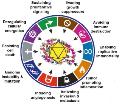
Human oncogenic viruses rely on persistence in host cells through their replication and immune evasion. Persistent infection can lead to cancer development by acquisition of cancer hallmarks as result of activation of anti-apoptotic and proliferative programs (Figure 1.1). Each cancer hallmark represents a biological consequence of oncogenic alterations by viral oncogenes that include:
1) Host Signaling: Viral proteins can modulate host-signaling mechanisms that regulate cell growth and survival;
2) DNA damage response (DDR): Viral replication induces DDR however host cells acquire genetic instability leading to increase of mutations rate and chromosomal alterations;
3) Chronic inflammatory responses to persistent viral infections: Inflammation leads to oxidative stress by reactive oxygen species generation that promotes acquisition of mutations.
The most cancer-related mutations are found in target genes which either can promote a gain of function of oncogenes or lead to the loss of function of tumor-suppressors genes (Hanahan and Weinberg, 2000).
2. Epstein Barr Virus (EBV)
2.1 Structure and characteristics
Epstein-Barr virus (EBV), also called Human Herpes Virus 4 (HHV-4), is a member of the Herpesviridae family. It is ubiquitous in nature and infects more than 90% of adult population worldwide. Primary infection normally occurs in childhood or early adulthood through salivary contact. Majority of children are asymptomatic, but some adolescents and young adults can develop infectious mononucleosis with harmless clinical manifestations (Grotto et al., 2003). As other members of Herpesviridae family, EBV is composed of a linear double-stranded DNA genome surrounded by an icosahedral nucleocapsid which encodes about 85 genes. The viral capsid contains 162 capsomeres, a protein tegument and an outer envelope with glycoprotein spikes (Thompson and Kurzrock, 2004).
EBV has been divided into two major types, EBV-1/EBV-A and EBV-2/EBV-B. Worldwide EBV-1/EBV-A is the most frequent type while EBV-2/EBV-B is more characteristic in Africa (Zimber et al., 1986). These variants show differences between nuclear antigens EBNA2, 3A, 3B, 3C, although the most sequence variation occurs in EBNA2-encoding gene, with only 50% of similarity (Cancian et al., 2011).
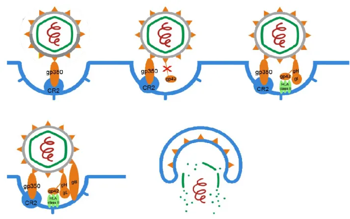
EBV exhibits dual tropism infecting both B-lymphocytes and some types of epithelial cells, especially those of nasopharynx. Cell entry is mediated through interaction of at least five envelope viral glycoproteins with host cell surface. The mechanisms of entry are different depending if EBV infects B-cells or epithelial cells (Shannon-Lowe and Rowe, 2014). In B-cells the virus enters via endocytosis (Figure 1.2). An initial interaction occurs between the major glycoprotein of the viral envelope (gp350/220) and B-cell specific complement receptor 2 (CR2 or CD21) (Tanner et al., 1987, Shannon-Lowe and Rowe, 2014). However, it has been referred that the entry into the cell is mediated by other viral glycoproteins (gp42, gH, gL and gB) which also interact with the host cell membrane. Gp42 binds to B-cell HLA class II and it is achieved through binding gH/gL complex that modify gp42 conformational form. Moreover, gH and gL glycoproteins are also responsible for the activation and recruitment of gB, another ligand of B-cell receptors (Molesworth et al., 2000, Hutt-Fletcher, 2007).
Applied Microbiology, MSc Andreia Fernanda Nora de Oliveira
Characterization of EBV-associated gastric cancers |6
Figure 1.2 - Model of EBV entry into B-lymphocytes. The first interaction occurs between gp350 and
CR2. Following this step, gp42 is cleaved to be possible the gp42/gH/GL complex formation. This complex can modify gp42 conformation to a form that is essential for HLA class II binding. Gp42-HLAII interaction is insufficient to establish full fusion. Thus, gH/gL complex is also responsible for activation, recruitment and binding of gB glycoprotein which promotes the fusion between the viral envelope and the B-cell endocytic membrane.
Regarding EBV infection of epithelial cells, some reports have described absence or restriction of CD21 expression and therefore the attachment of EBV in epithelial cells is less efficient (Kim et al., 1998, Burgos and Vera-Sempere, 2000, Jiang et al., 2008). The absence of CD21 in certain cells has led to study of others possible mechanisms that explain EBV entry (Shannon-Lowe and Rowe, 2014). One of these is explained by the virus coated with immunoglobulin A (IgA) specific to gp350/220 binds to the polymeric immunoglobulin A-receptor from surface membrane (Sixbey and Yao, 1992). Other mechanism is the interaction between gH/gL complexes with specific molecules present in epithelial cells (integrins) such, avb5, avb6 and avb8 (Chesnokova et al., 2009). Finally, on polarized epithelial cells, an interaction between a multispanvirus
membrane protein encoded by the BMRF2 openreading frame and integrins has also been demonstrated (Tugizov et al., 2003).
2.2 EBV “life” cycle
EBV infection cycle includes a lytic and a latent phase that describes a persistent infection. Latent infection is more advantageous for the virus to persist for long periods since it expresses a limited set of latent genes which can be recognized by the host immune system (Murata et al., 2014).
Lytic infection
During lytic infection, EBV expresses nearly 100 proteins, which play an essential role in viral replication, formation of virions as well as modulation of host immune response (Murata et al., 2014). Lytic cycle results in the production of infectious viral particles both after initial infection in oropharyngeal cells and reactivation from latently infected cells. Upon induction of lytic program, there are two key EBV immediate-early (IE) lytic genes (BZLF1 and BRLF1) which are expressed. BZLF1 and BRLF1 genes encode transactivators able to activate viral and certain cellular promoters triggering a coordinated cascade of viral gene expression. The early genes (E) of EBV are involved in DNA replication and metabolism whereas late genes (L) expression is associated with viral structural proteins. In contrast with latent cycle, the replication occurs through
viral DNA polymerase which is expressed with the other lytic genes (Tsurumi et al., 2005).
Latent infection
During latent phase, EBV remains persistently in infected cells and the viral DNA present in the nucleus acquires episomal or integrated form by fusing terminal repeats (TR, repetitive 500-bp structures) at both ends (Reisinger et al., 2006). EBV-terminal repeats have a specific structure contributing to evaluation of EBV clones, viral integration and the state of viral activation (Fukayama et al., 2008). The replication occurs in synchronization with the host genome (S-phase) and then EBV genome is delivered to daughter cells in mitosis (Murata et al., 2014).
Latent infection is characterized by a limited set of viral genes which lead to the expression of six nuclear proteins: EBNA1, EBNA2, EBNA3A, EBNA3B, EBNA3C,
Applied Microbiology, MSc Andreia Fernanda Nora de Oliveira
Characterization of EBV-associated gastric cancers |8
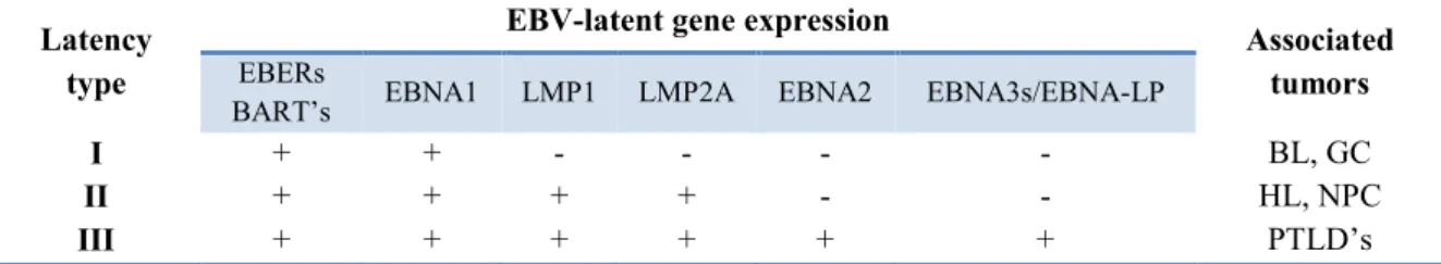
EBNA leader protein (EBNA-LP), and two latent membrane proteins: LMP1, LMP2. In addition, the non-polyadenylated EBER RNAs and rightward transcripts from the BamHI A region (BARTS) can be also detected (Young and Rickinson, 2004). The expression of EBV-latent genes can affect cell cycle, apoptosis and immune response contributing to tumor initiation and propagation (Seo et al., 2010). According expression pattern (Table 1.2), latency can be classified into three types (I, II, III) suggesting that EBV may affect cell growth by different ways (Lorenzetti et al., 2010).
Table 1.2 - Latency pattern of EBV according to latent gene expression and associated malignancies
(Lorenzetti et al., 2010)
BL: Burkitt’s lymphoma; GC: Gastric carcinoma; HL: Hodgkin’s lymphoma; NPC: Nasopharyngeal carcinoma; PTLD’s: post-transplant lymphoproliferative disorders.
Despite there are three latency patterns, all EBNAs are only expressed in latency III through differential splicing from a large primary mRNA. Complete expression of nuclear antigens is achieved due to initial activation of Wp promoter responsible for expression of EBNA2 and EBNA-LP following Cp activation able to express the other nuclear proteins (EBNA1, 3A, 3B) as well as Wp-initiated transcripts described above. Qp is another promoter that normally is not activated in latency III. However, in latency I and II, it is active to promote only EBNA1 expression (Shannon-Lowe et al., 2009). EBNA-1 contributes to maintenance and replication of episomal EBV genome through specific binding to the plasmid origin (OriP). In addition, EBNA-1 interacts with viral promoters contributing to transcriptional regulation of other EBNAs (including EBNA-1 itself) and LMP-EBNA-1. EBNA-2 and LMP-EBNA-1 are essential proteins for growth and transformation of both B-cells and epithelial cells. EBNA2 is a protein involved in transactivation of both cellular and viral genes. It up-regulates the expression of certain B-cell antigens such CD21 and CD23 as well as the viral proteins LMP1 and LMP2. LMP-1 induces the expression of cellular adhesion molecules, cytokines, anti-apoptotic
Latency type
EBV-latent gene expression Associated
tumors
EBERs
BART’s EBNA1 LMP1 LMP2A EBNA2 EBNA3s/EBNA-LP
I + + - - - - BL, GC
II + + + + - - HL, NPC
proteins as well as activation of transcription factors that lead to an uncontrolled cell proliferation (Young and Rickinson, 2004, Young et al., 2000).
Development of persistent infection
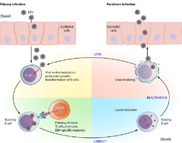
The life cycle of EBV (Figure 1.3) is initiated when the virus infects oropharyx involving squamous epithelial cells and possibly also locally-infiltrating lymphocytes. There, EBV develops a lytic replication with release of infectious viral particles able to spread throughout the lymphoid tissues and peripheral blood lymphocytes. Within B lymphocytes, the virus establishes an expression pattern designed by latency III which is characterized by expression of all six nuclear antigens (EBNA1, 2, 3A, 3B, 3C e -LP) three membrane proteins (LMP1, 2A e 2B) and two small molecules of RNA (EBER1 and 2). When EBV establishes latency III, it is able to promote B-cells transformation into lymphoblastoid cell lines (LCLs) leading to spontaneous proliferation (Odumade et
al., 2011).
Both lytic and latent infections, the viral antigens induce strong cell-mediated responses in the immunocompetent hosts, involving natural killer (NK) cells and CD4+/CD8 T cells, resting a limited EBV-infected B-cells as long term viral reservoir. However, some of these cells can reach germinal center where down-regulate the expression of growth-transforming proteins thereby escaping immune system recognition and establishing latent state (latency 0) in re-circulating memory B-cell pool. Latency 0 is characterized by restricted viral gene expression with only expression of protein-coding transcripts, (EBERs) and BamA rightward transcript (BART) (Young and Rickinson, 2004).
Occasionally, when immune system is compromised, these resting B lymphocytes can enter the lytic cycle where the virions will infect either new B-cells or epithelial cells (Sousa et al., 2011).
Applied Microbiology, MSc Andreia Fernanda Nora de Oliveira
Characterization of EBV-associated gastric cancers |10
2.3 EBV-associated diseases
EBV was the first virus related to neoplastic cells infection in humans with a proven role in carcinogenesis. It was discovered in 1964 when virus-like particles were identified in a cell line from Burkitt’s lymphoma biopsies through electron microscope (Epstein et al., 1964). Nowadays, it is well known that EBV can contribute to the development of several human diseases, including benign and malignant disorders. Infectious mononucleosis is a self-limiting benign and acute illness characterized by high fever, lymphadenopathy and pharyngitis or tonsillitis which occurs more frequently in adulthood and young adults, mainly when immune system is compromised. Oral hairy leukoplakia is another non-malignant disease that results in hyperplasia of epithelial cells in oral mucosa from immunocompromised patients with human immunodeficiency virus infection/acquired immunodeficiency syndrome (HIV/AIDS) or submitted to solid bone marrow transplantation (Tsuchiya, 2002).
Generally, EBV infection is well controlled by immune system. However, immune control can be disrupted leading to prolonged proliferation of EBV-infected cells and their malignant transformation. EBV latent infection has shown strong evidences in carcinogenesis of Burkitt’s lymphoma (BL), NK/T cell lymphomas, nasopharyngeal carcinoma (NPC), Hodgkin’s lymphoma (HL) and malignant lymphomas in immunocompromised patients such, post-transplant lymphoproliferative disease (PTLD). In contrast, in gastric and breast carcinomas its role in carcinogenesis is not well understood (Grywalska et al., 2013).
Recognition of EBV-infected cells in biological tissues is well achieved by detection of EBV-encoded RNAs (EBERs). These molecules are small nopolyadenylated noncoding RNAs which are the most abundant viral transcripts in latently EBV-infected cells. For this reason, EBERs are considered reliable markers when in situ hybridization (ISH) is used as detection method (Iwakiri, 2014).
Applied Microbiology, MSc Andreia Fernanda Nora de Oliveira
Characterization of EBV-associated gastric cancers |12
3. Gastric Cancer
3.1 Epidemiology
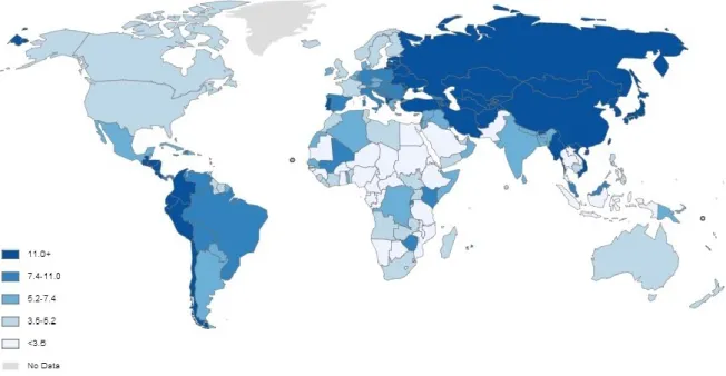
Gastric cancer (GC) is the sixth most common malignancy in both sexes worldwide with nearly one million new cases estimated in 2012 (952.000 cases). The incidence rates are twice higher in male (8.5%) than in female (4.8%) with about 631.000 and 320.000 new cases per year, respectively. Nevertheless, age standardized rates (ASR) show that carcinoma of stomach is the fourth most frequent in men and the sixth in women. Despite a significant reduction of incidence and mortality rates over the past few decades, GC still remains the third leading cause of death by cancer. Approximately, 8% of cancer-related mortality in world (723.000 deaths) is attributed to tumors of stomach. It is a major health problem with a distinct distribution according to geographical areas, socio-economic conditions and ethnic diversity. More than 70% of total cases occur in developing regions being the Eastern Asia (half cases in China), Eastern Europe and Latin America the areas with the highest age-standardized incidence rates (Figure 1.4). In contrast, the lowest incidence rates are observed in United States, Australia and some North European countries (Ferlay et al., 2013).
Figure 1.4 - Age-standardized incidence rates (World) per 100.000 habitants of gastric cancer in both
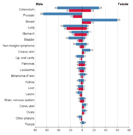
Portugal is one of the European countries with the higher incidence and mortality rates associated to gastric cancer. It represents the fifth most common cancer (Figure 1.5) in both sexes with about 3000 new cases per year. As in other countries, mortality rate is also high (9.5%) being this cancer the fifth most frequent, responsible for 1387 deaths in men and 898 in women (Ferlay et al., 2013).
Figure 1.5 - Numbers of new cases/deaths stratified by sex in Portugal (Ferlay et al., 2013)
3.2 Classification
Gastric tumors are classified anatomically and histologically. Anatomically, GC is divided into proximal and distal tumors depending on their localization of stomach.
Applied Microbiology, MSc Andreia Fernanda Nora de Oliveira
Characterization of EBV-associated gastric cancers |14
in the antrum/pyloric region. Histologically, tumors of stomach show high heterogeneity at both architectural and cytological level that difficult the establishment of well-defined classification system. Some classifications have been established to classify the histologic pattern of gastric adenocarcinomas: Ming, Carneiro and Goseki, but the most commonly used are those of World Health Organization (WHO) and Lauren (Lauren, 1965, Roy et al., 1998, International Agency for Research on Cancer, 2010, Hu et al., 2012).
Lauren’s classification is an essential system in gastric cancer history that over time have contributed to describe an association with several environmental factors, incidence trends and ethology. According to this classification, the two major histologic subtypes are intestinal and diffuse adenocarcinomas. The other types are classified to indeterminate type, when carcinoma is too undifferentiated and co-exist histological features, or uncommon variants (Lauren, 1965). The relative frequencies are approximately 54% for intestinal type, 32% for the diffuse type and 15% for the indeterminate type. In 2010, WHO referred five subtypes that have been correlated with Lauren’s classification as described in table 1.3 (International Agency for Research on Cancer, 2010, Hu et al., 2012).
Table 1.3 - Comparison of gastric cancer classifications between Lauren’s and WHO
classification systems WHO (2010) Lauren (1965) Papillary adenocarcinoma Intestinal type Tubular adenocarcinoma Mucinous adenocarcinoma Signet-ring cell carcinoma
Diffuse type Poorly cohesive carcinoma
Mixed carcinoma Indeterminate
Uncommon variants
-Lymphoepithelioma-like carcinomas or medullary carcinomas are described by WHO as an uncommon subtype but are not represented in the Lauren‘s classification. This specific tumor, which is characterized by uniform proliferation of cancer cells
throughout the lymphoid stroma, represents about 4% of all gastric carcinomas and more than approximately 80% of cases have EBV-infected cells (Herath and Chetty, 2008).
3.3 Risk factors
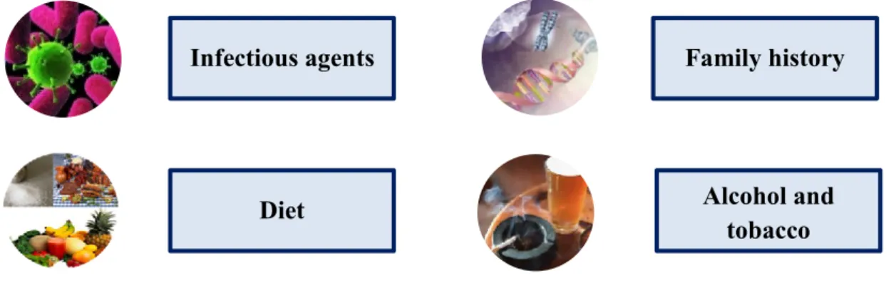
Gastric cancer is a multifactorial disease which has been etiologically associated with several genetics, environmental and lifestyle factors (Figure 1.6). Many causes have been recognized to initiation and progression of gastric carcinomas including modifiable factors (infectious agents, behavioral habits) and non-modifiable factors (age, sex). The risk factors may differ according to tumor location and histologic type whereas others are common for all types (Karimi et al., 2014). Some infectious agents and diet have demonstrated to be the most important risk factors to development of gastric cancer. Both Helicobacter pylori and diet are considered strong and well-established influences in intestinal-types (Yassibas et al., 2012). On the other hand, EBV has been studied for scientific community so that its presence in tumor cells can be definitely understood. Others risk factors such, family history, alcohol and tobacco have been associated to an increased risk of GC. (de Martel et al., 2013). Regarding to common features in overall tumors of stomach, male patients with older age are those, which have shown the greatest gastric cancers occurrence (Tural et al., 2012).
Helicobacter pylori
Since 1994, WHO has considered Helicobacter pylori as class I carcinogenic due its
Infectious agents
Diet
Family history
Alcohol and tobacco
Applied Microbiology, MSc Andreia Fernanda Nora de Oliveira
Characterization of EBV-associated gastric cancers |16
contributes to chronic inflammation and atrophy that can promote malignant transformation of the epithelium (Krejs, 2010, Zhu et al., 2014). Actually, it is clear that individuals with positive serology tests for H. pylori have shown an increased risk to development of GC (Kamangar et al., 2006). In contrast, several studies have described that early Helicobacter pylori eradication is associated with decreased risk of GC. (Wong et al., 2004, Take et al., 2005, Shiotani et al., 2010). It can reduce inflammation level preventing the progression of precancerous lesions such, gastric atrophy, intestinal metaplasia (IM) and dysplasia (Haziri et al., 2010). Furthermore, Fukase et al. (2008) provided evidences of a preventive effect of H. pylori eradication even in later events of carcinogenesis avoiding tumor recurrence (Fukase et al., 2008).
Epstein Barr Virus
During past decades, EBV has been associated to gastric cancer but it is not yet well-known if the presence of virus is a cause or consequence. A small percentage of cases has been related to EBV and it has also shown differential geographical distribution (Sousa et al., 2008). Several studies have described the presence of EBV in gastric tumor cells suggesting a role in carcinogenesis. Some of these studies have detected EBV infection exclusively in tumor cells, suggesting a late event in carcinogenesis (Truong et al., 2009, Zur Hausen et al., 2004). However, others investigations found EBV both in carcinoma cells and precursor lesions. Establishment of EBV in pre-malignant lesions suggests that virus may be initiating factor in early step of gastric carcinogenesis (Arikawa et al., 1997, Yanai et al., 1997). Despite these scientific evidences, clinicians are still requiring more investigation to clarify the mechanisms that may lead to initiation or promotion of EBV-associated gastric cancer.
Hereditary cancer
Genetic predisposition for gastric cancer is found in 10-15% of human population, however only 1-3% of these cases progresses to hereditary malignant diseases. Hereditary diffuse gastric cancer (HDGC) syndrome is the most common that leads to development of diffuse gastric adenocarcinoma cancer (Sereno et al., 2011). HDGC is autosomal dominant disease caused by germ line mutation in CDH1 gene that results in loss of expression of the cell adhesion protein (E-cadherin). Functionally E-cadherin is responsible for the maintenance of normal tissue morphology and cellular
differentiation. For this reason, CDH1 is considered a tumor suppressor gene and, when inactivating mutations occur, it contributes to the loss of cell adhesion, proliferation, invasion and metastasis (Fitzgerald and Caldas, 2004).
Diet
Epidemiological studies from different populations have shown a strong association between diet and gastric cancer, mainly in intestinal-type carcinomas (Fay et al., 1994, Bastos et al., 2010, Lunet et al., 2007). An adequate intake of fresh fruits and vegetables rich in polyphenols and vitamins may prevent GC. Their antioxidant effects inhibit formation of N-nitroso compounds (NOC) which are potential human carcinogens (Hernandez-Ramirez et al., 2009). In contrast, poor dietary habits and high salt consumption have associated with an increased risk. Moreover, in some populations smoked/cured meats or fish, pickled vegetables and chili peppers have established as a risk factor to occurrence of gastric cancer (Yassibas et al., 2012, Barad et al., 2014)
Others
Several studies have demonstrated that alcohol, tobacco and occupational exposures to nitrosamines and inorganic dusts are also risk factors of GC (Moy et al., 2010, Santibanez et al., 2012).
Applied Microbiology, MSc Andreia Fernanda Nora de Oliveira
Characterization of EBV-associated gastric cancers |18
4. Gastric carcinogenesis
The gastrointestinal tract has a great exposure to injury by infections and dietary toxins. Cellular metaplasia due to chronic inflammation, injury and repair are the most accepted processes in gastric carcinogenesis. However, additional mutations can be acquired conferring advantage to development of malignant phenotype.
4.1 Helicobacter pylori-associated GC
Helicobacter pylori infection is the leading cause of gastric cancer worldwide, mainly in
well differentiated tumors, such as intestinal types (Wang et al., 2014b). The carcinogenic model, which has been generally accepted, is based on development of precancerous lesions with potential progression to malignant disease. Intestinal-type gastric carcinomas are the most common type and its carcinogenesis includes a sequential steps where initially occurs a chronic active non-atrophic gastritis followed by multifocal atrophy, intestinal metaplasia, dysplasia and finally invasive carcinoma (Correa and Houghton, 2007).
Upon infection, H. pylori is responsible to activate multiple intracellular pathways in cells from gastric epithelium. This mechanism affects cellular signaling triggering an increased inflammatory cytokine production, altered apoptosis rate, epithelial cell proliferation and differentiation and consequently a malignant transformation. H. pylori strains that contain the cag pathogenicity island (CagPAI) induce direct effects by its virulence factors, such as the cytotoxin-associated antigen A (CagA), vacuolating cytotoxin (VacA) and other CagPAI-encoded proteins. However, indirect effects by inducing of gastritis and atrophy also play a critical role in initiating and progression of tumor. Inflammation leads to production of carcinogenic compounds as well as the hypermethylation of tumor suppressor genes (Ding et al., 2010).
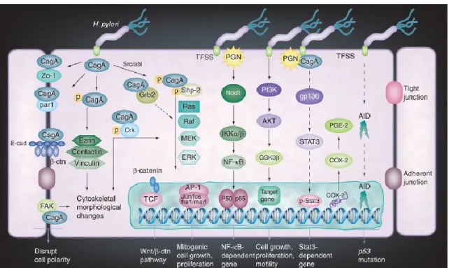
H. pylori CagA is the major oncogenic factor incorporated into host cells able to disrupt
epithelial cell functions. CagA as well as peptidoglycan are delivered into host cells through translocate the bacterial effectors (TFSS) of type IV secretion system. It confers an activation of multiple oncogenic pathways (Figure 1.7) in epithelial cells that includes NF-κB, activator protein-1, PI3K, signal transducers and activators of transcription 3, Wnt/β-catenin and cyclooxygenase 2. Persistent deregulation of these pathways triggers an important molecular mechanism toward the H. pylori-induced
carcinogenesis (Chang et al., 2004, Franco et al., 2005, Bronte-Tinkew et al., 2009, Nagy et al., 2009).
Figure 1.7 - H. pylori-induced host cell response and oncogenic signaling in gastric epithelial
cells
H. pylori is less virulent when it does not contain the cag pathogenicity island
(CagPAI), however these strains are also associated to development of cancer. Inflammatory response plays an essential role in tissue cells transformation due to an increased oxidative stress caused to release of reactive species from immune cells. The aberrant promoter methylation of tumor-related genes has been also considered (Matsusaka et al., 2014).
Furthermore, the establishment of H. pylori bacteria in gastric mucosa contributes to an increased pH of stomach. As consequence, anaerobic bacteria colonize stomach and produce active redutases which promote the transformation of nitrates from diet into nitrite (endogenous nitrosation). Nitrites are reactive molecules able to origin carcinogenic N-nitroso compounds that contribute to the accumulation of DNA damage (Hernandez-Ramirez et al., 2009).
Applied Microbiology, MSc Andreia Fernanda Nora de Oliveira
Characterization of EBV-associated gastric cancers |20
4.2 EBV-associated GC
Gastric adenocarcinomas are the most common tumor of stomach and EBV has been found in approximately of 2-20% that varies across the globe (Lee et al., 2009). Generally, EBV-associated gastric cancer (EBVaGC) occurs frequently in the upper part of the stomach (cardia and body region). It also shows a male predominance with relatively younger age when compared with EBV-negative gastric cancers (van Beek et
al., 2004, Truong et al., 2009).
The role of EBV in gastric carcinomas is still little known, however monoclonal proliferations and the presence of EBV in all tumor cells have suggested that EBV may act in pathogenesis of these tumors (Seo et al., 2010). Several studies have indicated that restricted pattern of viral gene expression categorizes EBVaGC as latency I. However, in some cases, EBV has acquired a modulated latency I because LMP-2A is also found in EBV-infected cells (Luo, et al., 2005).
Literature has shown that, DNA methylation of tumor-related genes, suppressor tumor genes silencing by viral miRNAs, loss of p53 activity and induction of growth cell factors by EBERs may be a contribute to carcinogenesis mechanism (Sivachandran et
al., 2012, Iizasa et al., 2012, Hino et al., 2009). DNA Hypermethylation
Aberrant methylation is clearly more frequent in EBVaGC than in EBV negative tumors (Iizasa et al., 2012). After EBV infection, methylation of viral promoters has been recognized as host defense mechanism. However, this mechanism does not only occur in viral genes but also in host genome (Uozaki and Fukayama, 2008). CpG-island methylation at promoter regions of tumor-related genes is clearly an epigenetic abnormality in cancer. In comparison with EBV negative tumors, EBVaGC have shown higher promoter hypermethylation able to silence gene expression of various tumor suppressor genes such, p16, p14, TP73, APC (Geddert et al., 2011).
The molecular mechanism of DNA hypermethylation observed in host genome is not completely understood. As viral factor of EBV, it has been described that LMP2A induces the phosphorylation of STAT3, triggering DNA methyltransferase 1 (DNMT1) transcription (Iizasa et al., 2012). Recently data reported that DNMT1 is responsible for CpG island methylation in promoter region of PTEN, a suppressor tumor gene (Hino et
al., 2009). Several studies have suggested that hypermethylation promoted by EBV
infection contributes significantly to gastric cancer development. However, further studies are necessary to elucidate the role of EBV in molecular mechanism (Iizasa et al., 2012).
Loss p53 activity
Latency I is the most common pattern which is found in EBVaGC and EBNA1 is the only viral nuclear protein expressed. Recent data have shown that EBNA1 contributes to decreasing promyelocytic leukemia (PML) nuclear bodies (NBs) which play important roles in apoptosis, tumor suppression, DNA repair as well as transcriptional regulation. In EBV-GC cells, it was found decreased levels of PML NBs with loss of p53 activity. It suggests that EBNA1 may affect cell cycle contributing to cell survival and proliferation (Sivachandran et al., 2012).
Viral miRNAs
miRNAs are endogenous small RNAs that play an important role in regulation of cellular differentiation, proliferation and apoptosis. Several miRNAs and non-coding RNAs, called “oncomirs” have been related with some types of cancer. These molecules act as oncogenes because they are responsible for degradation of various tumor suppressor genes. On the other hand, miRNAs are found in normal tissues as tumor suppressors when they target oncogenes (Nana-Sinkam and Croce, 2011). The levels of these molecules were found deregulated in different types of cancer, including EBVaGC. Down-regulation of the miR-200 family (host miRNAs) has been reported as common event in EBVaGC. It is associated to reduction of E-cadherin expression and loss of cell adhesion suggesting that these mechanisms may be involved in gastric carcinogenesis (Shinozaki et al., 2010). Several authors have also studied the association of viral microRNAs and gastric cancer. Actually, miR-BART5, a EBV-encoded miRNA, has shown to play a role in repression of cellular proteins (member of Bcl-2 family) promoting an uncontrolled proliferation (Choy et al., 2008).
Applied Microbiology, MSc Andreia Fernanda Nora de Oliveira
Characterization of EBV-associated gastric cancers |22
EBERs
EBERs have been linked to the malignant phenotypes in various EBV-infected cells. Previous studies demonstrated that transfection of EBERs genes into EBV-negative cells promotes growth cell in culture, tumor formation in immunocompromised mice and resistance to apoptosis (Yamamoto et al., 2000, Komano et al., 1999). Additionally, others investigations have described that EBERs promote the growth and proliferation of some epithelial cells. EBERs appear to induce insulin-like growth factor-1 (IGF1) which acts as an autocrine growth factor for NPC and GC cells. High levels of IGF1 were expressed in EBV-positive but not in EBV-negative NPC or GC tissues, suggesting that EBERs may contribute to epithelial carcinogenesis via the induction of IGF1 expression (Iwakiri et al., 2003, Iwakiri et al., 2005).
Despite the high incidence of gastric cancer in Portugal, there are no studies describing EBV-associated gastric carcinomas in our population. Therefore, the aims of this study were:
To determinate the prevalence of EBV infection in patients with gastric carcinomas in Portugal
1. Type of study
Retrospective cross-sectorial study
2. Population
The population of this study consists in 136 patients with confirmed diagnosis of gastric cancer attended at Portuguese Institute of Oncology of Porto (IPO Porto FG EPE) in the year of 2011. Inclusion criteria: 1) patients diagnosed with gastric cancer; 2) submitted to gastrectomy (total or partial), endoscopic mucosal resection or other surgical procedure; 3) with representative tumor blocks for adequate evaluation of EBV presence.
All tumors were submitted to histological examination by an experimented pathologist from our institution and classified according to the WHO and Lauren’s classification (International Agency for Research on Cancer, 2010, Lauren, 1965).
This study did not interfere with the routine procedures. Clinicopathological data was collected from individual clinical records and inserted on a database with unique codification. All procedures were submitted to approval of the IPO Porto FG EPE Ethical Committee.
3. Selection and Processing
Gastric carcinoma tissues were collected from gastrectomy (total or partial), endoscopic mucosal resection and other surgical procedures. Histological sections (3 µm slides) were obtained from formalin-fixed paraffin-embedded (FFPE) tissue blocks.
4. EBV detection
Epstein-Barr virus was identified by in situ hybridization (ISH) technique which detects EBV-encoded small RNA (EBER) in FFPE tissue blocks. Hybridization results in duplex formation of EBER sequences in the test material with the fluorescein/biotin-labeled DNA probes. This complex is indirectly detected using antibodies or streptavidin molecules that have high affinity to fluorescein and biotin, respectively. Then, a secondary enzyme-conjugated antibody is also used. The enzymatic reaction using a chromogenic substrate leads to the formation of a color precipitate that can be
Applied Microbiology, MSc Andreia Fernanda Nora de Oliveira
Characterization of EBV-associated gastric cancers|30
The ISH was performed using two protocols 1) BondTM Ready-to-use ISHEBER Probe and Anti-Fluorescein Antibody (Leica, Newcastle upon Tyne, UK) associated to
detection system Ultra Vision Large Volume Detection System Anti-Polyvalent, HRP (THERMO SCIENTIFIC, Fremont, USA) and 2) ZytoFast EBV Probe (Biotin-labeled) associated to ZytoFast CISH implementation kit HRP-AEC (ZytoVision, Bremerhaven, Germany).
Initially, all samples were performed using the first protocol described above which is considered the gold standard method of clinical diagnosis of EBV-associated tumors. Then, 10% of negative samples randomly selected and all positive cases were retested using ZytoFast Kit (ZytoVision, Bremerhaven, Germany).
4.1 EBV-ISH detection: protocol 1
Sample preparation: The samples were dewaxed in xylene for 2 x 3 minutes. After dewaxing, slides were sequentially hydrated in 100% v/v ethanol for 2 x 3 minutes, 96% v/v ethanol for 3 minutes and distilled water for 2 x 3 minutes. Proteolytic treatment was performed by addition of 15 mM proteinase K and incubation at 37ºC during 30 minutes. Finished the incubation time, the slides were immersed in distilled water for 2 x 3 minutes and then dehydrated in 96% v/v ethanol followed 100% v/v ethanol for 3 minutes to facilitate air drying.
Hybridization: Hybridization results in duplex formation of sequence present in EBV-infected cells (EBERs) and specific probe. The BondTM Ready-to-use ISHEBER Probe
(Leica, Newcastle upon Tyne, UK) was used with a volume of 20 µl for each slide. Slides were covered with coverslip and incubated at 37ºC for 2 hours.
Blocking: Endogenous peroxidase activity and nonspecific antibody binding were blocked by incubating the slides in 3% hydrogen peroxide (H2O2) solution and UV
block from UltraVision Large Volume Detection System Anti-Polyvalent, HRP (THERMO SCIENTIFIC, Fremont, USA), respectively. Each solution was incubated for 10 minutes at room temperature and washing was performed with TBS, 0.1% v/v Trinton X-100 (TBS-T) 2x 5 minutes.
Immunohistochemistry (IHC): In first step, IHC for EBERs detection was performed with Bond TM Anti-Fluorescein Antibody (Leica, Newcastle upon Tyne, UK) diluted
temperature for 30 minutes. After washing 2 x 3 minutes with TBS, UltraVision Large
Volume Detection System Anti-Polyvalent, HRP (THERMO SCIENTIFIC, Fremont,
USA) was used for revelation of hybrids. Biotinylated Goat Anti-Polyvalent Antibody (THERMO SCIENTIFIC, Fremont, USA) was addicted with incubation at room temperature for 10 minutes. The next step was washing with TBS-T 2 x 5 minutes following the addition of Streptavidin Peroxidase (THERMO SCIENTIFIC, Fremont, USA) with incubation for 10 minutes at room temperature. Streptavidin shows high affinity with several secondary antibody-conjugated biotin molecules providing a good revelation signal. Detection of hybrids is achieved by enzymatic reaction using a specific substrate to peroxidase. ImmPACTTM DAB, Peroxidase Substrate (VECTOR, Burlingame, CA USA) was used during 4 minutes at room temperature and diluted 3:100. The final washing was performed with distillated water 2 x 5 minutes.
Final preparation: Mayer’s hemalum solution (Millipore, Darmstadt, Germany) was used as counterstain for 10-20 seconds, depending of dye’s use. After coloration, slides were washed in running water for 5 minutes and the following step was sequential dehydration in 70% v/v ethanol for 2 x 4 minutes, 96% v/v ethanol for 2 x 4 minutes, 100% v/v ethanol for 2 x 4 minutes and xylene for 2 x 4 minutes. Mounting was performed with Microscopy Entellan (MERCK, Darmstadt, Germany).
4.2 EBV-ISH detection: protocol 2
Sample preparation: The samples were dewaxed in xylene for 2 x 5 minutes. After dewaxing, slides were sequentially hydrated in 100% v/v ethanol for 2 x 5 minutes, 96% v/v ethanol for 2 x 5 minutes and 70% v/v for 2 x 5 minutes. Proteolytic treatment was performed by addition of pepsin solution and incubation at 37ºC for 20-30 minutes. Finished the incubation time, the slides were immersed in distilled water.
Denaturation and hybridization: Hybridization step consists in addition of EBV probe that hybridizes to abundantly expressed Epstein virus encoded-RNA (EBERs) transcripts which are localized in the nucleus of latently infected cells. ZytoFast EBV
Probe (Biotin-labeled) (ZytoVision, Bremerhaven, Germany) was used with a volume
of 10 µl for each slide. Slides were covered with coverslip and then they were denatured at 75ºC for 5 minutes on a hot plate. After denaturation, slides were incubated at 55ºC for 60 minutes
Applied Microbiology, MSc Andreia Fernanda Nora de Oliveira
Characterization of EBV-associated gastric cancers|32
Post-hybridization and detection: Finished hybridization step, coverslips were removed by submerging in 1x Wash Buffer TBS at room temperature for 5 minutes. Two more washes were performed with 1x Wash Buffer TBS for 5 minutes. Then, it was used
ZytoFast CISH implementation kit HRP-AEC (ZytoVision, Bremerhaven, Germany)
and HRP-streptavidin was applied (3-4 drops) to the slides and incubated for 30 minutes at 37ºC. After washing with 1x Wash Buffer TBS 2 x 2 minutes at room temperature, the detection of hybrids was achieved by enzymatic reaction using a specific substrate to peroxidase. AEC substrate was used for 4 minutes at room temperature diluted 3:100. The final washing was performed with distillated water 2 x 5 minutes.
Final preparation: Mayer’s hemalum solution (Millipore, Darmstadt, Germany) was used as counterstain for 10-20 seconds, depending of dye’s use. After coloration, slides were washed in running water for 5 minutes and following step was sequential dehydration in 70% v/v ethanol for 2 x 4 minutes, 96% v/v ethanol for 2 x 4 minutes, 100% v/v ethanol for 2 x 4 minutes and xylene for 2 x 4 minutes. Mounting was performed with Microscopy Entellan new (MERCK, Darmstadt, Germany) to obtain permanent slides.
5. Quality control
As referred above, ZytoFast Kit was used to confirm the results obtained from BondTM
Ready-to-use Probe and Anti-Fluorescein Antibody. The internal positive and negative
controls provided by kits were also used. The internal positive control consists of poly-dT oligonucleotides targeting the poly (A) tails of mRNAs. A strong hybridization signals within the nuclei of cells verify the integrity of cellular mRNA in specimens. The internal negative control consists of a set of random sequence oligonucleotides with GC contents of 40-70% without known consensus to any naturally occurring sequences. These probes should not result in positive staining signals and are to be used to assess the unspecific background staining with specimens. As positive control for EBV detection were used EBV-positive FFPE tissues from nasopharyngeal carcinoma.
6. Statistical analysis
Statistical analysis was performed using the computer software IBM SPSS statistics for Macintosh, version 20.0 (IBM Corp, Armonk, NY, USA). Chi-Square (χ2) or Fisher Exact-test was used to compare categorical variables, with a significance level of 5%. The Odds Ratio (OR) was computed with its 95% confidence interval as a measure of the association between variables of interest.
1. Comparison of EBV ISH protocols
Detection of EBV for all samples was performed using protocol 1 and 10% of negative samples and all positive cases were retested using protocol 2. The results were completely concordant (100%). Both kits showed remarkable positivity however the first kit allowed better interpretation due to use of hematoxylin as contrast dye.
Figure 4.1 - Histological section from gastric
carcinoma. Positive result for EBV using protocol 1 (160x).
Figure 4.2 - Histological section from gastric
carcinoma. Negative result for EBV using protocol 1 (160x).
Figure 4.3 - Histological section from gastric
carcinoma. Positive result for EBV using protocol 2 (160x).
Figure 4.4 - Histological section from gastric
carcinoma. Negative result for EBV using protocol 2 (160x).
Applied Microbiology, MSc Andreia Fernanda Nora de Oliveira
Characterization of EBV-associated gastric cancers |38
2. Characterization of the study population
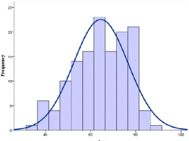
In this study, 136 cases were included, 82 male patients and 54 female with mean age of 64±12.0 (median age 65; range 34-91) (Figure 4.5). The clinicopathological characteristics of all patients are shown in table 4.1.
The majority of patients were submitted to gastrectomy (n=131), wherein 76 were total resections and 55 partial resections. Moreover, in this group of patients, 3 patients were submitted to mucosectomy and the other 2 to different surgical approaches (Table 4.1). Regarding the tumor localization in stomach, 92 carcinomas were present in distal region (antrum, pyloric, antro/pyloric or body/antro regions) 25 were from body and 19 from proximal regions (cardia, fundus, cardia/fundus, body/cardia or body/fundus regions) (Table 4.1).
All tumors were classified according to WHO (2010) and Lauren’s classifications. Regarding the WHO system, it was possible verify that the most common tumor was tubular adenocarcinoma (n=72) followed by poorly cohesive carcinoma (n=40), mixed carcinoma (n=18), mucinous adenocarcinoma (n=3) and papillary adenocarcinoma (n=1). According to Lauren, the classification revealed 76 intestinal-type carcinomas whereas diffuse and indeterminate types represented 40 and 18 of all gastric tumors, respectively. It was also found 2 carcinomas with lymphoid stroma, which are classified as uncommon variant by WHO and is included in others in Lauren’s classification (Table 4.1).
Table 4.1 - Characterization of study population Variables Gender, n (%) Male 82 (60.3) Female 54 (39.7) Age, (64 ± 12,0)
Surgical procedure type, n (%)
Gastrectomy 131 (96.3) Total 76 (55.9) Partial 55 (40.4) Mucosal resection 3 (2.2) Others 2 (1.5) Tumor localization, n (%) Proximal 19 (14.0) Body 25 (18.4) Distal 92 (67.6) Histology, n (%) WHO (2010) Papillary adenocarcinoma 1 (0.7) Tubular adenocarcinoma 72 (52.9) Mucinous adenocarcinoma 3 (2.2) Poorly cohesive carcinoma and signet-ring cell carcinoma 40 (29.4)
Mixed carcinoma 18 (13.2)
Carcinomas with lymphoid stroma 2 (1.5) Lauren (1965)
Intestinal types 76 (55.9)
Diffuse types 40 (29.4)
Indeterminate types 18 (13.2) Others (lymphoepithelioma-like carcinoma) 2 (1.5)
Invasion pattern, n (%) Infiltrative 64 (47.1) Expansive 51 (37.5) Mixed 2 (1.5) Global stage, n (%) IA 21 (15.4) IB 15 (11.0) IIA 18 (13.2) IIB 13 (9.6) IIIA 19 (14.0) IIIB 20 (14.7) IIIC 23 (16.9) IV 3 (2.2)
Applied Microbiology, MSc Andreia Fernanda Nora de Oliveira
Characterization of EBV-associated gastric cancers |40
Regarding tumor anatomic location of the stomach, the intestinal-type was the most frequent in any of the regions of stomach; the diffuse carcinomas were restricted to proximal and body regions with a percentage of 20% and 38%, respectively; the indeterminate types showed a similar distribution throughout the stomach with a percentage of 11% in proximal, 12% in body and 14% in distal region. The remaining cases, which represent lymphoepithelioma-like carcinomas, were localized in body (4%) and proximal region (5%) (Figure 4.6).
Relatively to the invasion pattern, there was similar distribution for infiltrative (n=64) and expansive (n=51) types. The mixed pattern was found in 2 samples and the information for remaining cases (n= 19) was not accessible.
Regarding TNM staging, the data was available for only 132/136 and it showed homogeneous distribution (Table 4.1). The TNM classification of malignant tumors is a cancer staging notation system for all solid tumors that gives codes to describe the stage of patient’s cancer. The TNM combinations correspond to one of five stages: 0, I, II, III or IV as described in Appendix I (Washington, 2010).
Figure 4.6 - Distribution of histological types according to their location in the stomach
Intestinal-type carcinomas Indeterminate-type carcinomas Diffuse-type carcinomas
3. EBV in GC
In our population, we have found 9/136 (6.6%) EBV-positive tumors. The majority of positive cases were found in males (8.5%) and with age >65 years old (8.6%) (Table 5). However, there were no statistically significant differences comparing the patient’s age (p=0.345) and gender (p=0.267). Considering tumor localization of stomach (proximal, body and distal), EBV was more often found in proximal and body regions (10.5% and 16.0%, respectively) while it was rare in distal regions (3.3%) (Figure 4.7). Nevertheless, the distribution showed no significant differences (p=0.058) (Table 4.2).
Comparing the distribution of EBV in the different histological types, we observed statistically significant differences either using the WHO or Lauren classifications (p<0.001). According to WHO classification, EBV was detected in 6.9% of tubular adenocarcinomas, 11.1% of mixed carcinomas and 100% of carcinomas with lymphoid stroma. Regarding the Lauren’s classification, it was found that 6.6% of intestinal-type carcinomas, 11.1% of indeterminate-type carcinomas and 100% of lymphoepithelioma-like carcinomas were EBV positive. There were no EBV-positive cases identified as diffuse-type carcinomas (Table 4.2).
Regarding invasion pattern, it was observed that tumors with expansive patterns have a higher prevalence of EBV when compared with infiltrative patterns (11.8% vs 4.7%, respectively), however these results have no significant differences (p=0.338) (Table
Figure 4.7 - Distribution of EBV according to its location in the stomach
EBV-negative cases EBV-positive cases
Applied Microbiology, MSc Andreia Fernanda Nora de Oliveira
Characterization of EBV-associated gastric cancers |42
4.2). As also shown in table 4.2, there were no statistically significant differences among clinical stages (p=0.426).
Table 4.2 - Description of EBV frequency in gastric carcinomas Negative n (%) Positive n (%) p Gender, n (%) Male 75 (91.5) 7 (8.5) 0.267 Female 52 (96.3) 2 (3.7) Age, (64 ± 12,043) < 65 63 (95.5) 3 (4,5) 0.345 ≥65 64 (91.4) 6 (8.6) Tumor localization, n (%) Proximal 17 (89.5) 2 (10.5) 0.058 Body 21 (84.0) 4 (16.0) Distal 89 (96.7) 3 (3.3) Histology, n (%) WHO (2010) Papillary adenocarcinoma 1 (100) -< 0.001 Tubular adenocarcinoma 67(93,1) 5 (6.9) Mucinous adenocarcinoma 3 (100) Poorly cohesive carcinoma 40 (100)
Mixed carcinoma 16 (88.9) 2 (11.1)
Carcinoma with lymphoid stroma - 2 (100)
Lauren (1965) Intestinal types 71 (93,4) 5 (6.6) < 0.001 Diffuse types 40 (100) Indeterminate types 16 (88.9) 2 (11.1) Others (lymphoepithelioma-like carcinoma) - 2 (100) Invasion pattern, n (%) Infiltrative 61 (95.3) 3 (4.7) 0.338 Expansive 45 (88.2) 6 (11.8) Mixed 2 (100) - Global stage, n (%) IA 20 (95.2) 1 (4.8) 0.426 IB 13 (86.7) 2 (13.3) IIA 16 (88.9) 2 (11.1) IIB 13 (100) - IIIA 16 (84.2) 3 (15.8) IIIB 19 (95.0) 1 (5.0) IIIC 23 (100) - IV 3 (100) -