IDENTIFICATION AND CHARACTERIZATION OF NEW
LISTERIA MONOCYTOGENES VIRULENCE FACTORS
by
Ana Cláudia Moutinho Gonçalves
IDENTIFICATION AND CHARACTERIZATION OF NEW LISTERIA
MONOCYTOGENES VIRULENCE FACTORS
Thesis presented to Escola Superior de Biotecnologia of the Universidade Católica Portuguesa to fulfill the requirements of Master of Science degree in Applied Microbiology
___________________________________
by
Ana Cláudia Moutinho Gonçalves
Place: Instituto de Investigação e Inovação em Saúde (i3S)
Supervision: Dr. Didier Cabanes Co-Supervision: Dra. Rita Pombinho
III
RESUMO
As doenças infeciosas são uma das principais causas de morte mundialmente, sendo a principal em bebés e crianças. Embora os tratamentos convencionais para combater infeções microbianas sejam muito eficazes, geram subpopulações bacterianas resistentes. O controlo da virulência bacteriana e a modulação da resposta do hospedeiro surgem como alternativas terapêuticas promissoras para combater as infeções. Listeria monocytogenes é uma ameaça recorrente para a saúde pública e para a indústria alimentar. É um patogénio Gram-positivo e intracelular de origem alimentar e um excelente modelo para o estudo da interação do hospedeiro com o patogénio. Esta bactéria tem a capacidade de atravessar as barreiras intestinal, hematoencefálica e materno-fetal e colonizar tecidos do hospedeiro podendo causar listeriose. Esta capacidade é alcançada através da expressão de inúmeros fatores de virulência que permite L. monocytogenes invadir, sobreviver e multiplicar dentro de células fagocíticas e não-fagocíticas. A análise do modo como este patogénio manipula as funções celulares do hospedeiro, leva à identificação de novos mecanismos que podem ser alargados a outros patogénios relevantes e ajuda na criação de novas estratégias terapêuticas. Deste modo, este trabalho teve como objetivo identificar e caracterizar novos mecanismos de virulência de L.
monocytogenes. A primeira análise transcriptómica in vivo de L. monocytogenes revelou que
vários genes são mais expressos no baço de morganhos infetados e que a maioria desses fatores de virulência são proteínas de superfície e proteínas secretadas. Neste trabalho, selecionámos três genes mais expressos in vivo, lmo2114, lmo2115 e lmo2522 com uma possível função na virulência de L. monocytogenes. Mutantes de deleção foram gerados para caracterizar o papel dos genes deletados no processo infecioso, analisando diferentes etapas do ciclo de infeção celular in vitro e virulência in vivo. Nós mostrámos que o transportador ABC, codificado pelos genes lmo2114 e lmo2115, é necessário para a entrada da bactéria em células eucarióticas e para a virulência in vivo. Além disso, observámos que uma proteína possivelmente de ligação à parede celular e codificada por lmo2522 é necessária para a capacidade de L. monocytogenes aderir e invadir células Caco-2 e importante para virulência
in vivo. De modo geral, este trabalho permitiu a identificação de novos genes de virulência de L. monocytogenes importantes para a infeção e, assim, e gerou possíveis e novos alvos para o
desenvolvimento de estratégias de anti-virulência.
Palavras-chave: Listeria monocytogenes, patogénico Gram-positivo, fatores de virulência,
V
ABSTRACT
Infectious diseases are one of the main responsible of deaths worldwide, being the prime cause of death in infants and children. Conventional approaches to combat microbial infections are very effective, however they generate strong resistant subpopulations. The control of bacterial virulence and the modulation of the host response appear as promising therapeutic alternatives to overcome infections.
Listeria monocytogenes is a recurrent problem in public health and food industry. It is a
foodborne Gram-positive intracellular pathogen and an outstanding model to study host-pathogen interactions. This bacterium has the ability to cross the intestinal, blood-brain and the placental barriers and colonize host tissues causing listeriosis. This capacity is achieved by numerous virulence factors that allow L. monocytogenes to invade, survive and multiply within phagocytic and non-phagocytic cells. Analysis of how this pathogen manipulates the host cell functions, leads to the identification of new mechanisms that could be extended to other relevant pathogens, and help designing new therapeutic strategies. Therefore, this work aimed to identify and characterize new L. monocytogenes virulence mechanisms.
The first in vivo transcriptomic analysis of L. monocytogenes revealed that a number of virulence genes are highly expressed during mouse infection and that the majority of these virulence factors are surface and secreted proteins. To identify new virulence determinants, we selected three up-regulated in vivo genes, lmo2114, lmo2115 and lmo2522 with a putative role in L. monocytogenes virulence. Deletion mutant strains were generated in order to characterize the role of the deleted genes in the L. monocytogenes infectious process by analysing different steps of cell infection cycle in vitro and virulence in vivo. We showed that the ABC transporter, which is encoded by lmo2114 and lmo2115, is required for bacterial entry in eukaryotic cells and full virulence in vivo. Moreover, we observed that the putative cell wall-binding protein encoded by lmo2522 is necessary for L. monocytogenes ability to adhere and invade Caco-2 cells and importantly for in vivo virulence. Altogether, this work allowed the identification of new L. monocytogenes virulence genes important for infection and thus provided new targets for the development of anti-virulence strategies.
VII
ACKNOWLEDGMENTS
This past year was filled with personal and professional growth and for that I would like to show my gratitude to some people that somehow contributed to this growth and to the development of this thesis:
To my supervisor Didier Cabanes for granting me this opportunity, for always being available, and for all the guidance and knowledge shared.
To my co-supervisor Rita Pombinho, thank you for all your patience, help and teachings, it was really a pleasure learn from you and I really hope you get the chance to be a university professor someday.
To Sandra Sousa, for the teachings, constructive criticism and great solutions that really helped the work to continue.
To all the Molecular Microbiology, Cell Biology of Bacterial Infections and lab colleagues, for making this journey more fun and enjoyable and also for all the help and advices given. A special thanks to Diana, it was really great sharing this experience with you, and to Joana, Déborah and Alexandra for your friendship and fun times. Also, to Ricardo for always being available to discuss ideas and to clarify my doubts.
I would like to thank my parents for granting me the opportunity to be in the higher education, for always believing in me and for being understanding and supportive. And also, to my big brother, for always being there for me when needed.
To all my friends, especially Marisa, Ricardina, Silvana and Nassa for giving me the best years of my life.
And finally, I would like to thank a special person, Diogo Filipe, that had to put up with me everytime I was tired, sad or upset about failed experiments, thank you for your patience, advices and for believing in me more than myself.
IX
CONTENTS
RESUMO ... III ABSTRACT ... V ACKNOWLEDGMENTS ... VII FIGURE INDEX ... XI TABLE INDEX ... XV LIST OF ABBREVIATIONS ... XVIIINTRODUCTION ... 21
1. Infectious Diseases ... 21
2. Listeria monocytogenes ... 21
2.1. A historical overview ... 21
2.2. Taxonomy, phylogeny and classification ... 22
2.3. Main features ... 24
2.4. Listeriosis ... 26
2.5. Intracellular infection cycle ... 27
2.6. Virulence arsenal ... 28
Adhesion ... 28
Invasion ... 29
Vacuole lysis ... 31
Cytoplasmic survival and multiplication ... 32
Intracellular motility and cell-to-cell spread ... 32
Virulence regulators ... 33
2.7. Surface proteins ... 34
Cell envelope composition ... 34
Anchoring mechanisms ... 35
Anchoring domains ... 35
2.8. ATP-binding cassette transporters ... 37
General structure and mechanism ... 37
Roles in bacterial virulence ... 39
PROJECT PRESENTATION ... 41
MATERIALS AND METHODS... 42
Bacterial strains, cell lines and growth conditions ... 42
Cloning ... 42
Mutants construction ... 42
Strains complementation ... 43
Growth analysis in vitro ... 43
Immunofluorescence ... 43
Adhesion, invasion and intracellular multiplication assays ... 44
Extraction of non-covalently cell surface-associated proteins ... 44
SDS-PAGE and Western Blot analysis ... 45
Animal infection ... 45
Statistical analysis ... 45
RESULTS ... 48
Selection of putative L. monocytogenes virulence genes ... 48
Construction of deletion mutants of the selected genes ... 52
Mutant complementation ... 55
Deletion of lmo2114, lmo2115 or lmo2522 do not impact bacterial growth in vitro ... 57
Lmo2114, Lmo2115 and Lmo2522 appear to have a role in Listeria adhesion and invasion of eukaryotic cell lines but not in intracellular multiplication within macrophages ... 59
Levels of surface-bound InlB protein are similar between the wt and the mutant strains ... 62
Lmo2115 and Lmo2522 play a role in virulence in vivo ... 63
DISCUSSION ... 65
CONCLUSION ... 70
FUTURE WORK ... 71
XI
FIGURE INDEX
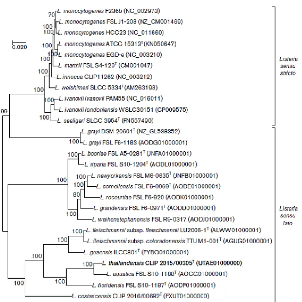
Figure 1 - Maximum-likelihood phylogenetic analysis based on the concatenated amino acid
sequences of 243 core genes present in Listeria species. Bar, 0.020 nucleotide substitutions per site. Adapted from Leclercq et al., (2018). ... 23
Figure 2 - Circular genome maps of L. monocytogenes EGDe and L. innocua CLIP 11262
(Glaser et al., 2001). ... 24
Figure 3 - Venn diagram showing the distribution of the up-regulated genes after 24, 48 and
72h post-infection (p.i.). Adapted from Camejo et al., (2009). ... 25
Figure 4 - Schematic representation of human listeriosis steps. Adapted from Radoshevich
and Cossart (2018). ... 27
Figure 5 – Schematic representation of the Listeria monocytogenes cell infection cycle. L.
monocytogenes is represented in dark blue and host actin in green. ... 27
Figure 6 – L. monocytogenes internalization into non-phagocytic cells, via InlA and InlB. .. 31 Figure 7 – Schematic representation of the listerial cell envelope structure. WTA, wall
teichoic acid; LTA, lipoteichoic acid; MurNAc, acetylmuramic acid; GlcNAc, N-acetylglucosamine; L-Ala, L-alanyl; glu, -glutamyl; m-Dpm, meso-diaminopimelyl; D-Ala, D-alanine. ... 35
Figure 8 – Schematic representation of the different classes of surface proteins of L.
monocytogenes and the respective prototypes (Bierne and Cossart 2007). ... 36
Figure 9 – Schematic representation of four Listeria monocytogenes LysM containing
proteins. AA, amino acids; SH3, Src homology 3; CHAP, cysteine, histidine-dependent amidohydrolase/peptidase; LPXTG, leucine-proline-unknown-threonine-glycine. Adapted from Bierne and Cossart (2007). ... 37
Figure 10 – Schematic representation of the general structure of an ABC importer in
Gram-positive bacteria. SBP, substrate-binding protein; NBDs, nucleotide binding domains; TMDs, transmembrane domains. ... 38
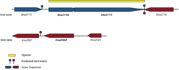
Figure 11 - Genomic organization in L. monocytogenes EGDe of the operon composed by
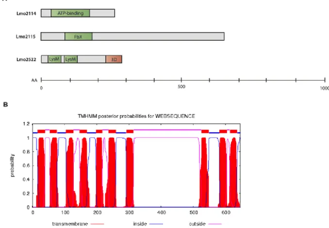
Figure 12 – Bioinformatic prediction of protein conformation. (A) Schematic representation
of conserved functional regions of the selected proteins, Lmo2114, Lmo2115 and Lmo2522. AA, amino acids; ATP-binding, ATP-binding domain of ABC transporters; LysM, lysin motif. (B) Number of transmembrane helices present in Lmo2115 (TMHMM Server). ... 49
Figure 13 - Alignment of the protein sequences of (A) Lmo2114, (B) Lmo2115 and (C)
Lmo2522 from different Listeria species with conserved sites highlighted in red. The sequence of the closest relative outside of the Listeria genus found by BLAST analysis is marked in grey for conserved site after alignment against L. monocytogenes EGDe (1/2a). .. 52
Figure 14 – Representation of the initial steps of lmo2522 deletion mutant construction. (A)
Schematic representation of the gene lmo2522 and the corresponding upstream (Up) and downstream (Dw) regions with the primers used to their amplification (AB and CD). (B) PCR products of the Up and Dw regions amplified with the primer pairs AB and CD respectively.
(C) Schematic representation of the Up and Dw fragments co-ligated in the MCS of the
plasmid pMAD and the pair of primers (SeqF, SeqR) used to verify fragments cloning into the vector. (D) PCR products of empty pMAD (1) and pMAD bearing target gene flanking regions, either fragment Up (2) or both fragments (Up and Dw), using primers SeqF/SeqR. . 53
Figure 15 – Colony PCR of the mutant strain and its isogenic wt to confirm gene deletion. (A) Schematic representation of L. monocytogenes EGDe wt and lmo2522 strains locus and
primers. (B) Agarose gel with wt and lmo2522 () both amplified with internal primers of the deleted gene lmo2522 EF, primers of the flanking regions of the gene lmo2522 AD and internal primers of a control L. monocytogenes gene, lmo1492 EF. ... 54
Figure 16 – Complementation of the mutant strain lmo2114lmo2115. (A) Schematic
representation of the construction of the complemented strain of L. monocytogenes (+lmo2114+lmo2115) and primers used. pIMK with lmo2114/lmo2115 and the corresponding promoter (P) and terminator (T) regions was inserted in lmo2114lmo2115 chromosomal locus at the attBB’ integration site. (B) E. coli DH5 colony transformed with the plasmid pIMK bearing the insert (P+lmo2114+lmo2115+T) and empty pIMK as negative control, using the pair of primers SeqF/SeqR. (C) PCR confirmation of a Listeria transconjugant (+) using primers lmo2114lmo2115 GH, lmo2114E lmo2115F and PL95/PL102. The mutant strain lmo2114lmo2115 was used as a control (). ... 56
Figure 17 – Growth profile of L. monocytogenes mutant and complemented strains in
XIII measurement. Growth curves of the wild type and (A) Δlmo2115 mutant strain, its complemented strain (B) Δlmo2114lmo2115 mutant strain and its complemented strain and
(C) Δlmo2522 and the respective complemented strain. Measurements of optical density
(OD600nm) were performed every 45 min. Results are mean ± SD of three independent
experiences. ... 58
Figure 18 – Morphological analysis of exponential phase bacteria. Immunofluorescence
staining of the mutant strain lmo2115 and the wt with DAPI. Images were acquired in Olympus BX53 microscope and processed using ImageJ. ... 59
Figure 19 - Adhesion of wt, lmo2115, lmo2114lmo2115, lmo2522 and the
corresponding complemented strains into the eukaryotic cell lines, HeLa and Caco-2 cells, upon 30 min of infection. Results are normalized to the wild type values, arbitrarily fixed to 100%. Data are means SD of at least three independent experiments. Significance is showed in relation to the wild type strain. *, p≤0.05; **, p≤0.01; ***, p≤0.001. ... 60
Figure 20 – Entry of wt, lmo2115, lmo2114lmo2115, lmo2522 and the corresponding
complemented strains into HeLa and Caco-2 cells, upon 1 h of infection and 1h30 in the presence of gentamycin. Results are normalized to the wt values, arbitrarily fixed to 100%. Data are means SD of at least three independent experiments. Significance is showed in relation to the wild type strain. *, p≤0.05; **, p≤0.01; ***, p≤0.001. ... 61
Figure 21 – Intracellular replication behavior of the wt, lmo2115, lmo2114lmo2115 and
lmo2522 strains in RAW 264.7 macrophages. Intracellular bacteria were quantified 2, 5, 7 and 22 h.p.i. Data are means SD of three independent experiments. ... 62
Figure 22 – Western blot of non-covalently cell surface-bound InlB obtained from the wt, the
mutant strains lmo2115, lmo2115lmo2115 and the corresponding complemented strains.
L. monocytogenes GAPDH (GAPDHLm) protein levels were used as a sample loading control.
Western blot is representative of two independent experiments. ... 63
Figure 23 – Role of lmo2115 and lmo2522 in the L. monocytogenes virulence in vivo.
BALB/c mice were intravenously infected with a sub-lethal dose of wt, lmo2115, lmo2522, and their complemented strains and 72 h.p.i. viable bacteria recovered from spleens and livers were quantified. Data are means SEM presented as scatter plot, with each animal represented by a dot and the mean indicated by a horizontal line. Significance is showed in relation to the wild type *, ρ≤0.05; **, ρ ≤0.01. ... 64
XV
TABLE INDEX
Table 1 – Strains and Plasmids. ... 46 Table 2 – Primers. ... 47
XVII
LIST OF ABBREVIATIONS
Fdfdf dfdfdfd
AA Amino acids
ABC ATP-binding cassette
ActA Actin assembly-inducing protein
ADP Adenosine diphosphate
agr Accessory gene regulator
Ala Alanine
AMP Antimicrobial peptide
Ap Ampicillin
Arp2/3 Actin-related proteins 2 and 3
ATP Adenosine triphosphate
BHI Brain heart infusion
BSA Bovine serum albumin
C-terminal Carboxyl-terminal
CAMPs Cationic antimicrobial peptides
CDC Cholesterol-dependent cytolysins
CFU Colony forming units
CHAP Cysteine, histidine-dependent amidohydrolase/peptidase
Cm Chloramphenicol
Crp Cyclic AMP receptor protein
CtaP Oligopeptide transport system
CWA Cell wall-anchoring
DAPI 4',6-diamidino-2-phenylindole
DltA D-alanine-activating enzyme
DMEM Dulbecco’s modified Eagle medium
DNA Deoxyribonucleic acid
Dpm Diaminopimelyl
Dw Downstream
EMEM Eagle’s minimum essential medium
ery Erytromycin
FbpA Fibronectin-binding protein
FBS Fetal bovine serum
FEA Flagellar export apparatus
Fnr Fumarate nitrate reductase
FPE Fimbrilin protein exporter
GAGs Glycosaminoglycans
GAPDH Glyceraldehyde-3-Phosphate Dehydrogenase
GlcNAc N-acetylglucosamine
Glu Glutamyl
GtcA Polypeptide wall teichoic acid glycosylation protein
GW Glycine-tryptophan
h.p.i Hours post-infection
Hpt Hexose phosphate transporter
HRP Horseradish Peroxidase
HRP Horseradish Peroxidase
Hsp60 Heat shock protein 60
HTH Helix-turn-helix
Inl Internalin
IR Inter-repeat
Kan Kanamycin
LAP Listeria adhesion protein
LB Luria-Bertani
Lgt Lipoprotein diacylglyceryl transferase
LLO Listeriolysin O
LPXTG Leucine-proline-unknown-threonine-glycine
LRR Leucine-rich repeats
LTAs Lipoteichoic acids
LysM Lysin motif
MCS Multiple cloning site
XIX
MOI Multiplicity of infection
MurNAc N-acetylmuramic acid
N-terminal Amino-terminal
NBD Nucleotide-binding domain
NlpC New lipoprotein C from Escherichia coli
OD Optical density
Opp Oligopeptide permease
ori Origin of replication
oriT Origin of transfer
P Promoter
P60 60 kDa extracellular protein
PAGE Polyacrylamide gel electrophoresis
PBS Phosphate-buffered saline
PCR Polymerase chain reaction
Pgl Phosphogluconolactonase
PI3-kinase Phosphoinositide 3-kinase
Plc Phospholipase
PrfA Positive regulatory factor A
PycA Pyruvate carboxylase
qRT-PCR Quantitative reverse transcription polymerase chain reaction
RecA Recombination protein recA
RNA Ribonucleic acid
RpoN RNA polymerase, nitrogen-limitation N
RT Room-temperature
SBP Substrate-binding protein
SD Standard deviation
SDS Sodium dodecyl sulfate
SEM Standard error of mean
SH3 Src homology 3
SrtA Sortase A
SvpA Surface virulence-associated protein
TMD Transmembrane domain
Ts Thermosensitive
Up Upstream
wt Wild-type
21
INTRODUCTION
1. INFECTIOUS DISEASES
Bacterial infections are major contributors to the high rate of mortality of infectious diseases, being one of the main responsible of deaths worldwide (Rasko and Sperandio 2010).
Although antibiotics are still the traditional choice of treatment in case of bacterial infections, they target the synthesis and assembly of essential components of bacterial processes leading to the disruption of bacterial growth (Walsh 2003). Even though this strategy has been very effective, it imposes substantial stress to the bacteria, leading to the development of resistant subpopulations, which may rapidly grow and become the dominant fraction of the population. Consequently, we are now exploring and dealing with the post-antibiotic era, with restricted treatment options for various bacterial infections (Rasko and Sperandio 2010). Hence, many alternative approaches are currently being studied that target bacterial virulence mechanisms instead of bacterial growth. By this way, anti-virulence strategies, interfere with virulence factors, thus compromising bacteria pathogenesis, being bacteria less able to colonize the host and cause diseases. The strategies are less likely to develop evolutionary resistance since most of the virulence factors are not essential for bacterial survival and thus, they are effective targets to the implementation of alternative treatments (Cegelski et al., 2008).
2. LISTERIA MONOCYTOGENES 2.1. A historical overview
In 1926, E.G.D. Murray was the first to describe the bacterium Listeria
monocytogenes (L. monocytogenes) due to unusual deaths of infected rabbits and guinea pigs
in the Department of Pathology in Cambridge. The organism was named Bacterium
monocytogenes due to a remarkable mononuclear leukocytosis in the blood of infected
animals (Murray et al., 1926). However, it is possible that this pathogen was actually isolated before, in 1891 by Hayem in France, in 1893 by Henle in Germany and in 1911 by Hulphers, which observed Gram-positive rods in tissue sections from dead patients and, at that time, named it Bacillus hepatis (Gray and Killinger 1966). In the following year of Murray’s
findings, Harvey Pirie isolated in the liver of gerbils the same microorganism responsible for the “Tiger River disease”, later known as listeriosis, and named it Listerella hepatolytica in honor to Lord Lister (Pirie 1927). In 1939, on The National Type Collection at the Lister Institute in London, it was found that Murray and Pirie identified the same microorganism, and since the name Listerella had already been applied to other organism, Pirie proposed the current name, Listeria monocytogenes (Pirie 1940).
In 1929, Nyfeldt isolated for the first time this bacterium in humans, specifically from three patients who had deceased from infectious mononucleosis like disease. However, the route of L. monocytogenes transmission was only elucidated in 1980 when a number of outbreaks associated with the ingestion of contaminated food occurred (Gray and Killinger 1966; Painter and Slutsker 2007). The first well documented foodborne outbreak of listeriosis was reported in 1983 in Canada upon the consumption of contaminated coleslaw (Schlech III
et al., 1983), and ever since then were reported several outbreaks in developed countries such
as United States of America, Japan and Europe, mainly associated to the consumption of contaminated meat and poultry products, dairy products, seafood and vegetables (Chen et al., 2009).
2.2. Taxonomy, phylogeny and classification
L. monocytogenes belongs to the Listeria genus, from the Listeriaceae family,
Bacillalles order, Bacilli class, Firmicutes phylum of the Eubacteria kingdom. Other genera of the Bacillalles order include Bacillus and Staphylococcus, which are closely related with
Listeria.
Currently, L. monocytogenes is one of 20 species from the Listeria genus belonging to the sensu stricto group, along with the species L. innocua (Seeliger 1981), L. seeligeri, L.
welshimeri (Rocourt and Grimont 1983), L. ivanovii (Seeliger et al., 1984) and L. martii
(Graves et al., 2010). The remaining 14 species, L. grayi (Larsen and Seeliger 1966), L.
rocourtiae (Leclercq et al., 2010), L. weihenstephanensis (Halter et al., 2013), L. fleischmannii (Bertsch et al., 2013), L. floridensis, L. aquatica, L. cornellensis, L. riparia, L. grandensis (den Bakker et al., 2014), L. booriae, L. newyorkensis (Weller et al., 2015), and
the most recently discovered, L. costaricensis (Núñez-Montero et al., 2018), L. goaensis (Doijad et al., 2018) and L. thailandensis (Leclercq et al., 2018) comprise the sensu lato group (Figure 1). From those, only two of them are considered to be pathogenic, L. ivanovii and L. monocytogenes, being the first one the cause of disease in livestock and the second one associated with severe illness both in animals and humans (Leclercq et al., 2018). Due to
23 rareness of L. ivanovii infection, only L. monocytogenes represents a worldwide public concern (Guillet et al., 2010; Leclercq et al., 2014).
Figure 1 - Maximum-likelihood phylogenetic analysis based on the concatenated amino acid
sequences of 243 core genes present in Listeria species. Bar, 0.020 nucleotide substitutions per site. Adapted from Leclercq et al., (2018).
Several methods have been used to classify L. monocytogenes strains (Liu 2006). L.
monocytogenes classification is based on the serotype, which comprises 13 serotypes based
on the immunoreactivity of the somatic (O) and flagellar (H) antigens: 1/2a, 1/2b, 1/2c, 3a, 3b, 3c, 4a, 4b, 4ab, 4c, 4d, 4e and 7 (Seeliger and Höhne 1979; Seeliger and Langer 1989; Gorski 2008). However, within this classification, many serotypes are known to represent genetically diverse groups of strains, and most of the reported cases of listeriosis in humans are from the serotypes 1/2a, 1/2b, 1/2c, and 4b (Farber and Peterkin 1991; Authority et al., 2014). Additionally, classification based on genotyping emerged, categorizing L.
monocytogenes serotypes in four lineages: lineage I (1/2b, 3b, 4b, 4d and 4e), lineage II (1/2a,
1/2c, 3a and 3c), lineage III (4a and 4c) and lineage IV (7) (Wagner 2008; Ward et al., 2008).
2.3. Main features
L. monocytogenes is a small rod-shaped bacterium (0.4-0.5 by 1-2 μm) usually found
in single cells or in short chains. It is a ubiquitous Gram-positive pathogen, non-spore forming, facultative anaerobe and a facultative intracellular bacterium. Listeria is motile at 20-30ºC due to the expression of peritrichous flagella but it is nonmotile at temperatures above 37ºC (Mauder et al., 2008; Wagner 2008). This bacterium cohabits different environments, such as the soil, water, sewage, animal faeces, vegetation and numerous types of raw, processed, cooked, and ready-to-eat foods, being able to survive and grow in a wide range of environmental conditions, with a pH between 5.2 and 9 (optimal at 7), temperatures ranging from <0 and 45ºC (optimal growth at 30-37ºC), high water activities and salt concentrations (Mauder et al., 2008; Strawn et al., 2013).
In 2001 it was published the first whole-genome sequencing of two Listeria species, comparing the genome of a pathogenic strain L. monocytogenes EGDe (serovar 1/2a), and the non-pathogenic strain L. innocua CLIP 11262 (serovar 6a). In this study, they showed that L.
monocytogenes has a low G+C content and a genome composed by 2 944 528 bp (Figure 2).
It was also revealed a great number of putative protein-encoding genes, both in L. innocua (represented in green) and L. monocytogenes (represented in red), encoding surface and secreted proteins, transporters, and transcriptional regulators (Glaser et al., 2001).
Figure 2 - Circular genome maps of L. monocytogenes EGDe and L. innocua CLIP 11262 (Glaser et
25 Importantly, the most crucial virulence genes encoded by L. monocytogenes are all absent from the homologous regions of the non-pathogenic species (Buchrieser 2007).
These comparative genomics studies were a step forward in the field of phylogeny and pathogenicity. Based on this, in 2009 Camejo and her colleagues developed the first in vivo transcriptome of L. monocytogenes, which approached the whole bacteria genome expression throughout mouse infection. A DNA macroarray technology was used to compare the transcriptional profile of Listeria growing under in vitro conditions (BHI, 37ºC with agitation) with its growth in vivo (mouse spleen at 24, 48 and 72 hours post-intravenous infection). This work revealed that during infection L. monocytogenes activates a number of genes related to virulence and subversion of the host immune system, being related to bacterial metabolism adaptation to host conditions. It was revealed that 457 genes were up-regulated during different stages of the in vivo infection (Figure 3).
Figure 3 - Venn diagram showing the distribution of the up-regulated genes after 24, 48 and 72 hours
post-infection (h.p.i.). Adapted from Camejo et al., (2009).
This approach is very useful on the identification of new genes involved in L.
monocytogenes virulence and also a powerful tool to better understand the complex strategies
used by pathogens to promote infections.
Along the years, L. monocytogenes has become one of the best well-studied foodborne pathogens and an outstanding model to understand host-pathogen interactions and bacterial adaptation to mammalian hosts due to: the fast bacterial growth capacity in different media, the possibility of being genetically manipulated, the co-existence of a close related non-pathogenic strain, the capacity to effectively infect tissue cultured cells and a variety of laboratory animal models frequently used to evaluate successive steps of L. monocytogenes infection (Liu 2008; Rolhion and Cossart 2017).
2.4. Listeriosis
Infection with L. monocytogenes can lead to a rare but deadly foodborne disease named listeriosis. Data from 2008 to 2015 show that listeriosis is a low incidence disease with about 1381 to 2206 confirmed cases; however it has a high fatality rate ranging from 12.7 to 20.5% in European countries (Authority et al., 2018). Immunocompromised individuals such as infants, elderly, pregnant women and patients receiving immunosuppressive agents include the high-risk groups. Whether healthy individuals infected by L. monocytogenes may experience a milder and self-limiting febrile gastroenteritis, among immunocompromised hosts listeriosis may manifest as a bacteremia that can evolve to septicaemia or to local organ infections, in particular the central nervous system and the placenta of pregnant women. In this case, listeriosis may cause meningoencephalitis, abortion and premature birth or stillbirth (Allerberger 2007; Allerberger and Wagner 2010). L. monocytogenes may also cause an extensive number of focal infections, such as conjunctivitis, skin infection, lymphadenitis, cholecystitis, peritonitis, osteomyelitis, myocarditis, arteritis and hepatic, splenic and brain abscesses (Allerberger 2007).
Listeriosis is generally acquired via the ingestion of contaminated food products and its progression rely on the capacity of L. monocytogenes to cross the intestinal, blood-brain and fetus-placental barriers. Following contaminated food consumption, L. monocytogenes is absorbed by the intestinal lumen and transverses the intestinal epithelial barrier into the lamina propria. Then it can disseminate via the lymph and bloodstream in case the immune system cannot control the infection reaching the primary target organs, liver and spleen, where it replicates within macrophages, endothelial and epithelial cells (Figure 4) (Lecuit 2007). The host defence mechanisms are dependent on the efficiency of the immune system, being the bacteria able to re-enter the bloodstream and possibility reach the brain or the placenta (Camejo et al., 2011).
27
Figure 4 - Schematic representation of human listeriosis steps. Adapted from Radoshevich and
Cossart (2018).
2.5. Intracellular infection cycle
L. monocytogenes has developed a virulence arsenal, which will be discussed below,
that provides to the bacteria the ability to infect both non-phagocytic cells (enterocytes, hepatocytes, fibroblasts and endothelial cells) and phagocytic host cells (macrophages, neutrophils and dendritic cells), in a cell cycle consisting of successive steps: adhesion, invasion, vacuole lysis, intracellular multiplication, intracellular mobility and cell-to-cell spread (Figure 5).
Figure 5 – Schematic representation of the Listeria monocytogenes cell infection cycle. L.
L. monocytogenes intimately adheres to the surface of the host cells and induces its
own internalization, involving host cytoskeletal rearrangement by actin nucleation and polymerization until being confined in a vacuole. The bacterium is then capable to acidify and lyse the vacuolar membrane escaping to the cytosol, where it can survive and multiply. Then,
L. monocytogenes also has the ability to spread from cell-to-cell by recruiting and
polymerizing host cell actin nucleators that form bundles of actin to propel the bacterium to neighbouring cells without being exposed to the extracellular milieu and thus creates a double membrane vacuole. Once bacteria escape from this secondary vacuole, it restarts a new infection cycle (Figure 5) (Cossart and Toledo-Arana 2008; Camejo et al., 2011; Radoshevich and Cossart 2018).
2.6. Virulence arsenal
The ability of L. monocytogenes to adapt to the host environment and successfully invade host cells requires the expression of a set of genes that encode a number of different virulence factors, which together compose its virulence arsenal (Camejo et al., 2011). Virulence genes and their encoded proteins are considered those that contribute for any step of the infection progress or disease transmission (Kazmierczak et al., 2005).
Adhesion
Adhesion is mediated by various bacterial proteins including FbpA, ActA, Ami, dlA, InlJ, InlF, RecA, CtaP, LAP and LapB (Camejo et al., 2011).
Listeria adhesion protein (LAP), previously known as surface protein p104, is a 104 kDa
alcohol acetaldehyde dehydrogenase, involved on the adhesion of Listeria strains to intestinal cells by the interaction with the epithelial receptor heat shock protein 60 (Hsp60). LAP is encoded by the gene lmo1634 and is present in all Listeria species with the exception of L.
grayi (Pandiripally et al., 1999; Jaradat et al., 2003; Wampler et al., 2004; Jagadeesan et al.,
2010; Jagadeesan et al., 2011). Recently it was also shown that LAP induces intestinal epithelial barrier dysfunction in order to promote bacterial translocation (Drolia et al., 2018).
Ami is an autolytic amidase protein with 102 kDa, containing an N‐terminal catalytic domain and a C‐terminal cell wall‐anchoring (CWA) domain. Whether its N-terminal catalytic domain is implicated in the cleavage of the amide bond between N-acetylmuramic acid and l-alanine residues of the peptidoglycan, the C-terminal CWA domain, which is composed by eight glycine-tryptophan (GW) modules, allow protein association to the cell
29 wall and is the main responsible for Ami’s role in L. monocytogenes adhesion to eukaryotic cells (Milohanic et al., 2001).
DltA is encoded by the gene dltA, which is integrated in an operon composed by three other genes (dltB, dltC and dltD) and is involved in the incorporation of D-alanine residues into lipoteichoic acids, thus leading to an increased sensitivity to cationic antimicrobial peptides (CAMPs) by increasing the overall cell surface charge (Perego et al., 1995; Abachin
et al., 2002).
Invasion
L. monocytogenes has the ability to invade both phagocytic and non-phagocytic cells. The
internalization into phagocytic cells is manly driven by the cell itself while invasion of non-phagocytic cells is triggered by several Listeria factors that co‐opt the cellular receptor mediated endocytosis machinery. This interaction is mediated by different virulence factors, such as, some proteins belonging to the internalin family (InlA, InlB, InlC, InlF, InlJ and InlP), Vip, Auto, p60, Lgt, GtcA, LpeA, MprF, ActA, LLO and RecA.
Internalins A (InlA) and B (InlB) are the two major proteins involved in this process (Camejo et al., 2011; Radoshevich and Cossart 2018). The members of the internalin family are characterized by the presence of a N-terminal domain containing a signal peptide sequence and a leucine-rich repeats (LRR) that promote the interaction with host cell ligands, a conserved inter-repeat (IR) domain followed by several other repeats and a variable C-terminal region.
InlA was the first internalin to be discovered. It is a 80 kDa acidic protein with 800 amino acids, constituted by 15 LRRs and a C-terminal sorting motif LPXTG followed by a hydrophobic membrane-spanning region (Gaillard et al., 1991). The LPXTG domain is responsible for the attachment of the protein to the cell wall, mediated by the enzyme sortase A (SrtA), a membrane-bound transpeptidase which catalyzes the covalent bond of the LPXTG motif between thereonine (T) and the glycine (G) residues (Gaillard et al., 1991; Bierne et al., 2002). The LRR domain of InlA interacts with the transmembrane glycoprotein E-cadherin, which is an adhesion molecule with a role in the formation of adherens junctions at the intestinal barrier, the blood–brain barrier, and the placenta (Mengaud et al., 1996; Lecuit et
al., 1997). Hence, the engagement of E-cadherin by InlA perturbs the usual E-cadherin
function, promoting cortical actin polymerization and rearrangement of the plasma membrane, thus leading to L. monocytogenes internalization (Figure 6) (Lecuit et al., 2001;
Lecuit et al., 2004). The interaction between InlA and E-cadherin is highly specific and requires the presence of a proline at position 16 in the extracellular domain of E-cadherin molecule. However, the mouse E-cadherin has a glutamic acid at this position avoiding InlA-dependent entry of Listeria (Lecuit et al., 1999). For the successfully internalization of L.
monocytogenes in InlA/E-catherin mediated entry, the PI3-kinase activity is required to
promote actin polymerization. In the intestinal barrier PI3-kinase is constitutively present, whereas in the placental barrier it is not, requiring also the expression of inlB for PI3-kinase activation (Gessain et al., 2015).
InlB, the second invasion protein identified for L. monocytogenes, it is a 67 kDa protein, with 630 amino acids, composed by 8 LRRs and a C-terminal domain with three GW modules (Gaillard et al., 1991; Dramsi et al., 1995; Braun et al., 1997). The GW modules mediate the binding of InlB to the cell wall through non-covalently interactions with lipoteichoic acids (LTAs) and peptidoglycan-bound wall teichoic acids (WTAs) (Jonquieres et al., 1999; Carvalho et al., 2018). InlB is able to interact with multiple cell receptors: the globular part of the complement component C1q (gC1qR), tyrosine kinase Met (hepatocyte growth factor) and glycosaminoglycans (GAGs). Through its N-terminal LRRs, InlB binds to the receptor Met, while soluble InlB directly interacts through its C-terminal GW repeats with the receptor gC1qR and with GAGs, in which internalization is predominantly mediated by InlB associated with bacteria (Figure 6) (Braun et al., 2000; Jonquieres et al., 2001; Ireton 2007). InlB has the capacity to induce Met autophosphorylation and the recruitment of adaptor proteins (Cbl, Shc, CrkII and Gab1) that consequently activate PI3-kinase. This signalling cascade induces Met ubiquitination, actin cytoskeleton rearrangements and bacterial internalization through clathrin-mediated endocytosis (Ireton et al., 1999; Basar et al., 2005; Sun et al., 2005).
31
Figure 6 – L. monocytogenes internalization into non-phagocytic cells, via InlA and InlB.
Vacuole lysis
Upon internalization, L. monocytogenes is confined in a membrane-bound vacuole. In goblet cells, the bacteria does not escape from the vacuole, being directly transcytosed to the lamina propria, where systemically disseminates (Nikitas et al., 2011). However, in most of other cell types, L. monocytogenes is capable to disrupt the vacuole and reach the host cell cytoplasm. This is accomplished mainly by the listeriolysin O (LLO), along with two phospholipases (PlcA and PlcB) (Birmingham et al., 2008). Other known virulence factors that contributes to this process are PrsA2, SvpA, SipZ, Lsp and ActA (Camejo et al., 2011).
LLO is encoded by the gene hly, that is part of a locus composed by the majority of crucial virulence genes of L. monocytogenes (prfA, plcA, hly, mlp, actA and plcB). LLO is a secreted pore-forming toxin that belongs to the family of cholesterol-dependent cytolysins (CDC) and oligomerizes in the vacuole membrane, in the presence of cholesterol, as ring-like pore complexes with optimal activity at acidic pH (Beauregard et al., 1997; Gilbert 2010; Ruan et al., 2016). LLO displays additional functions in bacterial infection such as mitochondrial fragmentation, alteration of intracellular calcium levels, compromission of lysosomal membranes, promotion of histone modifications and other nuclear processes, and can also suppression of host immune response, among others (Repp et al., 2002; Dramsi and
Cossart 2003; Ribet et al., 2010; Stavru et al., 2011; Samba-Louaka et al., 2012; Malet et al., 2017).
Cytoplasmic survival and multiplication
Within the cytosol L. monocytogenes must adapt its metabolism to the nutrients and metabolites available in the cytosol and must be able to evade host defenses. Important proteins involved in this process are Hpt, LplA1, PycA, Fri, RelA, PrsA2, OppA, PgdA, Pgl and InlH (Camejo et al., 2011).
The hexose phosphate transporter (Hpt) is the main protein expressed throughout the intracellular multiplication step, that mediates the uptake of hexose phosphates allowing L.
monocytogenes to use phosphorylated sugars such as glucose-1-phosphate within the host cell
cytosol, and therefore extending the range of carbon sources available for intracellular growth, in order to optimize the bacterial proliferation rate (Chico-Calero et al., 2002).
Intracellular motility and cell-to-cell spread
Actin-based motility allows L. monocytogenes cell-to-cell spread and organ dissemination, while avoiding host immunity. In this cellular infection step participate a number of different virulence factors including ActA, InlC, LLO, PlcA, PlcB and p60 (Camejo et al., 2011; Pizarro-Cerda and Cossart 2018).
The surface-anchored bacterial protein ActA contains a transmembrane hydrophobic tail region on its C-terminal domain that holds the protein at the bacterial cell membrane (Domann et al., 1992; Kocks et al., 1992). The N-terminal region of ActA contains an actin monomer-binding domain and 2 acidic regions that activates a host actin nucleator (Arp2/3 complex) (Welch et al., 1997; Welch et al., 1998; Campellone and Welch 2010). Therefore, ActA polymerases actin filaments at one pole of the bacteria, forming a comet tail that propels the bacteria through host cell cytosol, and to the neighbouring cells (Kocks et al., 1995). Besides its pivotal role in L. monocytogenes mobility, ActA was also implicated in L.
monocytogenes attachment and internalization into different cells, though the interaction with
glycosaminoglycans (Alvarez-Domínguez et al., 1997). This protein is also able to prevent autophagy in the cytosol of macrophages, either by conferring actin-based movement to the bacteria or by actin-masking of the bacteria, that no longer will be recognized by autophagy machinery (Dussurget 2008; Yoshikawa et al., 2009).
33 Virulence regulators
The first identified virulence regulator in L. monocytogenes was PrfA (Positive regulatory factor A), that regulates the expression of the main virulence related genes (hly, plcA, plcB,
actA, inlA and inlB) whose promoter regions contain a PrfA box (Leimeister-Wächter et al.,
1990; Chakraborty et al., 1992; Ripio et al., 1998; Dussurget et al., 2002; Milohanic et al., 2003; Raynaud and Charbit 2005; Marr et al., 2006; Scortti et al., 2007). PrfA is a 27 kDa protein with 235 amino acids that belongs to the family of cyclic AMP receptor protein (Crp)/fumarate nitrate reductase (Fnr) transcriptional regulators, composed by two major domains, the conserved N-terminal domain regulated by cyclic nucleotides and a C-terminal domain containing the DNA-binding helix-turn-helix (HTH) motif. The transcription of PrfA-dependent genes is activated by PrfA binding to the palindromic PrfA box, whose 14-bp canonical sequence is tTAACanntGTtAa (Leimeister-Wächter et al., 1990; Korner et al., 2003; Vega et al., 2004; Eiting et al., 2005; Pizarro-Cerdá et al., 2012). PrfA can exist in two functional states: weakly active in the native form and highly active after a conformational change. PrfA has the ability to undergo allosteric transition from weakly active to highly active conformations upon interaction with glutathione, a molecule present in the host cytosol (Reniere et al., 2015). The self-regulation of prfA expression and protein activity involves complex transcriptional, post-transcriptional and post-translational mechanisms (Port and Freitag 2007). The PrfA expression is thermoregulated by an RNA thermosensor which allows biggest expression of virulence genes at temperatures equal or above of 37°C (Renzoni
et al., 1997; Johansson et al., 2002).
Another characterized virulence regulator of L. monocytogenes is the two-component system virulence regulator, VirR/VirS, which is a response regulator found to be highly expressed during infection and regulates a number of genes involved in virulence. In this two-component system, an external signal is sensed by the histidine kinase protein (VirS), in which a conserved histidine residue is autophosphorylated releasing a phosphate group that activates the response regulator (VirR), allowing it to bind to a DNA sequence. This DNA sequence is predicted to be a palindromic sequence found in the promoter region of the regulated genes (Mandin et al., 2005).
Another important virulence regulator of L. monocytogenes is the transcription factor SigmaB (B) that regulates several genes that are predicted to be important in stress tolerance,
carbohydrate metabolism, transport and cell envelope processes (Hain et al., 2008). Importantly, some of the PrfA-regulated genes display potential σB promoter sequences
(Milohanic et al., 2003). Different studies have shown that B and PrfA co-regulate genes that
are important for L. monocytogenes to switch from an extracellular to an intracellular environment (Chaturongakul et al., 2008; Ollinger et al., 2008).
Most recently it was identified a novel virulence regulator, designated MouR, a dimeric DNA-binding transcription factor encoded by the gene lmo0651 (Pinheiro et al., 2018). MouR was shown to regulate the accessory gene regulator (agr) locus, a quorum sensing system, with a role in bacterial survival and competitive advantage in soil, adhesion to surfaces, biofilm formation, invasion of mammalian cells, virulence in vivo and global changes in gene expression (Autret et al., 2003; Rieu et al., 2007; Riedel et al., 2009; Vivant
et al., 2014; Vivant et al., 2015). It was demonstrated that MouR, through the Agr system,
regulates biofilm formation, invasion of mammalian cells and virulence in the gastrointestinal phase of infection (Pinheiro et al., 2018).
Other regulators of virulence determinants include MogR, DegU and GmaR (flagella production) and the RNA-binding protein Hfq (bacterial physiology) (Camejo et al., 2009).
2.7. Surface proteins
The majority of the virulence factors described above, include proteins located at the surface of the bacterial cell, either associated with the cell envelope or secreted to the extracellular milieu, allowing the direct interaction with the host cell. They are usually associated with crucial bacterial mechanisms that account for the successful colonization of the pathogen, including bacterial growth, adhesion, invasion and persistence within host cells.
Cell envelope composition
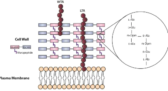
L. monocytogenes cell envelope a multilayered structure that not only provide to the
bacterium structural support and protection from the external environment, but also allows exchange of nutrients and waste products with the extracellular media. It is composed by a single plasma membrane and the cell wall. The cytoplasmic membrane is manly composed by proteins, lipids and carbohydrates and the cell wall displays a thick and highly polymerized macromolecule, the peptidoglycan layer. The peptidoglycan is composed of linear and parallel glycan strands of alternating units of acetylmuramic acid (MurNAc) and N-acetylglucosamine (GlcNAc) linked perpendicularly and cross-linked by (1,4) glycosydic bonds. These peptidic bridges (muropeptides), are composed by the sequenced peptides L
35 residue by L-alanine (Schleifer and Kandler 1972; Kamisango et al., 1982; Fiedler 1988; Dhar
et al., 2000). The cell wall is also decorated with polyanionic polymers designated teichoic
acids (TAs), which can be covalently bound to the peptidoglycan termed wall teichoic acids (WTAs), or embedded into the plasma membrane, the lipoteichoic acids (LTAs) (Figure 7) (Navarre and Schneewind 1999; Neuhaus and Baddiley 2003).
Figure 7 – Schematic representation of the listerial cell envelope structure. WTA, wall teichoic acid;
LTA, lipoteichoic acid; MurNAc, N-acetylmuramic acid; GlcNAc, N-acetylglucosamine; Ala, L-alanyl; D-glu, -D-glutamyl; m-Dpm, meso-diaminopimelyl; D-Ala, D-alanine.
Anchoring mechanisms
Bacterial surface proteins are synthesized in the cytoplasm, directed to the plasma membrane through an export signal and then translocated across the membrane through specialized secretion systems. Their transport can occur mainly by two different pathways, the canonical Sec-dependent pathway and the Tat pathway, both requiring a specific N-terminal secretion system (signal peptidases). Others secretion system have been identified such as the fimbrilin protein exporter (FPE) system, the flagellar export apparatus (FEA), the Esx-1/Wss system and prophage holins. Their final localization of the proteins will depend on the presence of specific domains or motifs (Desvaux and Hébraud 2006; Schneewind and Missiakas 2014).
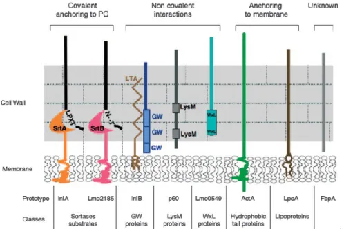
Anchoring domains
Protein association to the cell envelope are dependent on specific binding domains encoded in their sequence. Proteins can be whether associated to the cell wall or to the plasma membrane, with or without covalent interactions. Protein-cell wall association can be established either through a stable covalent bond between the peptidoglycan matrix and
specific protein sorting motif sequences, such as LPXTG and NXXTX proteins, or by a non-covalent interaction between cell wall components and cell wall-recognizing protein domains (LysM, WXL and GW proteins). Additionally, proteins anchored to the membrane may also contain lipobox motifs (lipoproteins) or hydrophobic residues (hydrophobic tail proteins). Other surface proteins either lack recognizable surface-targeting sequences or do not have predicted surface-domains (Figure 8) (Carvalho et al., 2014).
.
Figure 8 – Schematic representation of the different classes of surface proteins of L. monocytogenes
and the respective prototypes (Bierne and Cossart 2007).
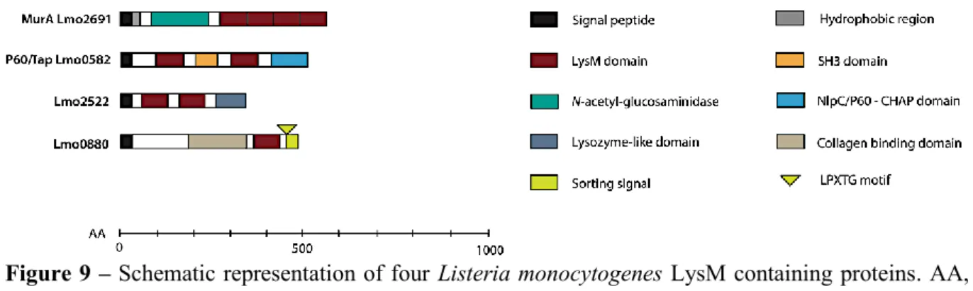
Lysin motif (LysM) domains are often present in bacterial lysins, in bacteriophage proteins and in some eukaryotic proteins, whereas can also be present in bacterial peptidoglycan hydrolases, peptidases, chitinases, esterases, reductases or nucleotidases (Desvaux et al., 2006). LysM domains contains a variable number of 40 to 80 residue repeats, separated by sequences rich in serine, threonine and asparagine or proline residues (Buist et
al., 1995; Buist et al., 2008; Ohnuma et al., 2008). In L. monocytogenes, six proteins contain
an LysM domains (Figure 9), among them, p60 and MurA, two autolysins involved in the bacterium virulence (Lenz et al., 2003; Bierne and Cossart 2007). The p60 protein is composed by a N-terminal region with two LysM domains separated by an Src homology 3 (SH3)-like domain that seems to mediate the specific regions of the peptidoglycan, that are important for p60 activity, and a C-terminal NlpC/p60 domain presumably related to peptidoglycan peptidase activity (Anantharaman and Aravind 2003; Bierne and Cossart 2007; Layec et al., 2008). On the other hand, MurA protein contains four LysM in the C-terminal that possibly contribute to position the MurA catalytic site differently from p60 to enhance its
37 activity (Carroll et al., 2003). Other four proteins containing LysM domains were more recently described: Lmo2522, Lmo1303, Lmo1947 and Lmo0880. The first two proteins contain two and one LysM domains, respectively, detected in supernatant fractions. In turn, Lmo1941 possesses one LysM and a transmembrane hydrophobic domain and is detected in the membrane fraction, and Lmo0880 contains a cell wall LPXTG domain (Pucciarelli et al., 2005; Trost et al., 2005; Wehmhöner et al., 2005).
Figure 9 – Schematic representation of four Listeria monocytogenes LysM containing proteins. AA,
amino acids; SH3, Src homology 3; CHAP, cysteine, histidine-dependent amidohydrolase/peptidase; LPXTG, leucine-proline-unknown-threonine-glycine. Adapted from Bierne and Cossart (2007).
2.8. ATP-binding cassette transporters
ATP-binding cassette (ABC) transporters are one of the largest protein superfamilies and are widespread among living organisms, including bacteria, archaea and eukaryotes (Holland et al., 2003). They are primary active membrane proteins involved in the transport of solutes (or allocrites) across the cell membrane (Zolnerciks et al., 2011). ABC transporters are involved into a number of crucial physiological processes, among them nutrient import, cellular detoxification, lipid homeostasis, signal transduction, antiviral defences and antigen presentation (Lewinson and Livnat-Levanon 2017).
In L. monocytogenes there are 331 predicted genes that encode diverse transport proteins, with the ability to colonize and grow in different environments that comprise 11.6% of all predicted proteins (Glaser et al., 2001).
General structure and mechanism
ABC transporters may function either as exporters by pumping toxins, drugs and lipids to outside of the cell, or as importers uptaking of nutrients and other molecules into the cell (Rees et al., 2009). ABC transporters, both exporters and importers, are usually constituted by four domains: two nucleotide-binding domains (NBDs), also known as ATPase domains or ATP-binding cassette, and two transmembrane domains (TMDs), also known as hydrophobic
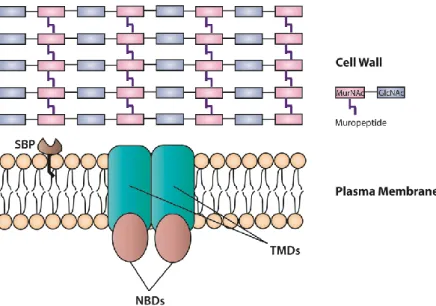
membrane-spanning domains or membrane domains. Bacterial ABC importers (permeases) can also include an additional protein partner, a substrate-binding proteins (SBPs), that in Gram-positive bacteria are lipoproteins anchored to the cell membrane able to bind to the substrate and delivers it to the TMDs (Figure 10) (Garmory and Titball 2004; Beis 2015; Wilkens 2015).
Figure 10 – Schematic representation of the general structure of an ABC importer in Gram-positive
bacteria. SBP, substrate-binding protein; NBDs, nucleotide binding domains; TMDs, transmembrane domains.
The NBDs are conserved among the known ABC transporters, being each of them constituted by two subdomains, a catalytic core domain and an -helical domain. The catalytic domain contains two conserved motifs, the Walker A motif and the Walker B motif, in which Walker A interacts with the phosphate groups of ATP and Walker B activates a water molecule for a nucleophilic substitution at the -phosphate of the nucleotide. In turn, the -helical domain contains a specific and highly conserved motif of the ABC superfamily, LSGGQ, involved in the binding of the nucleotide. The NBDs form dimers, which can assume open or closed conformations: in the absence of ATP molecules, the domains are separated (open conformation), but upon ATP binding, they form a “sandwich dimer” with two ATP molecules trapped inside. ATP is then hydrolysed, disrupting the “sandwich dimer” and thus releasing ADP and inorganic phosphate. The molecular motion is then transferred to the TMDs through the -helix, coupling helix, located in the cytoplasmic loops of the TMD (Garmory and Titball 2004; Beis 2015; Wilkens 2015).
39 Each TMD is composed by 5 to 10 transmembrane -helices, with a total of 10 to 20 transmembrane -helices depending on the transporter class (Davidson et al., 2008). These transmembrane -helices form a transmembrane pore that can be facing the outside or the inside of the cell. TMDs normally do not present significant sequence conservation but show similarities within a transporter type, possibly related to differences in the transporter substrate. For both importers and exporters, the transmembrane pore is lined up when the transport substrate interacts with residues of the transmembrane -helices (Wilkens 2015). Based on TMDs folds, ABC transporters can be subdivided in three types: Type I, Type II and Type III (or energy coupling factor) (Ter Beek et al., 2014). Type I ABC importers are involved in medium-affinity bacterial uptake of various nutrients, such as, ions, amino acids, short peptides and oligosaccharides (Locher 2016). Type II importers are involved in the uptake of metal chelates (e.g. vitamin B12), heme and oxanions and usually contain more transmembrane helices than Type I (Klein and Lewinson 2011; Beis 2015). Type III importers are normally associated with micronutrient uptake (Rice et al., 2014).
SBPs consist of two domains (C- and N-lobes), connected by a flexible hinge that enables the SBP to assume two different conformations: open-unliganded and closed-unliganded (Quiocho and Ledvina 1996; Lanfermeijer et al., 2000). These conformations have a high and a low affinity for the substrate, respectively (van der Heide and Poolman 2002). After substrate binding, the lobes close round the ligand and then interact with the translocator. It was previously suggested that the SBP has a role in the transmission of a signal to the NBDs, therefore increasing their affinity to ATP. Upon ATP binding and hydrolysis, the substrate is released from the SBP (Davidson et al., 1992).
New findings regarding ABC transporters, their mechanism of action and their biochemically, structurally and mechanistically characterization remain yet to be revealed (Rees et al., 2009).
Roles in bacterial virulence
ABC transporters are usually associated with nutrient uptake and drug resistance, however, there are strong evidences that they may be directly or indirectly involved in the virulence of the bacteria (Garmory and Titball 2004).
In L. monocytogenes, some ABC transporters with a role in virulence were already identified, such as the oligopeptide permease (Opp), that was shown to be necessary for bacterial growth at low temperature, for intracellular growth in macrophages and for early
phase of infection in a mice model (Borezee et al., 2000). It was also identified a substrate-binding component of an oligopeptide transport system, CtaP, associated with cysteine transport, whose the absence of the gene encoded-protein results in the reduction of bacterial adherence to host cells and virulence attenuation in intragastric and intravenously infected mice (Xayarath et al., 2009).
41
PROJECT PRESENTATION
Listeria monocytogenes is a facultative intracellular pathogen able to disseminate
throw out the host, colonize host tissues and cause listeriosis, upon the consumption of contaminated food products. Disease progression is achieved through the expression of a complex virulence arsenal that allows L. monocytogenes to invade, survive and multiply within phagocytic and non-phagocytic cells.
A previous study provided the first comprehensive view of the genome expression of
L. monocytogenes in infected mice spleen in comparison with gene expression of this
pathogen growing in standard culture conditions. This study revealed that 20% of the L.
monocytogenes genome is differentially expressed and among them 80% of the
protein-encoding genes were up-regulated in infected mice spleen. In addition, the great majority of known virulence factors are absent from non-pathogenic L. monocytogenes strains and highly expressed during infection of mouse organs. This analysis allowed also the identification of uncharacterized genes also absent from non-pathogenic strains and highly expressed during in
vivo infection. The aim of this project was to select some of these genes differentially
expressed during in vivo infection, construct mutants and analyse their phenotype in vitro and
in vivo. This approach had to allow the identification of new L. monocytogenes genes crucial
MATERIALS AND METHODS
Bioinformatic analysesTarget gene sequences were obtained from the ListiList database (http://genolist. pasteur.fr/ListiList/) and homologue searches were performed with the BLAST tool (Boratyn
et al., 2013). Search of conserved protein domains and protein function prediction were
performed with Pfam v. 32.0 (El-Gebali et al., 2018). The TMHMM Server v. 2.0 (http://www.cbs.dtu.dk/services/TMHMM/) was used to predict protein conformation and transmembrane helices domains. Multiple sequence alignment was performed by Clustal Omega (Madeira et al., 2019) and sequence similarities were identified with ESPript 3.0 (Robert and Gouet 2014).
Bacterial strains, cell lines and growth conditions
Listeria monocytogenes EGDe and Escherichia coli strains (Table 1) were grown in Brain
Heart Infusion (BHI, BD-Difco) and Lysogenic Broth (LB) medium, respectively, both at 37ºC with agitation. Bacterial strains harbouring the plasmids (Table 1) were grown in the presence of the following antibiotics: ampicillin 100 μg/ml (E. coli) and erythromycin 5 μg/ml (ery) (L. monocytogenes) in the case of pMAD; kanamycin 50 μg/ml (E. coli and L.
monocytogenes) for pIMK derivatives. Human cervical adenocarcinoma HeLa cells (ATCC
CCL-2), human epithelial colorectal adenocarcinoma Caco-2 (ATCC HTB-37), and RAW 264.7 murine macrophages (ATCC-TIB-71) were used for in vitro assays. HeLa cells and RAW 264.7 murine macrophages were cultured in Dulbecco’s modified Eagle medium (DMEM) supplemented with 10% fetal bovine serum (FBS) and Caco-2 cells were culture in Eagle’s minimum essential medium (EMEM), supplemented with 20% FBS, 1mM pyruvate, and 1% nonessential amino acids. The cells were grown without antibiotics at 37ºC in a 5% CO2 humidified atmosphere.
Cloning
Mutants construction
L. monocytogenes EGDe deletion mutants were generated by a double recombination process,
using pMAD thermosensitive shuttle vector as described previously (Carvalho et al., 2015). Briefly, ~1000 bp DNA fragments flanking upstream and downstream regions of the target genes were amplified by PCR from L. monocytogenes EGDe chromosomal DNA, with the