Universidade de Aveiro
2007
Departamento de Biologia
Pedro Daniel Coelho
dos Santos Betrão
Métodos Computacionais para o Estudo de Redes
Celulares
Computational Tools for the Study of Cellular
Networks
tese apresentada à Universidade de Aveiro para cumprimento dos requisitos necessários à obtenção do grau de Doutor em Biologia, realizada sob a orientação científica do Dr. Manuel Santos, Professor Associado do Departamento de Biologia da Universidade de Aveiro
Apoio financeiro da FCT e do FSE no âmbito do III Quadro Comunitário de Apoio.
o júri
presidente Reitora da Universidade de Aveiro
Doutor Amadeu Mortágua Velho da Maia Soares
Professor Catedrático da Universidade de Aveiro
Doutor Roderic Guigó Serra
Professor Associado da Universidade Pompeu Fabra - Barcelon
Doutor José Luís Guimarães Oliveira
Professor Associado da Universidade de Aveiro
Doutor Manuel António da Silva Santos
Professor Assoc da Univ de Aveiro (Co-Orientador)
Doutor Luis Serrano Pubull
Investigador Principal do Centre de Regulacio Genomica, (Orientador)
Doutor José Bartholo Pereira Leal
agradecimentos There is little space in typical science thesis to the human dimension.
Everything else that supports us when we go about doing our work. The writing is meant to convey scientific findings, discussion and future prospects but a PhD is so much more. I am left with a few lines to thank everyone that helped me during my stay in Heidelberg. Funnily enough this was one of the hardest parts of the thesis to start, exactly because it is so difficult to summarize the social aspects of these years into a couple of lines.
I would like to thank my thesis supervisor Luis Serrano for giving me support and above all the freedom to explore. I am thankful that I had my supervisor, along with all the people in the lab, especially Caroline Lemerle, Ignacio Sanchez and Marc Isalan, to discuss almost any silly idea that came to mind. More generally I would like to thank everyone in the lab for the fun times together in particular to Sandra Esteras and Barbara Di Ventura for showing me around Heidelberg in the first year.
I would like to thank my co-supervisor Manuel Santos and Catarina Gomes for all the help in dealing with the University of Aveiro.
I am grateful for having good friends around during these years. Thanks to Paulo Cunha, Catia Proenca, Ricardo Almeida and my family for listening. Finally I want to thank Silvia Santos for completing me so. To her I dedicate this thesis.
palavras-chave biologica computacional, bioinformática, evolucão, interacções entre proteínas, estruturas de proteínas, proteínas desordenadas
resumo Com a chegada da chamada era genómica tornou-se importante focar os estudos de fenómenos biológicas num ponto de vista de “sistema”. Isto deve-se ao facto de deve-ser fundamental compreender como é que as funções celulares emergem da interacção e integração dos muitos componentes celulares. Para tornar isto possível, muitos dos recentes desenvolvimentos tecnológicos têm sido focados na colecção de um grande números de dados sobre grande parte dos componentes celulares. A necessidade de desenvolver novos métodos computacionais, capazes de integrar e relacionar esta informação tornou-se por isso num imperativo. Estes novos métodos de análise devem permitir a criacao de modelos que consigam extender o conhecimento actual de forma a prever dados não conhecidos.
O trabalho apresentado nesta tese tem como foco o desenvolvimento de métodos computacionais para o estudo de interacções entre proteínas. Em particular, foi desenvolvido um método para prever a especificidade de interacção de domínios de proteínas que ligam péptidos utilizando informação estrutural. Para demonstrar este método, foram escolhidos domínios SH3 de
S. cerevisiae e domínios SH2 de H. sapiens. O trabalho aqui apresentado mostra que, conhecendo a especificidade de interacção, é possível usar genómica comparativa e o conhecimento da estrutura secondária das proteínas para prever quais os alvos de interacção destas proteínas no proteoma com mais de 75% de exactidão. Foi observado que a exactidão destas previsões aumenta quando se restringe a procura de locais de
interacção a zonas do proteoma previstos como desordenados, sugerindo que os locais de interacção de domínios que se associam a péptidos tendem a residir nestas zonas.
A análise dos actuais mapas de interacção de proteínas de várias espécies revelou que estas interacções apresentam considerável plasticidade evolutiva. O ritmo a que estas interacções mudam durante o processo evolutivo depende tanto da especificidade da interacção como dos processo biológico em que participam as proteínas. Como exemplo, o estudo do proteoma humano revelou que proteínas que participam na resposta imune, em funções de transporte e no estabelecimento de localização mostram sinais de selecção positiva para mudarem de interacções.
Em resumo, são apresentados nesta tese novos métodos para prever interacções entre proteínas assim como novas hipóteses sobre o processo evolutivo destas interacções.
keywords computational biology, bioinformatics, evolution, protein-protein interactions, protein structure, protein disorder
abstract With the developments of the so called Genomic era there has been an increasing awareness of the necessity to study biological phenomena from a systems view point. This is due to the importance of understanding how the interplay between the many cellular components brings about cellular functions. To this effect many of the most recent technological efforts in biology have been directed at collecting data that encompass most cellular components. Integration of these different experimental approaches and better
comprehension of the vast data available, urged for the development of computational methods in biological research. These computational tools should be able to search for patterns that can extend current knowledge by providing predictive models of biological events.
The work presented in this thesis focuses on computational methodologies to study protein-protein interactions. In particular, the results presented show that binding specificity of peptide binding domains can be obtained from protein structure. SH3 domains from S. cerevisiae and human SH2 domains were used to demonstrate this method. Also, this work demonstrates that, by knowing the binding specificity, it is possible to use comparative genomics and protein secondary structure information to accurately (>75% accuracy) predict the binding partners of a protein in the proteome. It was observed that restricting predictions to unstructured elements of the proteome increases the accuracy of the prediction.
The analysis of current protein interaction information of many different species has revealed that protein interactions are quite plastic in evolution and are determined both by binding specificity and biological function. It was observed, for example, that human proteins related with immune response, transport and establishment of localization, show signs of positive selection for change of interactions.
In summary, the work reported here, explores new methods to computationally predict protein interactions shedding light into the possible evolution of these interactions.
1 – Introduction 5
1.1 – Experimental methods to determine proteins interactions ………... 5
1.1.1 – Yeast-two hybrid and protein complementation methods …………... 5
1.1.2 – Microscopy detection methods ……… 6
1.1.3 – TAP-tag pull down, protein arrays and solid-phase detection ……… 6
1.1.4 – Molecular display methods ………. 7
1.2 - Computational methods to predict protein-protein interactions …………... 8
1.2.1 – Sequence based methods ………. 8
1.2.2 – Graph theory methods ………. 10
1.2.3 – Structural based methods ……… 10
1.3 – Large scale data integration methods ……….. 12
1.4 – Modular protein domains ………. 13
1.4.1 – SH3 domain ………. 13
1.4.2 – SH2 domain ………. 16
1.5 – Evolution of cellular networks ………. 17
2 – Manuscript 1: Comparative genomics and disorder prediction identify biologically relevant SH3 protein interactions 19 2.1 – Abstract ……… 19
2.2 – Non technical summary ……… 20
2.3 – Introduction ………... 21
2.4 – Results and Discussion ………. 23
2.4.1 – Identification and conservation of SH3 domains and selection of genomes ………... 23
2.4.2 – Evaluation of the “conservation” approach ……… 24
2.4.3 – Combining comparative genomics and disorder prediction ………… 26
2.4.4 – Determining an optimal divergence time for the genomes used when searching for conservation of target ligands of SH3 domains ……… 28
2.4.5 – Predictions of novel SH3 - linear peptide interactions ……… 30
2.5 – Summary ……….. 32
2.6 – Acknowledgements ……….. 33
2.7 – Methods ……… 33
2.7.1 – SH3 domain conservation ………... 33
2.7.2 – Positive and negative datasets ………. 34
2.7.3 – Accuracy and Coverage determination ………... 34
2.7.4 – Estimated divergence time from S. cerevisiae ……… 35
2.8 – Tables and Figures ………... 36
2.9 – Supplementary Results ………. 42
3 – Manuscript 2: Structure Based Prediction SH3-protein interaction 50 3.1 – Abstract ……… 50
3.2 – Introduction ………. 51
3.3 – Results and discussion ……… 51
3.4 – Methods ……… 56
3.4.1 – Homology modelling ………... 56
3.4.2 – Binding matrices and predicted binding energies ………... 57
3.5 – Tables and figures ……… 58
4 – Manuscript 3: Structure-based prediction of protein-phosphopeptide interactions using FoldX 68 4.1 – Abstract ……… 68
4.2 – Introduction ……….. 69
4.3 – Results and discussion ……….. 70
4.3.1 – Implementation of phosphorylated residues into FoldX …………. 70
4.3.2 – Validation of phosphate group energetics ………... 71
4.3.3 – FoldX predictions reproduce experimental consensus target sequences………... 72
4.3.4 – FoldX prediction of in vitro SH2 domain-phosphopeptide interactions ……….. 73
4.3.5 – FoldX prediction of in vivo SH2-mediated protein-protein interactions ……….. 75
4.3.6 – Combining FoldX with information on phosphorylation state, secondary structure and conservation……….. 76
4.3.7 – FoldX prediction prediction of in vivo SH2 binding sites.………….. 77 4.3.8 – High-confidence predictions of SH2-mediated protein-protein 77
interactions ……….. 4.3.9 – Comparison with current structure-based prediction methods for
phosphopeptide-mediated protein-protein interactions..……….. 78
4.4 – Conclusions and outlook ……….. 79
4.5 – Methods ……… 80
4.5.1 – Calculation of binding energies .……….. 80
4.5.2 – Conservation and phosphorylation filters ..………. 80
4.5.3 – High-confidence SH2-mediated network....………. 81
4.5.2 – Naïve Bayes predictor.... ……….……… 81
4.6 – Acknowledgements ……….. 82
4.7 – Figures and Tables ………... 83
4.8 – Supplementary material .………... 90
5 – Manuscript 4: Specificity and evolvability in protein interaction networks 102 5.1 – Abstract ……… 102
5.3 – Non technical summary ……… 102
5.2 – Introduction ……….. 103
5.4 – Results ……….. 105
5.4.1 – Recently duplicated proteins of eukaryotic species have a fast rate of interaction change ... 105
5.4.2 – The rate of change of interactions correlates with the number of binding partners of a protein in all eukaryotic species studied ... 106
5.4.3 – Domain binding characteristics influence the rate of change of interactions ... 107
5.4.4 – Human proteins related to Immune response, transport and localization have likely been under positive selection for change of their interactions in the recent evolutionary past ... 110 5.5 – Discussion ……… 111
5.5.1 – Calculation of the rate of change and potential caveats ... 111
5.5.2 – Binding properties and function determine the rate of change ... 113
5.5.3 – Link dynamics and cellular evolution ... 114
5.6 – Materials and Methods ………. 115
5.6.2 – Calculating the rate of change of interactions ... 116
5.6.3 – Preferential interaction change, protein domains and biological processes ... 116
5.7 – Acknowledgements ……….. 118
5.8 – Figures and tables ………. 119
5.9 – Supplementary results ……….. 124
6 – Concluding remarks and future prospects 128 6.1 – Future directions in computation methods to predict protein interactions ... 129
6.2 – From cellular interactions to cellular models ………... 132
6.3 – Evolutionary studies of cellular networks ……… 134
6.4 – Mutational robustness at the cellular level ………... 136
6.5 – Towards realistic in silico evolutionary studies ………... 137
7 – Manuscripts not included in the thesis ………... 139
1 -
Introduction
Cellular functions and complexity emerges from the interaction of cellular components. To understand cellular complexity, scientists need to continue the efforts to map and comprehend these cellular interactions. In the past decade several technological developments have created tools that allowed us to explore these interactions at a much larger pace than in the early days of molecular biology. The need to deal with larger amounts of information has triggered a boom in the use of computers in biological research. This PhD was done in this context, of an increase need to establish computational methods that are able to take the incoming information and to extend it into predictive models. I have focused my research in the computational study of protein-protein interactions, in particular on methods to predict these interactions and to study their evolutionary dynamics.
1.1 -
Experimental methods to determine proteins interactions
I will briefly review here some of the most commonly used experimental methods to determine protein interactions. Most of information on protein-protein interactions that I have used in my studies has been obtained with one of these methods.
1.1.1 - Yeast-two hybrid and protein complementation methods
In vivo detection of protein interactions on a large scale was made possible with the development of the yeast-two-hybrid (Y2H) method [1]. This method was inspired on the modular nature of some transcription factors that contain a separate DNA binding domain (DBD domain) and a transcription activation domain (TAD domain). The protein of interest (called “bait” protein) is fused to a DBD domain and the potential target proteins (called “prey” proteins) are fused to TAD domains. If a bait interacts with a prey the DBD and TAD domains will act together to drive the expression of a reporter gene.
The Y2H method is one of the most popular methods for detection of protein interactions and it has been used is several attempts to map large parts of the possible interactome (all protein interactions) in several species [2-7].
The success of the Y2H method has guided other scientist to explore similar strategies of domain complementation. Instead of the DBD and TAD domains, other domains (or domain fragments) are used. Upon interaction of prey and bait proteins, the domains or domain fragments that were fused complement each other signaling a positive interaction. Examples of this include the split ubiquitin, split lactamase, split galactosidase, split YFP and split luciferase methods (see review [8]). These methods overcome one the biggest caveats of the Y2H, that the interaction can only be detected in the nucleus. The Y2H method is also not very appropriate to study the impact of post-translational modification (PTMs) on the interaction being studied.
1.1.2 - Microscopy detection methods
Protein-protein interactions can also be studied in vivo by microscopy with the use of spectroscopy methods (for review see [9]). The most widely used is Florescence Resonance Energy Transfer (FRET) that can measure the interaction of two chromophores if they are closer than 80 angstroms and in an appropriate orientation. The protein and its binding partner are tagged with two different chromophores with overlapping emission/absorption spectra and the interaction can be observed in the living cell with millisecond time resolution. Commonly the chromophores used are fluorescent protein domains as the green fluorescent protein (GFP) or other color variants that are fused to the proteins of interest. Given the limited emission/absorption spectra of these proteins and the relatively large size there has been a lot of interest in developing chemical chromophores to be used in microscopy [10].
Another related powerful technique to detect protein interactions is fluorescence cross-correlation spectroscopy (FCCS). This sensitive method focuses on a very small volume of the cell (ca. 1um3) and tries to determine if the protein and it’s putative target, both labeled with two different chromophores, have simultaneous occurring fluctuations in the fluorescence intensity. This correlated change signals that the proteins are likely traveling together trough the small volume studied.
1.1.3 - TAP-tag pull down, protein arrays and solid-phase detection
In contrast to the above mentioned methods, there are several techniques that detect interactions outside of the cell (in vitro). One commonly used approach is to tag the bait protein, express it in the cell and to purify it from a cell extract trying to enrich the purification with binding targets. The isolated targets are then identified with the use of
mass spectrometry (for a review see [9]). Several different tags have been developed [11] for this propose and currently the most commonly used tag for large scale mapping of protein complexes is the TAP tag [12]. TAP stands for Tandem Affinity Purification and it allows for a two step purification of protein complexes. Recent examples of large scale usage of TAP-tag pull down include the mapping of protein complexes in S. cerevisiae [13-15]. Some would argue that a pull-down experiment is probing interactions that occur inside the cell and therefore should be described as an in vivo method but given that the detection is made outside the cell I have grouped it with other in vitro methods.
A promising technology to determine protein binding in a high-throughput manner is protein-arrays. In this approach purified proteins or peptides are fixed in an array format and the whole array is probed for potential interactions (for review see [16]). One of the advantages of this approach is that it also gives information on relative binding affinities of the probed interaction with all the spotted proteins or peptides in the array. Some interesting examples of this technology include the detection of SH2 and PTB domain interactions with phosphopeptides derived from the ErbB receptors [17], SH3 domain interactions with spotted peptides [18] and the detection of MAP kinase substrates within a set of spotted Arabidopsis proteins [19].
Knowing that proteins might interact is informative but it does not say much about properties of the interaction observed like the kinetics and affinity of binding. More detailed studies are possible with the use of surface plasmon resonance (SRP) where one of the interaction partners is immobilized and binding of a flowing putative target is detected in real-time. The interaction is detected as a change of mass in the layer of medium near to the sensor surface. This approach can be used to detect protein-protein interactions but it can also be used to detect other type of interactions (for review see [20]).
1.1.4 - Molecular display methods
A group of methods, collectively referred to as molecular display, aim to define specificity or improve the binding affinity of a protein to a ligand. These approaches are based on the presentation of a large library of variants of the protein ligand to the binding protein/domain. The variants are displayed to the binding protein in a form that is always linked to the coding genotype allowing for easy identification of the selected ligand. The oldest and most commonly used form of molecular display developed is the
Phage display [21, 22]. In this method, the protein variants are displayed in the phage coat. The best ligands are recovered by probing against the immobilized binding protein and amplified by bacterial infection. More recently, other display vectors like bacteria, yeast and ribosomes have also been tried (see Review [23]). Most commonly, molecular displayed is used to evolve proteins with redesigned binding specificities [24] or binding affinities [25] but it can also be used to study the binding specificity of a binding domain [26]. For example, phage display has been used to study the binding specificity of most S. cerevisiae SH3 domains to proline rich peptides [27]. For each domain a motif was defined according to the peptides that were enriched in the selection procedure. These motifs establish, for each position of the binding ligand, the preferred residues that determine binding to the SH3 domain. Although phage display allows the exploration of many different peptides it is still unlikely that all possible amino acid combinations of even a small binding ligand can be adequately sampled. For this reason the libraries are usually biased using previous knowledge of the binding specificity of these binding domains. For these reasons it is possible to miss some of the binding determinants using this approach. Also, characterizing the binding specificity of a domain will only tells us what in the cell is likely to bind to the domain but more information is required (i.e availability of the binding determinants, protein concentration and localization) to define the binding partners in the cell.
1.2 -
Computational methods to predict protein-protein interactions
The wealth of information regarding protein interactions should be studied for extraction of rules and models that may be used in a predictive manner. The search for these predictive patterns is a main goal of current computational biology. I have tried to review here most of the approaches for computational prediction of protein-protein interactions developed so far, trying to highlight some of the known advantages and drawbacks. I have also collected some of the attempts to unify the different methods into a common integrative predictive model.
1.2.1 - Sequence based methods
The large scale genome sequencing was arguably the start of the “-omics” boom, by which vast amounts of data are produced in some automated fashion. Comparative
studies of genome sequences (comparative genomics) were one of the initial tasks of bioinformatics, along with all of the data management problems associated with large amounts of information. Some of the early attempts to predict protein-protein association came from these comparative genomics analysis. For example, it was observed that conserved proximity of two open reading frames correlates with increase in likelihood of protein interaction between the coded proteins [28]. More precisely, the approach identifies functional association between proteins and it is particularly useful for bacterial species that tend to code functional units in operons.
In similar fashion, it was shown that phylogenetic association of protein pairs also signals functional linkage. That is, if two proteins are always present or absent together (not necessarily in close vicinity in the genome) in many different species then the two proteins are likely part of the same complex/pathway [29]. Another sequence based method relies on the determination of protein fusion events. In 1999, Marcotte and colleagues [30] showed that if two proteins are sometimes seen in some species fused into one contiguous protein, then these are very likely to be related in function and therefore also more likely to interact.
These methods have the advantage that only require the simple analysis of a large number of genomes but they do not predict directly protein interactions but instead functional association. Another disadvantage is that these methods are not very effective in eukaryotic species, given that they have a more complex genome structure and fewer of these genomes are available to study.
A different method to predict protein interaction from sequence information was developed by Pazos and colleagues [31, 32]. Analyzing alignments of interacting proteins the authors showed that correlated mutations, between the two proteins, are identifiable signals for protein-protein interaction. This method not only identifies directly protein-protein association but it also determines the protein binding regions. Unfortunately the approach requires that the interaction be conserved in many other species, a problem that I have also faced and discuss further in manuscripts 1 and 4.
Another analysis that can be done, knowing only the sequence of the proteins, is to study the domain composition of each protein of interest. From the analysis of the different interaction networks experimentally determined one can extract the likelihood that any two given protein domains might interact [33] and extend this to any protein pair under analysis. Although this is a relative week predictor it can be used in
conjunction with many other methods to increase the overall accuracy of most approaches.
1.2.2 - Graph theory methods
One of the first approaches to study the large cellular interaction maps was to simplify the information into a graph form. Each component was symbolized as a node and each interaction as an edge in the graph [34]. This graph abstraction is in many respects too large of a simplification from what we already know of proteins and cellular functions but it allowed for a vast number of studies regarding interactions networks [34-37]. One interesting observation coming from these graph studies is that protein interactions, or edges, can be predicted just by analyzing the graph structure of current incomplete interactomes [38, 39]. In my opinion some of the most interesting recent advances in protein-protein interaction prediction have come from trying to do comparative graph analysis between the interactomes of different species [40, 41]. This type of approach can be though as the analogy to comparative genomics mentioned above and I will cover this in more detail when I review some of the studies of protein network evolution.
1.2.3 - Structural based methods
Instead of simplifying information about protein interactions one could look in even more detail, into how the protein’s structure might give us some insight into its interactions. Trying to understand and predict how two proteins interact in a complex has been the challenge of structural computational biology for more than two decades now. The initial attempt to understand protein-interaction from computational analysis of structural data (what is known today as docking) was published by Wodak and Janin in 1978 [42]. In this seminal study, the authors established a computational procedure to reconstitute a protein complex from simplified models of the two interacting proteins. In the twenty-years that have followed the complexity and accuracy of docking methods has steadily increased but still faces difficult hurdles (see reviews [43, 44]). Docking methods start from the knowledge that two proteins interact and aim at predicting the most likely binding interfaces and conformation of these proteins in a 3D model of the complex. Ultimately, docking approaches might one day also predict new interactions for a protein by exhaustively docking all other proteins in the proteome of the species, but at the moment this is still not feasible.
It should still be possible to use the 3D structures of protein complexes to understand at least particular interactions types. In a recent study, Russel and Aloy have shown that it is possible to transfer structural information on protein-protein interactions by homology to other proteins with identical sequences [45]. In this approach the homologous proteins are aligned to the sequences of the proteins in the 3D structure. The changes in the new sequences are evaluated with an empirical potential to determine the likelihood of binding. A similar approach was described soon after by Lu and colleagues [46] and both have been applied on large scale genomic studies [47, 48]. As any other functional annotation by homology this method is limited by how much the target proteins have diverged from the templates. Alloy and Rusell estimated that interaction modeling is reliable above 30% sequence identity [49].
This method also depends on the structural coverage of interactions types that is currently very low. It was however recently postulated that there is a limited number of possible interactions types, used in the cell, so structural genomics initiatives targeting uncharacterized interaction types will greatly increase our ability to predict new interactions in this way [50].
Instead of determining binding by looking at aligned proteins and evaluating changes with statistical potentials as described by Alloy and Russel a more sophisticated approach requires homology modeling of the aligned sequences on the structural template of the complex followed by accurate energy evaluation. This was first done by Kiel and colleagues [51] with the use of FoldX [52] a protein design algorithm developed in the lab I have done my PhD in.
All of these studies focused on trying to predict domain-domain interactions and ignore domain-peptide interactions. That is, the interaction is assumed to be between two folded structures with relatively large binding interfaces instead of interactions with linear peptides has is the case of several protein domains (ex SH2, SH2, WW domains). To my knowledge the first attempt to predict domain-peptide interaction with the aid of some structural information was developed by Brinkworth and colleagues in 2003 [53]. In this work, the authors analyzed the structures of several protein kinases, for which they had experimental information on binding specificity, in complex with substrates. The authors then determined the kinase residues important for binding and observed a set of rules correlating observed residues at important positions with the known binding specificity. With these rules it is possible to take any protein kinase domain, with similar enough sequence, and match key residues with predicted binding specificity.
This approach depends on very detailed human structural analysis and it is therefore not very easily extendable. As described by Kiel and colleagues [51] and in this thesis in manuscripts 2 and 3 it is possible to take any domain-domain or domain-peptide structure and an empirical force field to predict the specificity of interactions by automatic in silico mutagenesis. Even in the cases where no structure is available it is possible to use predicted domain-peptide complex from homology modeling to determine the binding specificity of SH3 domains (see Manuscript 2 and [51]).
In 2006 one other group has used a similar in silico mutagenesis strategy to predict the binding specificity and binding targets of the Abl SH3 domain [54].
1.3 -
Large scale data integration methods
The proliferation of experimental and computational methods to study protein-protein interactions has prompted comparative studies of the different approaches [55]. These analyses have shown that the different experimental and computational approaches are not overlapping and all suffer from low accuracy and low coverage when benchmarked against a trusted data-set. Also, it was demonstrated that interactions that were observed in more than one of the analysis was more likely to be a true interaction. From these first efforts to compare the different methods came then the idea that more reliable information can be obtained from the combination of different experimental and computational observations. One year after the comparative analysis from von Mearing and colleagues [55], two different groups provided the first examples of a statistical combination of the different approaches with a Bayesian framework [56, 57]. This strategy was used in 2005 to integrate different information sources to predict human protein interactions with considerable success (10 000 predictions with a 20% false positive rate) [58]. Given the interdependences that occur between the different datasets used there is a limit to the benefit obtained from this type of integration [59]. Nevertheless, an effort should be made by the community to establish prediction servers that are constantly updated to integrate meaningful datasets and computation methods. A very good example of this type of server is the STRING database [60] that integrates datasets and computational methods to predict functional association between proteins.
1.4 -
Modular protein domains
Most of my efforts, in predicting protein-protein interactions, have been directed at domain-peptide interactions. These peptide interacting domains are part of a large family of what are called modular protein domains. These are parts of proteins that form independently folding units that can be studied and viewed as autonomous units inside the proteins [61]. This modular nature of proteins was first observed in studies of oncogenes like v-Fps in the lab of Tony Pawson [62]. In 1986 Sadowski and colleagues in Pawson’s lab observed that by inserting a dipeptide into different parts of the v-Fps tyrosine kinase they could impair different functions of the protein separately. From this and from looking at the sequence of different tyrosine kinases they postulated that these proteins were composed of independent folding units with separate functions. One of these domains that they identified by insertion and homology with other sequences was the Src homology domain 2 normally referred today as SH2 domain. At that time the kinase domain was the only other known Src domain but latter, with increase in the number of sequenced proteins [63, 64] they identified a common domain that was also present in Src that was called the Src homology domain 3 (still called today the SH3 domain). Since then many other domains have been identified and studied in detail. Many of these domains either modify residues of other proteins by inserting or removing post-translational modification (phosphorylation, ubiquitination, sumoylation, etc) or are able to recognize other proteins and their modification states [65].
Given the historical perspective outlined above it is not surprising that those initially discovered protein domains like the SH2, SH3 are among the most well characterized domains to date. The large amount of structural and binding information available for these domains allows us to more readily develop computational methods that can be tested. This is why I have chosen SH2 and SH3 domains as the focus for my computational prediction efforts in manuscripts 1, 2 and 3.
1.4.1 - SH3 domains
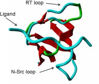
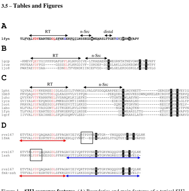
The first two structures of SH3 domains were both reported in 1992 by two separate groups, one by X-ray crystallography in the lab of Serraste [66] and the other by NMR in the lab of Schreiber [67]. These domains are relatively small, around 60 amino-acids long, and their secondary structure is composed of five antiparallel beta strands with three connecting loops: the RT-loop connecting strands 1 and 2; the N-Src
loop connecting strands 2 and 3; and the distal loop connecting strands 3 and 4. Strands 4 and 5 are separated by a 3(10) helix (see Figure 1).
Figure 1 – A visual representation of an SH3 domain in complex with a ligand. Ligand, RT and N-Src loops are highlighted. The 3D representation was prepared with the Yasara package.
The SH3 domain participates in protein-protein interactions by binding to proline rich peptides trough a relatively flat interface with three shallow binding pockets defined by a series of conserved aromatic residues (see Figure 2). The interaction with proline rich regions was first observed in studies with a GST-tagged SH3 domain from Abl in 1992 [68] and further characterized in 1994 with the aid of randomized sequences either bound to beads [69] or in screens of phage display libraries [70, 71]. These studies of randomized peptides were instrumental in describing the binding specificity of SH3 domains. They uncovered a motif of PxxP (were x is any amino-acid) common to all binding peptides. Further structural studies revealed a more complex picture of SH3 binding specificity with at least two well established binding modes: class 1 consensus peptides ([+]xxPxxP) that is bound with a basic residue N-terminal to the PxxP core and a class 2 motif (PxxPx[+]) where the basic residue is C-terminal in respect to central PxxP [70, 71]. In both cases, the peptide adopts an extended polyproline-2 helix (also know as PPII helix) with thee residues per turn and left-handed. The PPII helix interaction with the SH3 domain can be visualized as a triangular prism with a base sitting on the SH3 interface (see Figure 2).
A detail study of most of S. cerevisiae SH3 domains using phage display has shown that although most SH3 domains conform to the previously defined rules, they also have individual motifs that are more specific than the general binding pattern [27]. That is, each SH3 domain of S. cerevisiae binds a set of proline rich regions in the proteome and these regions overlap but not completely. The same study then showed that it is possible to use the specificity patterns derived from phage display in combination with yeast-two-hybrid studies to derive a set of high confidence SH3-target interactions. I will show in Manuscript 1 that to make the most of these binding motifs we should use secondary structure information and comparative genomics to search for conserved putative targets that are accessible for binding. Also, in Manuscript 2 I will show that there is indeed a large structural variability in peptide binding conformation and possible changes in loop length and loop conformation in the SH3 domains. This structural variability observed is important in binding specificity and should be taken into account when trying to predict protein-interactions from structural information.
Figure 2 – Typical binding modes of SH3 domains. Crystal structures of two SH3 domains in complex with peptides are displayed to exemplify the two most common modes of binding of SH3 domains. In the figure, to the left, are shown the peptide motifs most typically associated with those binding modes (X corresponds to any amino acid) and the peptide orientation. The structures were visualized using the Yasara package.
1.4.2 - SH2 domains
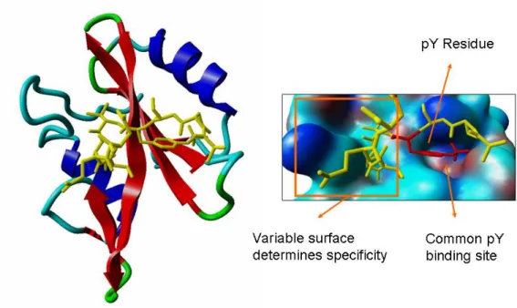
SH2 domain, are small modular domains constituted by around 100 amino acids. Like the SH3 domains, these also bind short peptide sequences. However they specifically recognize phosphorylated tyrosine residues and don’t usually bind to the un-phosphorylated form of the same peptide [72, 73]. The SH2 domain can then serve as a sensor for the cell reporting on the phosphorylated state of proteins. The prototypic example of this sensing mechanism occurs in the initial steps of a signal transduction cascade: the cell is exposed to a ligand molecule; 2) interaction of the ligand with the membrane receptor leads to phosphorylation of the cytoplasmatic part of the receptor; 3) phosphorylated tyrosine residues are recognized by SH2 domains leading to the recruitment of more signaling molecules.
Randomized peptide studies have shown that individual SH2 domain further restrict their binding by accepting different residues at positions +1 and +3 C-terminal to the phosphorylated tyrosine residue (pTyr) [74, 75]. Structural analysis of the SH2 domain showed that it general consists of two a-helices surrounding a large b-sheet containing four strands (see Figure 3). The structure contains two separate binding sites, one to each side of the beta-sheet (see Figure 3). The pTyr residue binds the more conserved of the two binding pockets contacting several positively charged residues. The second binding pocket is more divergent and contributes more for the discrimination between different phospho-peptides.
Figure 3 - The SH2 domain and its typical binding characteristics. A structure of an SH2 domain in complex with a phosphorylated peptide is shown. To the right the binding pockets are shown in more detail. The structure was visualized using the Yasara package.
Given the ability to discriminate between different phosphorylation states, these domains participate in many of the cellular pathways involved in response to extra-cellular signals and in the control of extra-cellular states. To understand these processes it is therefore important to study and to comprehend the underlying rules that determine SH2 specificity. In manuscript 3 I show that structural information can also be used to predict the specificity of binding of different SH2 domains.
1.5 -
Evolution of cellular networks
The development of some of the experimental methods described above increased the amount of information on protein-protein interactions available for several different model organisms [2-7, 13-15]. It is tempting to try to compare these interactomes very much like one would compare genomes (referred to as comparative genomics), trying to look for similarities and differences that might tell us something about the cellular function and evolution. The attempt at what could be called comparative interactomics was first tried by Cesareni [76] with very limited success. The main conclusion from this analysis was that the current coverage of these maps is so low that it is pre-mature to embark on these comparative efforts. A more recent
attempt to compare the interaction networks of different species also suggested that very little could be concluded from the small overlap observed [77]. Both these studies have however ignored the large evolutionary distance between the species studied. Given that little is nown about the evolutionary rate of protein interaction one other possible explanation for the small overlap could be evolutionary divergence. In fact, Andreas Wagner proposed in 2001 that in S. cerevisiae around 50 new interactions might be formed every million years [37]. This would also explain the apparent limited success in extrapolating interactions across different species, attempted by Lisa R. Matthews and colleagues in Vidal’s lab [78]. Functional constraints should lead to conservation of complexes and pathways but not necessarily of the exact binary interactions or individual proteins involved. Therefore, a more promising direction in comparative interactomics is to look for conservation of network structures and not conservation of exact binary interactions. This approach was developed in the Ideker lab in the form of the PATHBLAST tool [40, 41] and recently also by Flannick and colleagues [79]. In Manuscript 4 I have tried to study the evolutionary rate of protein interaction networks as well as the protein’s characteristics that shape the rate of change in their interactions.
2 –
Manuscript 1: Comparative genomics and disorder prediction
identify biologically relevant SH3 protein interactions
Pedro Beltrao & Luis Serrano
EMBL Structural & Computational Biology. Meyerhofstrasse 1.
Heidelberg D-69012 Germany
e-mail:[email protected]
2.1 –
Abstract
Protein interaction networks are an important part of the post-genomic effort to integrate a part-list view of the cell into system-level understanding. Using a set of eleven yeast genomes we show that combining comparative genomics and secondary structure information greatly increased consensus based prediction of SH3 targets. Benchmarking of our method against positive and negative standards gave 83% accuracy with 26% coverage. The concept of an optimal divergence time for effective comparative genomics studies was analysed, proving that genomes of species that diverged very recently from S. cerevisiae (S. mikatae, S. bayanus and S. paradoxus), or a long time ago (N.crassa and S. pombe) contain less information for accurate prediction of SH3 targets than species within the optimal divergence time proposed. It was shown here that intrinsically disordered SH3 domain targets are more probable sites of interaction than equivalent sites within ordered regions. Our findings highlight several novel S. cerevisiae SH3 protein-interactions, the value of selection of optimal divergence times in comparative genomics studies and importance of intrinsic disorder for protein interactions. Based on our results we propose novel roles for the S. cerevisiae proteins Abp1p in endocytosis and Hse1p in endosome protein sorting.
2.2 –
Non-technical summary
How can we tackle the complexity of a living cell? It is commonly said that living organism are complex and display “emergent” properties. Emergence is perceived in this context as the process of deriving behaviours at the system level that are not observable at the level of the system’s components. In the cell this would be equivalent to saying that the cellular complexity could be understood if we could understand the interplay between the cellular components. Not only describe the “parts” that make up a cell but understand how they interact with each other to perform the necessary tasks. A big step on the road of understanding cellular complexity will be a complete list of all relevant interactions between the cellular components. Although a lot of progress as been made in this direction, we are often dependent on experimental methods that are costly and time consuming. Computational Biology has here a big challenge to process the current available knowledge and to propose new ways of predicting the interactions between cellular components.
We have studied, in this work, protein interactions that are mediated by small linear peptide motifs. An example of this type of interactions is the one between an SH3 domain and its targets, usually small peptide stretches containing a PXXP motif (where X is any amino acid). The results showed that the putative target motifs that are conserved in ortholog proteins and are within regions that do not have a defined secondary structure are more likely to be relevant binding sites.
Besides proposing a way to combine secondary structure information with comparative genomics to predict protein-protein interactions, we have highlighted a possible role of intrinsically disordered proteins in SH3 protein interactions. The results also showed that when looking for conservation of these motifs it is important to carefully select the species to use in the study. Some species will be more informative than others and our work proves that there is an optimal divergence time for the species to include.
2.3 – Introduction
Important advances have been made on mining the ever growing quantity of experimental results with computational methods in order to derive predictions of protein-protein interactions. For such interactions there are methods that explore sequence and structure analysis, like gene fusion [30, 80], gene order [28], phylogenetic profiling [29, 81-83], correlated mutations [31, 84] and multimeric threading [46, 48]. It as also been shown that it is possible to combine different experimental and functional data to predict protein interactions, especially when weighted using Bayesian networks [56]. The accumulation of validated interactions can also be mined by interolog mapping in order to transfer protein interaction annotations across species [2, 85].
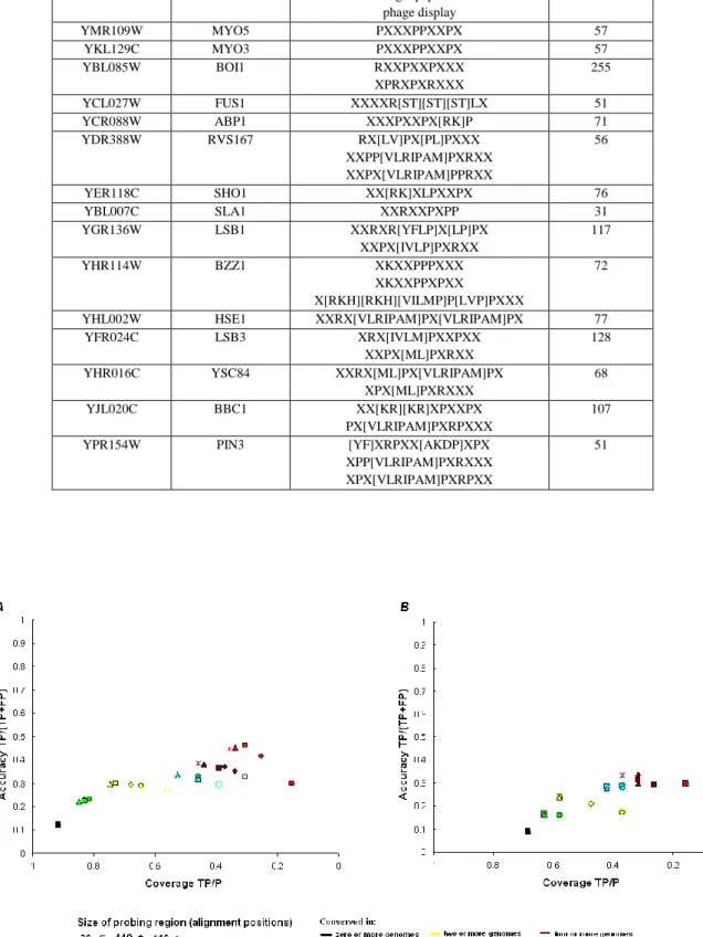
The work described here deals with the prediction of protein interactions mediated by recognition modules that target small linear motifs [86, 87] and more specifically to SH3 domains. This type of asymmetric binding between globular domains and linear peptides was first reported in the work on Src kinase [62, 64, 68, 88] and many other domains have now been shown to have similar properties [86, 87]. In a previous study [89], knowledge from phage display experiments was used to derive a position specific scoring matrix (PSSM) for a particular SH3 domain which was then used to predict putative target ligands. Later, Tong et al, devised a strategy where two-hybrid and PSSM were combined to derive a high confidence network [27]. It was reasoned that an interaction identified by two-hybrid was more likely to be biologically relevant if the target protein had a high scoring linear peptide according to the PSSM of the bait SH3 domain.
In this work we set out to obtain a high-confidence, biologically relevant, protein interaction network, starting from the consensus information and using computational methods. The study showed that it is possible to greatly increase the accuracy of consensus based predictions of protein-linear sequence interactions by considering that biological relevant target ligands of SH3 domains were more likely to be within disordered regions and conserved in orthologs. The method’s performance was improved by selection of species within “an optimal divergence time” from the species of interest.
It has been proposed that intrinsic disorder may play a role in protein interactions [90-93] and there are documented cases where binding is coupled to folding [94, 95] (reviewed in [96]). It is also been observed that small linear motifs tend to
accumulate in protein regions predicted to be intrinsically disordered [97] and that proline rich regions are usually devoid of secondary structure [98]. In most structures that we are aware of, the SH3 domain is in complex only with short target peptides, and not with full proteins. In all cases the ligands adopted a non-regular secondary structure but there is little information one can take from these, in respect to the order/disorder of target sites in the context of the whole target protein. Although there is currently no experimental evidence to support that the SH3 domains preferentially bind to intrinsically disordered regions the results presented here showed that binding motifs within disordered protein regions are more likely to be biologically relevant binding sites than equivalent sites within ordered regions.
We used the method developed to suggest novel SH3 interactions for S. cerevisiae with information of the binding sites within the target proteins.
2.4 – Results & Discussion
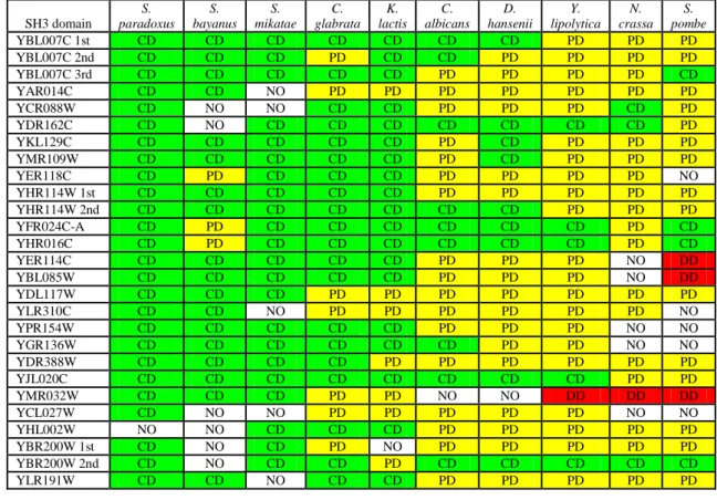
2.4.1 – Identification and conservation of SH3 domains and selection of genomes. Using profile hidden Markov models (see methods and Table 1), all putative SH3 domains, and their key binding positions (see methods) were determined in S. cerevisiae and in a set of thirteen yeast species: C. glabrata, D. hansenii, K. lactis, Y. lipolytica [99], C.albicans [100], S. paradoxos, S. bayanus, S. mikatae [101], S. castellii, S. kudriavzevii, S. kluyveri [102], N. crassa [103] and S. pombe [104].
In S. castellii, S. kluyveri and S. kudriavzevi no orthologs for the majority of the S. cerevisiae SH3 domains could be identified (results not shown). However, these genomes had only been sequenced with a two to three fold coverage [102] which may have led to some genomic regions being poorly sequenced. . As a result of this, these three genomes were not included in our work.
The ortholog SH3 domains were split into three groups: “conserved domain”, “possibly divergent” (if the putative ortholog SH3 domain is in the same branch of the phylogenetic tree and has more than two conservative changes in the binding positions - see methods) or “divergent domain” (if the putative ortholog SH3 domain was not in the same branch of the phylogenetic tree) (Table 1). As expected the percentage of “conserved” domains was higher in genomes of species that diverged recently from S. cerevisiae.
Intuitively we can think that there should be an optimal divergence time, for the species to be used in a particular comparative genomic study. In recently divergent species most protein sequence is conserved and the statistical power for comparative genomics of biological features is therefore smaller. Inter-species conservation becomes less meaningful in a background of low evolutionary divergence. On the other hand, finding a conserved consensus in a very divergent genome might be more significant but only if there was no major change in the specificity of the domain. This change will be more probable the more divergent the specie is from the specie of interest.
To test the improvement of consensus based predictions with a comparative genomics approach an initial set of genomes was chosen based on the conservation analysis of the SH3 domains across the different yeast species (Table I). N. crassa and S. pombe were excluded because the SH3 domains in these two species might be too
divergent to observe conservation of the S. cerevisiae motifs. Very close relatives (S. paradoxus, S. bayanus and S. mikatae) were excluded as these species would have lower statistical power. Therefore the first group analysed consisted of five yeast genomes that broadly covered the hemiascomycete phylum, containing the four recently reported genomes of C. glabrata, D. hansenii, K. lactis and Y. lipolytica that we grouped with the C.albicans’ genome.
2.4.2 – Evaluation of the “conservation” approach.
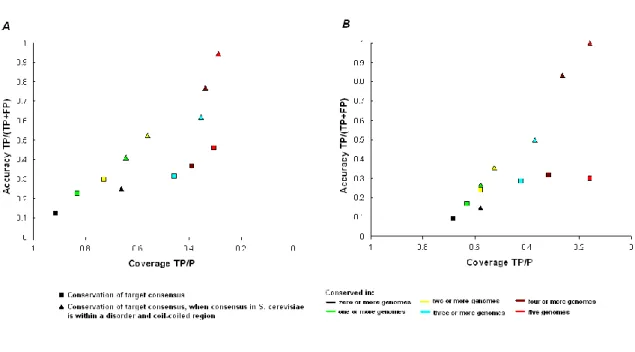
To evaluate the predictive power of our method two positive datasets, containing experimentally verified SH3-linear peptide interactions, and one negative dataset, containing non interacting protein pairs were defined (see methods). The binding motifs of the SH3 domains of S. cerevisiae included in the two sets of positive standards (15 SH3 domains in the gold set and 10 in the platinum set) were taken from the data published in Tong et al [27]. Table II shows the consensus used in the study and also, for each SH3 domain, the total number of peptides found with these consensus in the S. cerevisiae proteome. From this a measure of accuracy and coverage (see methods) based on the positive and negative datasets was calculated. For simple pattern matching of consensus the accuracy (defined as TP/(TP+FP), where TP means True Positives and FP, False Positives) for predicting protein interaction was 12% and the coverage (defined as TP/P) was 92% when using the gold positives set (see fig 1a).
Using T-Coffee [105], an alignment of all putative orthologs (obtained using the BLAST reciprocal best hit method [106]) of S. cerevisiae proteins containing sequences matching a consensus for an SH3 domain was carried out. This alignment was then used to determine the level of conservation of putative target ligand sites by searching for sequences matching the same consensus in the orthologs. We did not search for conservation of putative target motifs in genomes without an ortholog for the domain in study. If there is no ortholog SH3 domain in the comparing specie then the conservation of the motif, in the ortholog of the putative target, is not biological relevant and should not be counted to increase our confidence in the putative interaction. Having said this it should be noted that there could be several technical reasons why the ortholog of an SH3 domain could be missed in a genome. There might be errors for example in the genome assembly, genome annotations, domain annotation and ortholog assignment. For all these reasons we also tried to calculate conservation scores without disregarding genomes with no ortholog for the domain in study. While this did not change
significantly the results (data not shown) we felt that the first approach was more stringent.
In the orthologs, the search was restricted to a window surrounding the putative target ligand in the S. cerevisiae sequence and we called this the probing region. In Figure 1, accuracy versus coverage for increasing probing regions are plotted and it can be seen that by searching in a wider region of the alignment both coverage and accuracy are increased, especially for higher conservation scores (the complete analysis with the number of hits and false and true positives for each positive set is given in supplementary table 1). Optimal results were obtained when using a probing region of 210 alignment positions. It is important to emphasize that these were not necessarily amino acids, but 100 gaps or amino-acids on each side of a motif of 10 amino acids. This result could be due to poor alignment of some proteins, especially those rich in Pro sequences. In fact most of the gain in coverage was due to interactions with proline rich proteins that were difficult to align and had multiple gaps (i.e. Las17p, App1p and Vrp1p). Also, this data may suggest that these small target ligands may be easily moved in primary sequence space during evolution, due to compensatory mutations in proteins that are already proline rich in nature. For both sets of positives a big improvement in accuracy was observed when we selected for consensus conserved in the 5 genomes used (3.8 fold with the “gold” positives and 3.3 fold with the “platinum” positives). There was, however, a similar fold reduction in the coverage, 3 fold for the gold and 4.3 fold for the platinum set.
Since most known target proteins in the SH3 interaction network are proline rich and a large probing window was used, it is possible that the hits found in orthologs were due to chance and lacked biological meaning. To eliminate this possibility two “decoy” proline rich patterns were analysed: PxxxPxxxP and ExxPxxP (where x is any amino-acid), different from the consensus sequences. Both patterns were found with high frequency (>400 hits) on S. cerevisiae proteins. Using these two patterns a loss in accuracy and coverage was observed (an average of 1.4 times less accuracy and 1.2 times less coverage for the PxxxPxxxP motif and an average of 3.4 times less accuracy and 2.5 times less coverage for the ExxPxxP motif). Thus we can eliminate that the results were generated by chance and confirm that the observed phenomenon was the conservation of specific SH3 binding motifs and not of Pro-rich tracks.
However, the accuracy obtained with conservation alone was still poor (using the gold set: accuracy 46%; coverage 31% and the platinum set: accuracy 30%; coverage
16%). A hyper-geometric test allowed us to say that that the improvement in both positive sets and for all conservation scores was significant (p<0.05) and not due to random sampling.
2.4.3 – Combining comparative genomics and disorder prediction
Since SH3 domains generally bind linear amino acid stretches we tried to improve the accuracy of our consensus based method by extracting secondary structure information about the sequences containing the target motifs. It has been argued that there might be biological advantages in presenting binding sites within unstructured regions [90-93]. It has also been observed that small linear motifs tend to accumulate in protein regions predicted to be intrinsically disordered [97] and that proline rich regions are usually devoid of secondary structure [98]. To our knowledge there is no clear experimental evidence to support that SH3 domain target sites are generally unstructured before binding, but since SH3 domains bind small linear peptide motifs that are proline rich we hypothesised that SH3 domain targets might be mainly found in unstructured regions of the polypeptide chain. Therefore we used GlobPlot [97] in combination with coil-region predictions [107] to identify and study all consensuses found within disordered protein regions.
Combining disorder prediction with comparative genomics resulted in a significant (p<0.01, using a hyper-geometric test) increase in the accuracy of protein target prediction (there was a 2 fold average increase in both sets) (Figure 2). The decrease in coverage was 1.4 for the gold and 1.1 fold for the platinum set. For consensus conserved in five or more genomes we obtained 94% accuracy with 28% coverage for the gold set. For consensus conserved in four or more genomes 83% accuracy with 26% recovery for the platinum set were obtained. These results argue that intrinsic disorder plays an important role in SH3 protein-interactions, however further experimental work is needed to verify this observation.
Since the platinum positive set was independent (see methods) the values obtained with this set may be used as a score for the performance of our method compared to others. Higher values of coverage and accuracy with the golden positive set were observed when using our method, but it should be noted that this could be due to a possible bias (see methods). A detailed table with the number of hits, false and true positives for each conservation level in both positive sets can be found in supplementary table 2.
Using the methods described in this work, we show proof of concept on how to integrate secondary structure prediction with comparative genomics to increase the accuracy of consensus based prediction of peptide recognition modules. However, the method employed involves a clear trade-off between accuracy and coverage.
Of the 59 interactions in the final high confidence interaction presented in the Tong et al. paper the method was able to predict 20 interactions when restricting for consensus within disorder and found in 4 of the 5 genomes used. We then tried to look for distinguishing features within these 20 interactions, when compared to the remaining 39 that the method does not predict. There were no statistical differences in the average size of protein targets (p=0.32 with a T-Test), average proline content of protein targets (p=0.12 with a T-Test), usage of Class II motif (p=0.21 with a Hypergeometric distribution test) and conservation of SH3 domain (p=0.82 with a T-Test). There was a statistically significant difference in the average conservation of the target proteins (p=0.03 with a T-Test). The protein targets the method was able to predict were on average conserved in 8.7 of the 10 species, while the targets not recovered were conserved in 7.6 species. This small but significant difference highlights the bias this method has for conserved interactions. A higher level of confidence can be placed in any putative target motif found conserved in most yeast species analysed, but this level of conservation will only happen for essential interactions. It is important to note that for this reason this method will always miss species-specific protein interactions. By requiring high conservation we also lower our coverage and probably bias our predictions to essential interactions. However adding more genomes of species within an appropriate divergence time should alleviate this problem, a concept discussed in more detail below.
Another possible cause of loss in coverage could come from interactions that are mediated by currently uncharacterized motifs or through non-canonical SH3 binding (i.e. through globular regions of the target protein).
As shown by other authors (reviewed in [108]), it should be possible to further improve the reliably of a protein interaction network and therefore our method, by adding information from other sources of data (i.e. RNA expression, essentiality and function information). This is especially true if the information is efficiently combined, for example employing a Bayesian network [56]. It was our intention to develop a method that could be used in species where these sources of information were not
available, but in the future we will try to develop weighting schemes to include such sources for prediction of interactions mediated by small linear motifs.
2.4.4 – Determining an optimal divergence time for the genomes used when searching for conservation of target ligands of SH3 domains
Included in our initial hypothesis, was the notion that there might be an optimal time of divergence to efficiently use the comparative genomics approach. To test this, phylogenetic data [99, 109, 110] with approximate values for the divergence times of the yeast species from S. cerevisiae (see Methods), was used to create seven groups of four genomes with increasing average divergence time. Using the gold positives, the highest accuracy obtained for a small range of coverage values was determined for each of these groups. For different coverage ranges the highest accuracy was generally obtained with groups of genomes that diverged from S. cerevisiae on average around 400My to 950My (Fig. 3).
To explore this issue further we tried to find out which genomes might be more or less informative to our consensus based predictions. For each possible combination of two or more genomes we calculated the highest accuracy obtained for eleven small windows of coverage (with intervals of 5% of coverage from 15% to 70%). Figure 4 shows the average of the individual genome representation in all possible groups, in the groups scoring in the highest 20% accuracies and in the groups scoring within the lowest 20% accuracies, over all the coverage windows studied. For each species, a t-test determination was carried out to see if the average frequencies within the highest and lowest combinations were significantly different from the frequency in all possible combinations. From the analysis of the results the more informative genomes are: C. albicans, D. hansenii, C. glabrata, K. lactis and Y. lypolitica. We can also see that N. crassa and S.pombe are not over represented in highest scoring groups, suggesting that they have less informative genomes. More importantly it is clear that including the genomes of S. bayanus, S. mikatae or S. paradoxus leads to a decrease in the accuracy of predictions. These observations correlate well with the degree of divergence observed for the SH3 domains (Table 1) and with our proposed range for optimal divergence time.
In a very recent report [111] Sean R. Eddy used a theoretical model to study the statistical power of comparative genome sequence analysis. The model showed that, at