UNIVERSIDADE DE TRÁS-OS-MONTES E ALTO DOURO
ANATOMICAL AND IMAGING STUDIES OF
THE HUMEROULNAR CONGRUITY IN DOGS
PHD THESIS IN VETERINARY SCIENCES
MARIA SOFIA RODRIGUES ALVES PIMENTA
Advisor: Professor Doutor Bruno Jorge Antunes Colaço Co-Advisor: Professor Doutor Mário Manuel Dinis GinjaUNIVERSIDADE DE TRÁS-OS-MONTES E ALTO DOURO
ANATOMICAL AND IMAGING STUDIES OF
THE HUMEROULNAR CONGRUITY IN DOGS
PHD THESIS IN VETERINARY SCIENCES
MARIA SOFIA RODRIGUES ALVES PIMENTA
Advisor: Professor Doutor Bruno Jorge Antunes Colaço Co-Advisor: Professor Doutor Mário Manuel Dinis GinjaJury Composition:
Professor Doutor Vivente de Seixas e Sousa (President) Professor Doutor António José Almeida Ferreira Professora Doutora Graça Pires Lopes de Melo Professora Doutora Maria Dulce Cordeiro Madeira
Professora Doutora Maria Isabel Ribeiro Dias Professor Doutor Bruno Jorge Antunes Colaço Professor Doutor Carlos Alberto da Silva Venâncio
Original thesis presented by Maria Sofia Rodrigues Alves Pimenta at the University of Trás-os-Montes and Alto Douro, to obtain the doctor’s degree
This work is supported by: European Investment Funds by FEDER/COMPETE/POCI– Operacional Competitiveness and Internacionalization Programme, under Project POCI-01-0145-FEDER-006958 and National Funds by FCT - Portuguese Foundation for Science and
vii
À minha família
em especial aos meus Pais, irmão, e aos meus filhos
Aos alunos da UTAD
ix
“It had long since come to my attention that people of accomplishment rarely sat back and
let things happen to them. They went out and happened to things.”
Leonardo da Vinci
Ao Professor Doutor Bruno Jorge Antunes Colaço
Ao Professor Doutor Mário Manuel Dinis Ginja
xi
Agradecimentos
A realização da presente tese só foi possível devido à colaboração, dedicação, apoio e esforço de muitas
pessoas. A todas elas expresso o meu mais sincero agradecimento, em particular:
À Universidade de Trás-os-Montes e Alto Douro, na pessoa do seu Magnífico Reitor Professor
Doutor Fontainhas Fernandes, o meu reconhecimento pelos apoios concedidos.
Ao Professor Doutor Vicente de Seixas e Sousa, presidente da Escola de Ciências Agrárias e
Veterinárias, por ter criado as condições académicas que permitiram a minha dedicação à presente
tese.
Ao Professor Doutor José Carlos Marques de Almeida, diretor do Departamento de Zootecnia, pelo
apoio académico essencial à conclusão da presente tese, pela disponibilização de meios e
infraestruturas para a realização de parte da componente prática do trabalho e pela simpatia com que
sempre me recebeu.
Ao Professor Doutor Bruno Jorge Antunes Colaço, cuja orientação foi fundamental para a realização
desta tese. Obrigada por aceitar a orientação, por todos os conhecimentos transmitidos sobre ciência,
investigação e ensino da Medicina Veterinária. O seu enorme exemplo de carácter, de humildade, de
honestidade, de altruísmo e profissionalismo, fizeram-me crescer, profissionalmente e pessoalmente.
A sua determinação e persistência nos momentos menos bons ao longo desta caminhada fizeram-me
acreditar que na vida todas as dificuldades e obstáculos são ultrapassáveis. Obrigada pelos conselhos
sempre sábios e pela sua amizade. Obrigada por estar sempre presente quando precisei.
Ao Professor Doutor Mário Manuel Dinis Ginja, coorientador da presente tese, manifesto o meu
sincero agradecimento por aceitar a coorientação, pelos conhecimentos em imagiologia transmitidos e
valiosas sugestões apresentadas. Agradeço-lhe a oportunidade que me deu de realizar uma parte da
componente prática desta tese em cães das associações de criadores com quem mantém protocolos há
vários anos, colocando à disposição todos os meios e infraestruturas. Agradeço-lhe ainda por me ter
apresentado o Professor Doutor Pedro José de Melo Teixeira Pinto. Obrigada pela coragem,
disponibilidade, partilha de ideias, e pela amizade.
Ao Centro de Investigação e Tecnologia de Ciências Agroambientais e Biológicas, na pessoa do
Professor Doutor Eduardo Augusto dos Santos Rosa.
xii
Professor Doutor Pedro José de Melo Teixeira Pinto, do Departamento de Engenharias e ao Professor
Doutor Armando Fernandes, do INOV - INESC Inovação pela idealização e conceção do software
de análise de curvas.
À direção do Hospital Veterinário da Universidade de Trás-os-Montes e Alto Douro, na pessoa da
Professora Doutora Maria Isabel Ribeiro Dias, pela disponibilização de meios e infraestruturas para
a realização de parte da componente prática do trabalho.
Ao Professor Doutor Jorge Colaço, pela inestimável ajuda na análise estatística, ensinamentos e pela
simpatia com que sempre me recebeu.
Ao Professor Doutor António Ferreira e à Professora Doutora Sandra Jesus, da Faculdade de
Medicina Veterinária da Universidade de Lisboa, pela disponibilização dos meios para a realização
do estudo por tomografia computorizada.
Ao Professor Doutor Lio Gonçalves do Departamento de Engenharias pelo imprescindível contributo
na interpretação de dados.
À Professora Doutora Aura Antunes Colaço, pela confiança e entusiasmo, conselhos e amizade.
Ao Professor Doutor Carlos Alberto e Silva Venâncio, pelo seu apoio e compreensão ao longo deste
percurso, pelo profissionalismo, retidão e amizade.
Ao Sr. João Gonçalves e Sr. Filipe Fontinha pela valiosa assistência na preparação anatómica das
peças, auxílio durante todos estes anos na preparação de aulas práticas, pela amizade e bons
momentos passados no Laboratório de Anatomia.
Às Associações de Criadores e aos proprietários dos cães das Raças Autóctones Portuguesas Cão da
Serra da Estrela e Perdigueiro Português, pela colaboração no trabalho realizado.
Ao corpo docente e não docente dos Departamentos de Zootecnia e de Veterinária da Escola de
Ciências Agrárias e Veterinárias da Universidade de Trás-os-Montes e Alto Douro. Aos meus
Professores, aos amigos e companheiros desde que entrei na UTAD como estudante, a todos expresso
a minha enorme gratidão pela formação pessoal e profissional, pelo apoio e bons momentos passados.
Ao Grupo de Animais de Laboratório do Centro de Investigação e Tecnologia de Ciências
Agroambientais e Biológicas.
xiii
À Professora Doutora Paula Oliveira, pelo entusiasmo, amizade e simpatia com que sempre me
recebeu.
Professor Doutor Luís Lucas Cardoso pelo apoio e amabilidade.
À Professora Doutora Maria de Lurdes Pinto pela colaboração académica nas aulas de Anatomia.
Ao Professor Doutor Paulo Salgado, pelos conhecimentos transmitidos na área da programação.
Ao Professor Doutor Luís Maltez pelos sábios conselhos e amizade.
À Professora Doutora Filomena Adega a amizade e disponibilidade para revisão do trabalho.
À Professora Doutora Cátia Teixeira pelo auxílio de linguística.
À Professora Doutora Tânia Sofia Cordeiro Martins, e à Doutora Ana Faustino pelos conselhos e
pela disponibilidade para revisão do trabalho.
Ao corpo clínico do Hospital Veterinário de Trás os Montes e Grupo OneVet pelo apoio prestado.
Aos alunos de Anatomia, pelos ensinamentos e inspiração para melhorar em cada dia.
Às companheiras Antonieta e Catarina pela companhia, sentido de humor e bons momentos passados.
À Mané, ao Zé Pedro e ao Márcio pelo apoio gráfico e amizade.
À minha família e amigos. Aos meus Pais Mário e Dulce e irmão Gonçalo. Ao meu Pai, em especial
pelo seu exemplo de empreendedorismo, perseverança, otimismo e humildade! À minha mãe por ser o
pilar da família e proporcionar bons momentos em família! Ao meu sogro Artur pelo seu exemplo de
vida e apoio na educação dos netos. Ao meu marido Paulo, por decidir em cada dia partilhar comigo
os bons e maus momentos, pelo seu sentido de família, exemplo de empenho e resiliência, e por toda a
compreensão. Aos meus filhos Artur e Pedro por me ajudarem a olhar todos os dias para o Mundo
com uma perspetiva nova. Aos meus sobrinhos Vasco e Tomás. A todos, pelo amor e apoio ao longo
da minha vida. À Maria João e Margarida pela amizade e apoio.
xv
R
ESUMOA displasia do cotovelo é uma doença frequente em ortopedia veterinária, afetando cães jovens de raças grandes, com elevado impacto quer económico quer na qualidade de vida dos animais afetados. Os modelos pré-clínicos da articulação do cotovelo são uma ferramenta fundamental na pesquisa de métodos de diagnóstico mais precisos. A incongruência articular tem um papel importante no desenvolvimento, tratamento e prognóstico da displasia. O seu diagnóstico é desafiante e foco de intensa pesquisa nos últimos anos. A proximidade das estruturas articulares e a normalização do posicionamento do cotovelo são apontados como os principais obstáculos a uma boa interpretação. A medição dos espaços articulares é influenciada pelo posicionamento. Os avanços biomecânicos melhoraram o conhecimento da geometria úmero ulnar, permitindo a descrição de uma incongruência fisiológica.
Utilizando articulações de cadáveres (>20kg) e amostras de despiste da displasia do cotovelo em cães das raças Cão da Serra da Estrela (EMD) e Perdigueiro Português (PPD), pretendemos: validar um software e metodologia para análise das curvaturas da incisura troclear ulnar (UTN) e da tróclea do úmero (HT), independentemente do ângulo de flexão do cotovelo, usando as técnicas de diagnóstico de imagem mais comuns na prática clínica; obter uma perspetiva geral da forma das curvaturas presentes em cães com peso superior a 20Kg; caracterizar o padrão de curvatura existente numa raça média e grande, e as diferenças entre raças que pudessem indicar uma maior propensão da raça grande para desenvolver incongruência.
As curvaturas UTN/HT foram estudadas separadamente nas articulações normais de cadáveres, usando as projeções medio lateral em extensão (MLE) e em flexão (MLF). Para o estudo sem sobreposições radiográficas foi utilizada uma técnica anatómica de desarticulação e obtidas as projeções medio laterais adicionais rádio ulnar (MLRU) e do úmero (MLH). O
software criado foi aplicado para medir os raios de curvatura a nível subcondral das UTN/HT.
Os tipos de revestimento cartilaginoso articular foram também investigados. A fiabilidade das medidas foi estabelecida por comparação dos valores obtidos nas diferentes projeções radiográficas.
Para investigar do ajuste entre a UTN/HT nas articulações normais, foram analisados os raios de curvatura nas projeções radiográficas e em exames de tomografia computorizada (CT) do cotovelo, em animais com exame CT normal. Para implementar a metodologia nas raças média e grande e pesquisar as diferenças, foram utilizadas as projeções radiográficas MLE
xvi
de cães das raças PPD e EMD, e as projeções radiográficas MLF de cães da raça EMD, com exames ortopédico e radiográfico normais.
O software e metodologia propostos permitiram uma análise dos raios de curvatura das UTN/HT adequada nas radiografias e nas imagens de CT estudadas. As medições apresentaram repetibilidade e reprodutibilidade, com uma boa concordância entre os valores obtidos (p>0.05). As tipologias das curvaturas (UTN/HT) foram semelhantes, no entanto os valores na UTN foram superiores aos valores na HT. A partir da observação do comportamento das curvaturas encontrado, foram estabelecidos sete pontos de interesse, P1
a P7, correspondentes aos extremos (proximal e distal), dois picos intermédios e três valores
mínimos intermédios, para serem investigados mais detalhadamente. As maiores diferenças entre UTN/HT em cadáveres foram encontradas no ponto inicial (P1) e no segundo pico
intermédio (P5). A forma da curvatura encontrada foi relacionada com o padrão de transmissão
de cargas bicêntrico através da articulação do cotovelo, descrito em raças grandes. Os dois picos intermédios observados na UTN (P3 e P5) correspondem a pontos de aplanamento
relativo do osso subcondral e podem ser o resultado de uma transição entre áreas onde ocorre maior carga articular, em ambos os extremos da UTN, e uma zona de menor carga articular, no centro da UTN. Foram encontradas áreas articulares livres de cobertura cartilaginosa, lateralmente ao nível central na UTN.
Em relação às duas raças estudadas, os raios de curvatura da UTN foram superiores aos da HT. Também os valores de ambas as curvaturas (UTN/ HT) foram superiores na raça EMD do que na raça PPD. Ambas as raças mostraram curvaturas UTN/HT com forma semelhante, no entanto a raça EMD apresentou menor proximidade nos raios de curvatura das superfícies úmero ulnar. Os picos intermédios na UTN (P3, P5) foram mais pronunciados na raça grande,
correspondendo a um maior aplanamento do osso subcondral.
A curvatura da UTN encontrada foi semelhante às descritas anteriormente na literatura usando diferentes metodologias. Do conhecimento dos autores, este é o primeiro trabalho sobre medições do raio de curvatura da HT, e o primeiro estudo da congruência úmero ulnar através da determinação dos raios de curvaturas. Os resultados representam um avanço na caracterização da congruência úmero ulnar nas raças estudadas e também no uso da projeção MLF do cotovelo para avaliação da congruência.
PALAVRAS-CHAVE: anatomia clínica, incongruência articular, displasia do cotovelo no cão, imagiologia, radiografia, tomografia computorizada.
xvii
A
BSTRACTElbow dysplasia is a common disease in veterinary orthopedics, affecting young large-breed dogs, with a high economic and life quality impact of affected dogs. The use of elbow joint preclinical models is an important tool in the research for more accurate diagnostic methods. Joint incongruity has an important role in the development, treatment and prognosis of dysplasia. Its diagnosis can be challenging and has been the focus of intensive research in recent years. The proximity of joint structures and the elbow positioning standardization are pointed as the main obstacles to a good interpretation. Measurement of joint spaces is influenced by its positioning. Advances in the biomechanical field improved our knowledge on humeroulnar geometry, allowing the description of a physiological incongruity.
Using joints from dog cadavers (>20Kg) and samples from elbow dysplasia screening in the Estrela Mountain Dog (EMD) and Portuguese Pointing Dog (PPD) breeds, this thesis aimed to validate a software and methodology for the ulnar trochlear notch (UTN) and humeral trochlear (HT) curvatures assessment, independent from the elbow flexion angle, using the most common imaging tools in clinical practice; to obtain an overview of the curvature shape presented in dogs >20Kg; to characterize the curvature pattern present in a medium and a large-breed in vivo, and the differences between breeds that could indicate a greater propensity for the large-breed to develop incongruity.
The UTN/HT curvatures were studied firstly in separate, on the cadaveric normal joints, using mediolateral extended (MLE) and flexed (MLF) radiographic views. An anatomical technique of disarticulation was performed to study the anatomical articular surfaces without radiographic overlapping. Additional mediolateral radioulnar (MLRU) and humeral (MLH) radiographic views were acquired. To study and measure the curvature radii, the dedicated software was applied at the subchondral level from the UTN/HT. The types of articular cartilage covering were also investigated. The reliability of measurements was established by comparison of the values obtained using the different radiographic views.
In the combined fit investigation between UTN/HT in normal joints, curvature radii were analyzed on elbow radiographic and computed tomographic (CT) exams. Elbow joints were considered normal when a normal CT exam was observed. To implement the methodology in the medium and large-breed dog and evaluate the differences between breeds, the MLE radiographic views from the PPD/EMD, and the MLF views from the EMD, with normal orthopedic and radiographic exam were used.
xviii
The software and the methodology proposed enabled an adequate curvature radii analysis from the UTN/HT in the radiographic views and CT images studied. Repeatability and reproducibility of measurements were observed, with a good agreement between values achieved (p>0.05). The UTN/HT curvature radii typologies were similar, yet the UTN values were higher than the HT values. From the curvature behaviour found, the extremes (proximal and distal), two intermediate peaks and three intermediate minimum values were detected, allowing the establishment of seven points of interest, P1 to P7 to be investigated in further
detail. Higher differences between the UTN/HT were detected at the most proximal aspect of the joint (P1) and in the second intermediate peak (P5) in cadavers. The curvature shape found
is related to the pattern of bicentric load transmission through the elbow joint described in large-breeds. The two intermediate peaks observed at the UTN (P3 and P5) correspond to
relative flattener subchondral points and may be the result of a transition between the more loaded articular areas, at both UTN extremes, to a less loaded articular area, at the central UTN. Areas free of articular cartilaginous coverage were found lateraly at the central level at the UTN.
In the two breeds studied, the curvature radii values were higher in the UTN than in the HT. Furthermore, both UTN/HT curvature radii values were higher in the EMD than in the PPD breed. Both breeds showed UTN/HT curvatures with similar shape, however EMD breed presented less proximity of the curvature radii on the humeroulnar surfaces. The intermediate peaks (P3, P5) observed at the UTN were more pronounced in the large-breed corresponding
to points of greater flattening from the subchondral bone.
The UTN curvature found had a similar behaviour to the reported in previous literature using different methodologies. Concerning the curvature radii measurements from the HT, from the authors’ knowledge, this is the first report and also the first description of humeroulnar congruity research through curvature radii evaluation. The results from this thesis represent an advance on the humeroulnar congruity characterization in the breeds investigated and in the use of a MLF radiographic view to assess congruity.
KEY-WORDS: clinical anatomy, joint incongruity, elbow dysplasia in dogs, imaging, radiography, computed tomography.
xix
T
ABLE OF CONTENTSAGRADECIMENTOS xi
RESUMO xv
ABSTRACT xvii
LIST OF FIGURES xxiii
LIST OF TABLES xxv
LIST OF ABREVIATURES, SYMBOLS AND ACRONYMS xxvii
CHAPTER 1 1
1. GENERAL INTRODUCTION 3
1.1 Elbow dysplasia 3
1.1.1 Etiopathogenesis 3
1.1.2 Diagnosis and screening 4
1.1.3 Prevalence 5
1.1.4 Treatment 6
1.1.5 Current challenges in dysplasia screenning research 6
1.2 REFERENCES 7
CHAPTER 2 13
2.AIMS 15
CHAPTER 3 17
3.JOINT INCONGRUITY ON ELBOW DYSPLASIA IN DOGS, A REVIEW 21
3.1 Introduction 21
3.2 Elbow incongruity 23
3.2.1 Radioulnar incongruity 24
3.2.2 Humeroulnar incongruity 25
3.2.3 Soft tissue-bone mismatch 26
3.3 Elbow incongruity diagnostic tools 27
3.3.1 Imaging methods 27
3.3.1.1 Radiography 27
3.3.1.2 Computed tomography 29
3.3.1.3 Arthroscopy 31
3.3.1.4 Magnetic resonance imaging 32
3.3.2.5 Ultrasonography 33
3.3.2 Biomechanical studies 35
3.3.3 Genetic research 39
3.4 Conclusion 40
xx
CHAPTER 4 55
4.CURVATURE RADIUS MEASUREMENTS FROM THE ULNAR TROCHLEAR NOTCH IN LARGE DOGS 59
4.1 Introduction 59
4.2 Materials and methods 60
4.2.1 Animals 60 4.2.2 Measurement protocol 61 4.2.3 Statistical analysis 63 4.3 Results 64 4.4 Discussion 65 4.5 References 67 CHAPTER 5 69
5.CURVATURE RADIUS MEASUREMENTS FROM THE HUMERAL TROCHLEA IN LARGE DOGS 73
5.1 Introduction 73
5.2 Material and methods 73
5.3 Results 75
5.4 Discussion 75
5.5 References 76
CHAPTER 6 79
6. COMPUTED TOMOGRAPHIC AND RADIOGRAPHIC ASSESSMENT OF CONGRUITY BETWEEN ULNAR TROCHLEAR NOTCH AND HUMERAL TROCHLEA IN LARGE-BREED DOGS
83
6.1 Introduction 83
6.2 Materials and methods 84
6.2.1 Animals included 84
6.2.2 Image acquisition 84
6.2.3 Computer imaging analysis 85
6.2.4 Anatomical preparation 85
6.2.5 Statistical analysis 87
6.3 Results 88
6.3.1 Comparison between radiographic and CT measurements 88
6.3.2 Comparison between UTN and HT 89
6.4 Discussion 91
6.5 References 93
CHAPTER 7 97
7. RADIOGRAPHIC ASSESSMENT OF HUMEROULNAR CONGRUITY IN A MEDIUM AND A LARGE BREED OF DOG
101
xxi
7.2 Material and methods 102
7.2.1 Selection of subjects 102 7.2.2 Data acquisition 102 7.2.3 Statistical analyses 103 7.3 Results 104 7.4 Discussion 107 7.5 References 109 CHAPTER 8 113 8.GENERAL DISCUSSION 115 8.1 References 122 CHAPTER 9 129 9.CONCLUDING REMARKS 131 SUPPLEMENTS 133
xxiii
L
IST OF FIGURESFigure 3.1 Different types of elbow incongruity suggested and the relation with other
manifestations of elbow dysplasia.
23
Figure 3.2 Positive radioulnar step incongruity and fragmented medial coronoid process
in a right elbow joint.
24
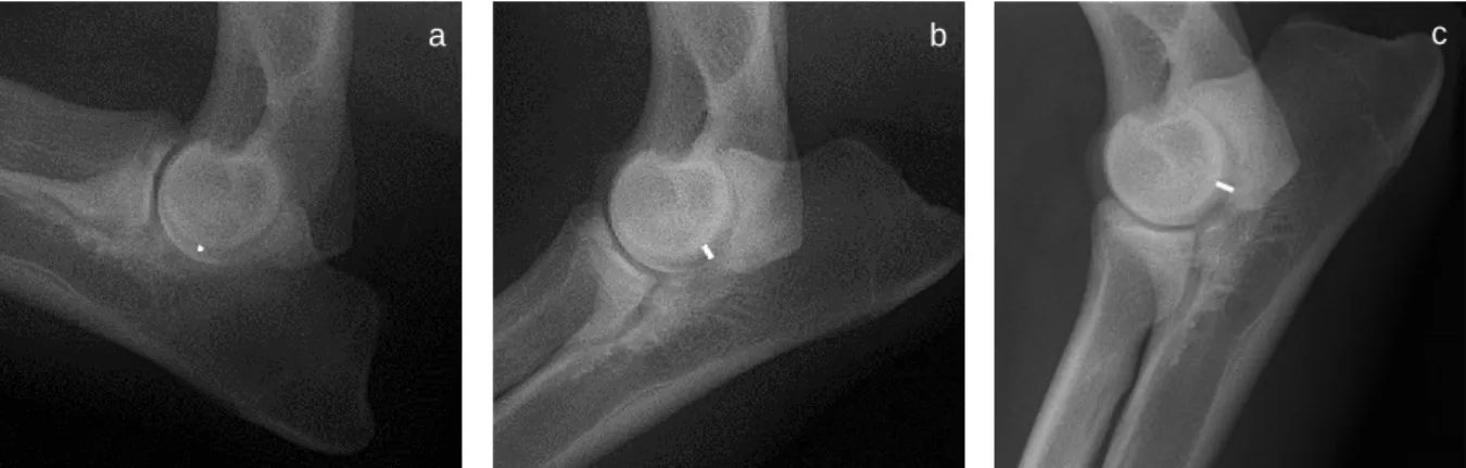
Figure 3.3 Humeroulnar joint space variation on elbow radiographic views from the same
right joint at different angle positionings.
28
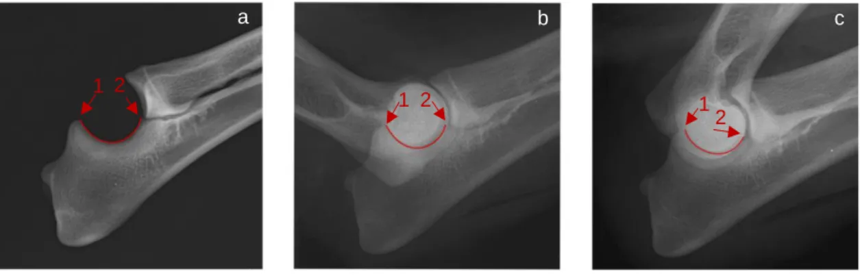
Figure 3.4 Assessment of humeroulnar congruity based on the UTN and the HT curvature
radii.
29
Figure 3.5 Humeroradial, humeroulnar and radioulnar joint spaces assessed at CT
sagittal and dorsal reconstructed images for elbow incongruity evaluation.
31
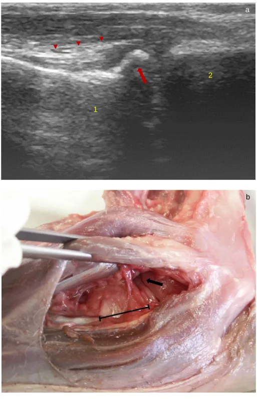
Figure 3.6 Relation between the ultrasound image and anatomical dissection from biceps
brachii / brachialis muscles ulnar insertion tendons and the medial coronoid disease.
34
Figure 3.7 Relation between elbow loading distribution and type of UTN cartilage
covering.
36
Figure 3.8 Relation between the elbow joint load transmission and the UTN curvature
radii.
38
Figure 4.1 Series of data points selected at the subchondral level from the UTN central
ridge in the different views studie using the dedicated software.
61
Figure 4.2 Schematic representation of the rotated points from the UTN curvature radii. 62
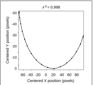
Figure 4.3 Curvature radii expressed as a function of the normalized natural coordinate
for each data set, along the UTN central ridge.
63
Figure 4.4 UTN curvature radii obtained using the MLRU, the MLE and the MLF
radiographic views.
xxiv
Figure 5.1 Elbow radiographic views and the HT sagittal groove analyzed. 74
Figure 5.2 HT curvature radii obtained using the MLH, the MLE and the MLF radiographic
views.
76
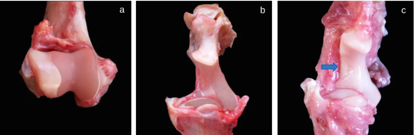
Figure 6.1 Illustration of the UTN and HT curvatures assessed on a medial view from the
proximal radius and ulna and on a caudal view from distal humerus.
86
Figure 6.2 Location of the sagittal reconstruction CT used to study humeroulnar congruity
on a left elbow.
86
Figure 6.3 MLE ~132º radiographic view and sagittal reconstructed CT image from the
elbow.A set of points was marked at the UTN central ridge and on adjacent HT groove.
87
Figure 6.4 MLE ~132º radiographic view and sagittal reconstructed CT image from the
elbow. The curves were traced by a curve fitting process on the UTN and on the HT groove.
87
Figure 6.5 Relationship between the curvature radii found along the UTN and the HT
curvatures, at the MLE view.
90
Figure 6.6 Illustration of the seven points of interest (P1 to P7) defined at the UTN and HT, on the elbow MLE radiographic view; and sagittal reconstructed CT image.
91
Figure 7.1 MLE elbow radiographic views from a medium-breed, the PPD; and a
large-breed, the EMD; and MLF view from the EMD. A set of points were marked at the subchondral level from the UTN and the HT groove in contact with the UTN on extension.
103
Figure 7.2 Curvature radii from the UTN and the HT found along the curves on a
medium-breed, PPD; and on a large-breed dog, EMD, at the MLE radiographic views.
106
Figure 7.3 Schematic representation of the points of interest anatomical position in the
UTN and HT, at MLE views from PPD and EMD; and at MLF view from EMD.
108
Figure 7.4 Schematic representation of curvature radii length in the seven points of interest studied from the EMD at the UTN, and at the HT.
xxv
L
IST OF TABLESTable 3.1 Advantages and disadvantages of the most commonly used imaging methods
to study elbow incongruity and dysplasia development.
41
Table 3.2 Summary of selected references related with radioulnar and humeroulnar
measurement methods to characterize the elbow joint congruity.
42
Table 3.3 Advances obtained through the biomechanical methods most commonly used
to study elbow incongruity and dysplasia development.
47
Table 4.1 Comparison of curvature radii measurements along the UTN obtained at the
MLRU, MLRU1, MLE and MLF elbow radiographic views.
64
Table 6.1 Comparison between curvature radii measurements obtained at the MLE
radiographic view and at the sagittal reconstructed CT image.
89
Table 6.2 Mean differences and 95% CI observed between the UTN and HT curvature
radii, at the seven points of interest selected, on the MLE views and on the sagittal reconstructed CT images.
90
Table 7.1 Comparison between curvature radii measurements obtained at the MLE and
MLF radiographic views from the UTN and the HT.
104
Table 7.2 Difference (mm) between UTN and HT curvature radii measurements in the
points of interest (P1 to P7), by breed.
xxvii
L
IST OF ABREVIATURES,
SYMBOLS AND ACRONYMSAAP - Associação Anatómica Portuguesa
Adv. - Advantage
APMVEAC - Associação Portuguesa de Médicos Veterinários Especialistas em Animais de Companhia
CT - Computed tomography
CI - Confidence interval
DAdv. - Disadvantage
DNA - Deoxyribonucleic acid
e.g. - Example. From Latin exempli gratia
EMD - Estrela Mountain Dog
HT - Humeral trochlea
IEWG - International Elbow Working Group
ICC - Intraclass correlation coefficient
i.e. - It is. From Latin id est
Kg - Kilogram
xxviii
LA - Limits of agreement
mAs - Milliamperes
MCD - Medial coronoid disease
MG - Mário Ginja
MLE - Mediolateral extended
MLF - Mediolateral flexed
MLH - Additional mediolateral humeral view
MLRU - Additional mediolateral radioulnar view
MLRU 1 - Second session of measurements in the MLRU view
mm - Millimeter
ms - Milliseconds
n - Number of joints
OFA - Orthopedic Foundation for Animals
PPD - Portuguese Pointing Dog
P1 to P7 - Points 1 to 7 selected along the curves
SAP - Sociedade Anatómica Portuguesa
xxix
SEM - Standard error of the mean
SP - Sofia Pimenta
SPSS - Statistical Packge for the Social Sciences
T2 - Transverse (or spin-spin) relaxation time
UTN - Ulnar trochlear notch
WSAVA - World Small Animal Veterinary Association
3D - Three dimension
d - Mean difference
𝒇′ - First derivative of the polynomial fit to the points
ρ - Radii of curvature
r2 - Correlation coefficient
s* - Normalized natural coordinate
𝒇′′ - Second derivative of the polynomial fit to the points
~ - Approximately
< - Inferior to
C
HAPTER
1
General introduction
3
1.
G
ENERAL INTRODUCTION1.1ELBOW DYSPLASIA
Elbow dysplasia is the term used to describe an important group of developmental abnormalities affecting the canine elbow joint of young rapidly growing dogs (Temwichitr et al., 2010), and remains the most frequent cause of non-traumatic forearm lameness in large-breed dogs (Farrell et al., 2014), affecting also a few medium and chondrodystrophic breeds (Michelsen, 2013; Coggeshall et al., 2014). The rate in males can reach twice the females’ rate, and the clinical manifestation is more often observed at young (1 year) and at older ages (>6 year) (Meyer‐Lindenberg et al., 2006). The clinical signs involve lameness, which may remain subtle for long periods of time, external rotation of the extremity, moderate joint distension, crepitation during movement, and in advanced cases decreased range of motion (Burton et al., 2008; Temwichitr et al., 2010; Cuddy et al., 2012; Goodrich et al., 2014; Coppieters et al., 2015). Elbow dysplasia is challenging both to diagnose and to treat, presents frequent bilateral involvement and considerable loss of function in a highly mobile joint, resulting in a high clinical impact (Samoy et al., 2012a; 2013). When choosing the dogs’ breed to acquire, pet owners may become apprehensive with the possibility of dysplasia development and costs associated to its treatment (DeCamp et al., 1993; Proks et al., 2011; Caron et al., 2014).
1.1.1ETIOPATHOGENESIS
The medial coronoid disease, osteochondrosis of the humeral trochlea, ununited anconeal process and joint incongruity were defined as specific primary lesions making up this disorder (Hazewinkel, 2016). Locations within the elbow joint for secondary osteophyte production are nonspecific and probably represent typical changes of elbow osteoarthritis (Farrell et al., 2014).
The etiophatogenesis is unclear, although several hypotheses have been proposed (Hazewinkel, 2016). The mechanisms postulated include disturbed development of endochondral ossification with the development of osteochondrosis (Tirgari, 1974; Olsson, 1983; Ekman and Carlson, 1998); as well as anomalies in the coronoid subchondral bone (Burton et al., 2007; Temwichitr et al., 2010) and abnormality of the trabecular bone (Danielson et al., 2006; Mariee et al., 2014; Fitzpatrick et al., 2016). Differences in the distribution of shear stresses and forces within the joint alignments have also been hypothesized (Wolschrijn and Weijs, 2004; Hulse et al., 2010; Lau et al., 2013). In most cases, influences on the development
General introduction
4
and growth play an important role and various forms of joint incongruity are considered as the most likely mechanism involved in the elbow dysplasia development (Wind and Packard, 1986; Preston et al., 2001; Samoy et al., 2012b; 2012c; Böttcher et al., 2013; Fitzpatrick et al., 2013; Nemanic et al., 2016). Elbow joint incongruity is a fitting disturb between the ulnar trochlear notch, radial head and humeral condyle, first reported by Wind in 1982, on radiographs from Bernese Mountain Dogs with medial coronoid fragmentation process (Wind, 1986; Samoy et al., 2006). Incongruity types described in the literature include radioulnar incongruity, humeroulnar incongruity and soft tissue-bone mismatch (Wind, 1986; Fitzpatrick and Yeadon, 2009; Samoy et al., 2012b). Humeroulnar incongruity research is less developed and the pathway to the signs of disease remains unclear (Proks et al., 2011; Samoy et al., 2012b; Michelsen, 2013).
Recent research showed that inherited polygenic traits can occur independently or together, yet the pattern of inheritance differs between breeds and needs to be elucidated (Mäki et al., 2002; Temwichitr et al., 2010). Pointed as a complex multifactorial disease, the onset of clinical signs is also influenced by many environmental factors such as the weight gain rate (Mäki et al., 2002; Lavrijsen et al., 2012).
1.1.2DIAGNOSIS AND SCREENING
The imaging tools most frequently described to screen and diagnose elbow dysplasia in the clinical practice are radiography (Samoy et al., 2006; 2012a; Lau et al., 2015), CT (Kramer et al., 2006; Böttcher et al., 2009; Burton et al., 2013), arthroscopy (Samoy et al., 2013; Skinner et al., 2015), magnetic resonance imaging (Janach et al., 2006; Probst et al., 2008), and ultrasonography (Lamb et al., 2005; Hulse et al., 2010). Molecular techniques to genetically screen animals for the genes responsible for the disease are in development (Temwichitr et al., 2010).
Radiography is the most reliable and affordable method to screen elbow dysplasia and the first approach in clinical cases (Samoy et al., 2006; Lau et al., 2015). Early detection of dogs affected diminishes the transmission of the responsible genes to offspring (Mäki et al., 2002; Hazewinkel, 2016). To compare the prevalence and to realize the epidemiology of the condition, it is necessary to standardize the classification (Hazewinkel, 2016). The most consensual protocol among the scientific community was developed and is regularly updated by the International Elbow Working Group (IEWG) (Stock and Distl, 2010). In Portugal the elbow dysplasia screening program started in 2014, by the Associação Portuguesa de Médicos
General introduction
5 Veterinários Especialistas em Animais de Companhia (APMVEAC)1 in association with the
Portuguese Kennel Club, according to the IEWG standards. Two radiographic perpendicular views: a mediolateral extended (MLE) and a craniocaudal 15º pronated views are required. However, in some countries, as the United States of America elbow dysplasia is screen based only on a mediolateral projection with the elbow flexed, taking into account only the osteoarthritic changes as recommended by the Orthopedic Foundation for Animals (OFA) (Kunst et al., 2014). Some subjectivity remains in the classification, since some specific changes are not easily measurable (Burton et al., 2008; Lappalainen et al., 2009; Fitzpatrick et al., 2009a) and even within the normal grade, differences in joint conformation are reported (Mäki et al., 2000; Collins et al., 2001; Preston et al., 2001; Lappalainen et al., 2009). To ensure a common identification system and provide a uniform and accurate language, the Federation Cynologique Internationale, the IEWG and the World Small Animal Veterinary Association (WSAVA) have worked together to create an international certificate based on the existing IEWG protocol.
1.1.3PREVALENCE
The true prevalence of elbow dysplasia in the populations is unknown, as studies are based on non-representative samples of the population (Janutta et al., 2006; Malm et al., 2008). Yet, prevalence values reported in the most frequently affected breeds were of 38.7-54.2% in the Rottweiler (Beuing et al., 2000; Mäki et al., 2001; Malm et al., 2008), 33% in the Newfoundland Dog (Grøndalen and Lingaas, 1991), 30.5% in the Bernese Mountain Dog (Malm et al., 2008), 23% in the Golden Retriever (Mäki et al., 2001), 16.5% in the Estrela Mountain Dog (Alves-Pimenta et al., 2013), 14.11-19.4% in the German Shepherd Dog (Remy et al., 2004; Janutta et al., 2006) and 17.1% in the Labrador Retriever (Mäki et al., 2001). The discrepancies found in the range of published data may derive from different protocols of elbow dysplasia scoring (Janutta et al., 2006), besides the efforts that some countries are doing (for several years) to decrease the disease incidence. Elbow dysplasia must be considered part of the breeding decisions, and intentional inbreeding should be avoided in the future (Mäki et al. 2001; Lewis et al., 2011). A correlation of 0.23 to 0.41 with hip dysplasia was described (Malm et al., 2008; Cachon et al., 2010; Woolliams et al., 2011) and therefore the selection against hip dysplasia is expected to decrease the prevalence of elbow dysplasia, and vice versa (Mäki et al., 2000; Ginja et al., 2008).
General introduction
6
1.1.4TREATMENT
The treatment of elbow dysplasia is another important field of veterinary intervention, beyond the subject of the present thesis. Both medical and surgical approaches were described (Fitzpatrick and Yeadon, 2009). Briefly, different medical treatment options of secondary osteoarthritis have been extensively reported in the literature; and surgical options were proposed acting directly inside the joint (Fitzpatrick at al., 2009b), or focused on the dynamic axial radioulnar adjustment when incongruity was deemed to be present (Mason et al., 2008; Böttcher et al., 2013; Fitzpatrick et al., 2013), and more recently, in cases of medial coronoid disease, axial to abaxial load shift from medial to lateral joint compartments (Fitzpatrick et al., 2009c; 2015; McConkey et al., 2016). Those techniques are based on the correct biodynamical principal of removing mechanical weight away from the medial compartment, frequently affected, to the lateral compartment of the joint (Fitzpatrick et al., 2009c; Böttcher et al., 2013). Some extra-articular surgical procedures revealed a good outcome, but all await quantitative objective appraisal as to their long-term efficacy (Fitzpatrick et al., 2015; Burton et al., 2016; McConkey et al., 2016). Several prosthetic elbow designs have been attempted over the years to improve the life quality in the severe cases, yet thus far it has not enjoyed the same high success rate that has been shown in the total hip arthroplasty, and a long way needs to be traveled until it becomes a routine procedure (Franklin et al., 2014; Lorenz et al., 2015).
1.1.5CURRENT CHALLENGES IN DYSPLASIA SCREENNING RESEARCH
Extensive advances were recently made in the elbow geometry research field, through biomechanical and imaging studies, allowing the identification of joint components associated to development of elbow incongruity (Wolschrijn et al., 2003; Wagner et al., 2007; Breit et al., 2010; Villamonte-Chevalier et al., 2015). Although the huge efforts made in the diagnostic field, a reliable, affordable and well accepted protocol to early detect the presence and severity of humeroulnar incongruity is claimed, and it is crucial to find new diagnostic approaches (Collins et al., 2001; Samoy et al., 2006; House et al., 2009; Werner et al., 2009; Skinner et al., 2015). Unfortunately, the investigation of humeroulnar incongruity is still scarce (Wind, 1986; Samoy et al., 2012b). Radiographic examination is often considered difficult, due to the overlaping of various anatomical structures, and different complementary projections are required (Mason et al., 2002; Stein et al., 2012). On the other hand, clinically oriented elbow anatomy in its complexity earned a high importance with new imaging and surgical features in the benefit of diagnosis and treatment of canine elbow disorders, allowing the emergence of new diagnostic
General introduction
7 and therapeutic approaches (De Rycke et al., 2002; Breit et al., 2005; Constantinescu and
Constantinescu, 2009; Villamonte-Chevalier et al., 2015).
Other area of research in the elbow diagnosis and pathogenesis is the coronoid tissue examination and development of medial compartment disease, including investigation of the cartilaginous and subchondral tissues (Künzel et al., 2004; Wolschrijn and Weijs, 2005; Burton et al., 2010; Lau et al., 2013). Finally, researchers also suggest that far beyond the search for new effective diagnostic techniques, it is extremely important to find molecular methods to effectively know if the dog is free of the genetic condition before breeding (Mäki et al., 2002). However, because of the complexity of inheritance and the effects of environmental variables in the disease expression, it is unlikely that genetic testing for elbow dysplasia will be possible in the foreseeable future (Temwichitr et al., 2010).
1.2REFERENCES
Alves-Pimenta, S., Colaco, B., Silvestre, A.M., Ginja, M.M., 2013. Prevalence and breeding values of elbow dysplasia in the Estrela Mountain Dog. Veterinarni Medicina 58, 484-490.
Böttcher, P., Werner, H., Ludewig, E., Grevel, V., Oechtering, G., 2009. Visual estimation of radioulnar incongruence in dogs using three-dimensional image rendering: an in vitro study based on computed tomographic imaging. Veterinary Surgery 38, 161-168.
Böttcher, P., Bräuer, S., Hinnerk, W., 2013. Estimation of joint incongruence in dysplastic canine elbows before and after dynamic proximal ulna osteotomy. Veterinary Surgery 42, 371-376.
Breit, S., Künzel, W., Seiler, S., 2005. Postnatal modelling of the humero antebrachial contact areas of radius and ulna in dogs. Anatomia, Histologia, Embryologia 34, 258-264.
Breit, S., Pfeiffer, K., Pichler, R., 2010. Use of a 3D laser scan technique to compare the surface geometry of the medial coronoid process in dogs affected with medial compartment disease with unaffected controls. The Veterinary Journal 185, 285-291.
Beuing, R., Mues, C.H., Tellhelm, B., Erhardt, G., 2000. Prevalence and inheritance of canine elbow dysplasia in German Rottweiler. Journal of Animal Breeding and Genetics 117, 375-383.
Burton, N.J., Comerford, E.J., Bailey, M., Pead, M.J., Owen, M.R., 2007. Digital analysis of ulnar trochlear notch sclerosis in Labrador Retrievers. Journal of Small Animal Practice 48, 220-224. Burton, N.J., Dobney, J.A., Owen, M.R., Colborne, G.R., 2008. Joint angle, moment and power
compensations in dogs with fragmented medial coronoid process. Veterinary and Comparative Orthopaedics and Traumatology 21, 110-118.
Burton, N.J., Perry, M.J., Fitzpatrick, N., Owen, M.R., 2010. Comparison of bone mineral density in medial coronoid processes of dogs with and without medial coronoid process fragmentation. American Journal of Veterinary Research 71, 41-46.
Burton, N.J., Warren-Smith, C.M.R., Roper, D.P., Parsons, K.J., 2013. CT assessment of the influence of dynamic loading on physiological incongruency of the canine elbow. Journal of Small Animal Practice 54, 291-298.
General introduction
8
Burton, N.J., Parsons, K.J., Cunliffe, M., Warren-Smith, C.M.R., Ness, M.G., Fenton, G., 2016. Canine elbow realignment osteotomy (CERO): validation of the accuracy of acute radial lengthening in a cadaveric incongruency model. Veterinary Surgery 45, 642-650.
Cachon, T., Genevois, J.P., Remy, D., Carozzo, C., Viguier, E., Maitre, P., Arnault, F., Fau, D., 2010. Risk of simultaneous phenotypic expression of hip and elbow dysplasia in dogs: a study of 1,411 radiographic examinations sent for official scoring. Veterinary and Comparative Orthopaedics and Traumatology 23, 28-30.
Caron, A., Caley, A., Farrell, M., Fitzpatrick, N., 2014. Kinematic gait analysis of the canine thoracic limb using a six degrees of freedom marker set. Study in normal Labrador Retrievers and Labrador Retrievers with medial coronoid process disease. Veterinary and Comparative Orthopaedics and Traumatology 6, 461-9.
Coggeshall, J.D., Reese, D.J., Kim, S.E., Pozzi, A., 2014. Arthroscopic-guided ulnar distraction for the correction of elbow incongruency in four dogs. Journal of Small Animal Practice 55, 46-51.
Collins, K.E., Cross, A.R., Lewis, D.D., Zapata, J.L., Goett, S.D., Newell, S.M., Rapoff, A.J., 2001. Comparison of the radius of curvature of the ulnar trochlear notch of Rottweilers and Greyhounds. American Journal of Veterinary Research 62, 968-973.
Constantinescu, G.M., Constantinescu, I.A., 2009. A clinically oriented comprehensive pictorial review of canine elbow anatomy. Veterinary Surgery, 38, 135-143.
Coppieters, E., Gielen, I., Verhoeven, G., Van Vynckt, D., Van Ryssen, B., 2015. Erosion of the medial compartment of the canine elbow: occurrence, diagnosis and currently available treatment options. Veterinary and Comparative Orthopaedics and Traumatology 28, 9-18.
Cuddy, L.C., Lewis, D.D., Kim, S.E., Conrad, B.P., Banks, S.A., Horodyski, M., Fitzpatrick, N., Pozzi, A., 2012. Contact mechanics and three-dimensional alignment of normal dog elbows. Veterinary Surgery 41, 818-828.
Danielson, K.C., Fitzpatrick, N., Muir, P., Manley, P.A., 2006. Histomorphometry of fragmented medial coronoid process in dogs: a comparison of affected and normal coronoid processes. Veterinary Surgery 35, 501-509.
DeCamp, C.E., Soutas-Little, R.W., Hauptman, J., Olivier, B., Braden, T., Walton, A., 1993. Kinematic gait analysis of the trot in healthy greyhounds. American Journal of Veterinary Research 54, 627-634.
De Rycke, L.M., Gielen, I.M., van Bree, H., Simoens, P.J., 2002. Computed tomography of the elbow joint in clinically normal dogs. American Journal of Veterinary Research 63, 1400-1407.
Ekman, S., Carlson, C.S., 1998. The pathophysiology of osteochondrosis. Veterinary Clinics of North America: Small Animal Practice 28, 17-32.
Farrell, M., Heller, J., Solano, M., Fitzpatrick, N., Sparrow, T., Kowaleski, M., 2014. Does radiographic arthrosis correlate with cartilage pathology in Labrador Retrievers affected by medial coronoid process disease? Veterinary Surgery 43, 155-165.
Fitzpatrick, N., Yeadon, R., 2009. Working algorithm for treatment decision making for developmental disease of the medial compartment of the elbow in dogs. Veterinary Surgery 38, 285-300.
Fitzpatrick, N., Smith, T.J., Evans, R.B., Yeadon, R., 2009a. Radiographic and arthroscopic findings in the elbow joints of 263 dogs with medial coronoid disease. Veterinary Surgery 38, 213-223. Fitzpatrick, N., Smith, T.J., Evans, R.B., O'Riordan, J., Yeadon, R., 2009b. Subtotal coronoid ostectomy
General introduction
9
Fitzpatrick, N., Yeadon, R., Smith, T., Schulz, K., 2009c. Techniques of application and initial clinical experience with sliding humeral osteotomy for treatment of medial compartment disease of the canine elbow. Veterinary Surgery 38, 261-278.
Fitzpatrick, N., Caron, A., Solano, M.A., 2013. Bi-oblique dynamic proximal ulnar osteotomy in dogs: reconstructed computed tomographic assessment of radioulnar congruence over 12 weeks. Veterinary Surgery 42, 727-738.
Fitzpatrick, N., Bertran, J., Solano, M.A., 2015. Sliding humeral osteotomy: medium-term objective outcome measures and reduction of complications with a modified technique. Veterinary Surgery 44, 137-149.
Fitzpatrick, N., Garcia, T.C., Daryani, A., Bertran, J., Watari, S., Hayashi, K., 2016. Micro-CT structural analysis of the canine medial coronoid disease. Veterinary Surgery 45, 336-346.
Flückiger, M., 2010. Radiographic diagnosis of elbow dysplasia in the dog: Requirements for the IEWG standardized screening procedure. 25th Annual Meeting International Elbow Working Group, Bologna, Italia 8-13.
Franklin, S.P., Schulz, K.S., Karnes, J., Cook, J.L., 2014. Theory and development of a unicompartmental resurfacing system for treatment of medial compartment disease of the canine elbow. Veterinary Surgery 43, 765-773.
Ginja, M.M., Silvestre, A.M., Colaço, J., Gonzalo-Orden, J.M., Melo-Pinto, P., Orden, M.A., Llorens-Pena, M.P., Ferreira, A.J., 2008. Hip Dysplasia in Estrela Mountain Dogs - prevalence and genetic trends 1991-2005. The Veterinary Journal 182, 275-282.
Goodrich, Z.J., Norby, B., Eichelberger, B.M., Friedeck, W.O., Callis, H.N., Hulse, D.A., Kerwin, S.C., Fox, D.B., Saunders, W.B., 2014. Thoracic limb alignment in healthy Labrador Retrievers: evaluation of standing versus recumbent frontal plane radiography. Veterinary Surgery 43, 791-803.
Grøndalen, J., Lingaas, F., 1991. Arthrosis in the elbow joint of young rapidly growing dogs: a genetic investigation. Journal of Small Animal Practice 32, 460-464.
Hazewinkel, H.A., 2016. Mode of inheritance of elbow dysplasia and principles of screening methods. 30th Annual Meeting International Elbow Working Group, Vienna, Austria 8-13.
House, M.R., Marino, D.J., Lesser, M.L., 2009. Effect of limb position on elbow congruity with CT evaluation. Veterinary Surgery 38, 154-160.
Hulse, D., Young, B., Beale, B., Kowaleski, M., Vannini, R., 2010. Relationship of the biceps-brachialis complex to the medial coronoid process of the canine ulna. Veterinary and Comparative Orthopaedics and Traumatology 23, 173-176.
Janach, K.J., Breit, S.M., Künzel, W.W.F., 2006. Assessment of the geometry of the cubital (elbow) joint of dogs by the use of magnetic resonance imaging. American Journal of Veterinary Research 67, 211-217.
Janutta, V., Hamann, H., Klein, S., Tellhelm, B., Distl, O., 2006. Genetic analysis of three different classification protocols for the evaluation of elbow dysplasia in German Shepherd Dogs. Journal of Small Animal Practice 47, 75-82.
Kramer, A., Holsworth, I.G., Wisner, E.R., Kass, P.H., Schulz, K.S., 2006. Computed tomographic evaluation of canine radioulnar incongruence in vivo. Veterinary Surgery 35, 24-29.
Kunst, C.M., Pease, A.P., Nelson, N.C., Habing, G., Ballegeer, E.A., 2014. Computed tomographic identification of dysplasia and progression of osteoarthritis in dog elbows previously assigned OFA grades 0 and 1. Veterinary Radiology and Ultrasound 55, 511-520.
Künzel, W., Breit, S., Probst, A., 2004. The subchondral split line patterns of the medial coronoid process in canine ulnae. Anatomia, Histologia, Embryologia 33, 339-343.
General introduction
10
Lamb, C.R., Wong, K. 2005. Ultrasonographic anatomy of the canine elbow. Veterinary Radiology and Ultrasound 46, 319-325.
Lappalainen, A.K., Mölsä, S., Liman, A., Laitinen-Vapaavuori, O., Snellman, M., 2009. Radiographic and computed tomography findings in Belgian shepherd dogs with mild elbow dysplasia. Veterinary Radiology and Ultrasound 50, 364-9.
Lau, S.F., Wolschrijn, C.F., Siebelt, M., Vernooij, J.C., Voorhout, G., Hazewinkel, H.A., 2013. Assessment of articular cartilage and subchondral bone using EPIC-microCT in Labrador Retrievers with incipient medial coronoid disease. The Veterinary Journal 198, 116-121.
Lau, S.F., Theyse, L.F.H., Voorhout, G., Hazewinkel, H.A.W., 2015. Radiographic, computed tomographic, and arthroscopic findings in Labrador Retrievers with medial coronoid disease. Veterinary Surgery 44, 511–520.
Lavrijsen, I.C., Heuven, H.C., Voorhout, G., Meij, B.P., Theyse, L.F., Leegwater, P.A., Hazewinkel, H.A., 2012. Phenotypic and genetic evaluation of elbow dysplasia in Dutch Labrador Retrievers, Golden Retrievers, and Bernese Mountain Dogs. The Veterinary Journal 193, 486-492.
Lewis, T.W., Ilska, J.J., Blott, S.C., Woolliams, J.A., 2011. Genetic evaluation of elbow scores and the relationship with hip scores in UK Labrador Retrievers. The Veterinary Journal 189, 227-33. Lorenz, N.D., Channon, S., Pettitt, R., Smirthwaite, P., Innes, J.F., 2015. Ex vivo kinematic studies of a
canine unlinked semi-constrained hybrid total elbow arthroplasty system. Veterinary and Comparative Orthopaedics and Traumatology 28, 39-47.
Mäki, K., Liinamo, A.E., Ojala, M., 2000. Estimates of genetic parameters for hip and elbow dysplasia in Finnish Rottweilers. Journal of Animal Science 78, 1141-1148.
Mäki, K., Groen, A.F., Liinamo, A.E., Ojala, M., 2001. Population structure, inbreeding trend and their association with hip and elbow dysplasia in dogs. Animal Science 73, 217-228.
Mäki, K., Groen, A.F., Liinamo, A.E., Ojala, M., 2002. Genetic variances, trends and mode of inheritance for hip and elbow dysplasia in Finnish dog populations. Animal Science. 75, 197-207.
Malm, S., Fikse, W.F., Danell, B., Strandberg, E., 2008. Genetic variation and genetic trends in hip and elbow dysplasia in Swedish Rottweiler and Bernese Mountain Dog. Journal of Animal Breeding and Genetics 2008, 403-412.
Mariee, I.C., Gröne, A., Theyse, L.F., 2014. The role of osteonecrosis in canine coronoid dysplasia: arthroscopic and histopathological findings. The Veterinary Journal 200, 382-386.
Mason, D.R., Schulz, K.S., Samii, V.F., Fujita, Y., Hornof, W.J., Herrgesell, E.J., Long, C.D., Morgan, J.P., Kass, P.H., 2002. Sensitivity of radiographic evaluation of radio-ulnar incongruence in the dog
in vitro. Veterinary Surgery 31, 125-132.
Mason, D.R., Schulz, K.S., Fujita, Y., Kass, P.H., Stover, S.M., 2008. Measurement of humeroradial and humeroulnar transarticular joint forces in the canine elbow joint after humeral wedge and humeral slide osteotomies. Veterinary Surgery 37, 63-70.
McConkey, M.J., Valenzano, D.M., Wei, A., Li, T., Thompson, M.S., Mohammed, H.O., van der Meulen, M.C.H., Krotscheck, U., 2016. Effect of the proximal abducting ulnar osteotomy on intra-articular pressure distribution and contact mechanics of congruent and incongruent canine elbows ex vivo. Veterinary Surgery 45, 347-355.
Meyer‐Lindenberg, A., Fehr, M., Nolte, I., 2006. Co‐existence of ununited anconeal process and fragmented medial coronoid process of the ulna in the dog. Journal of Small Animal Practice 47, 61-65.
Michelsen, J., 2013. Canine elbow dysplasia: aetiopathogenesis and current treatment recommendations. The Veterinary Journal 196, 12-19.
General introduction
11
Mota-Veiga, S., 2002. Novo guia prático do cão da Serra da Estrela. Mem-Martins, Portugal, Spoepress. Nemanic, S., Nixon, B.K., Baltzer, W., 2016. Analysis of risk factors for elbow dysplasia in giant breed
dogs. Veterinary and Comparative Orthopaedics and Traumatology 29, 369-377.
Olsson, S.E., 1983. The early diagnosis of fragmented coronoid process and osteochondritis dissecans of the canine elbow joint. Journal of the American Animal Hospital Association 19, 616-626
Preston, C.A., Schulz, K.S., Kass, P.H., 2001. In vitro determination of contact areas in the normal elbow joint of dogs. American Journal of Veterinary Research 61, 1315-1321.
Probst, A., Modler, F., Künzel, W., Mlynarik, V., Trattnig, S., 2008. Demonstration of the articular cartilage of the canine ulnar trochlear notch using high-field magnetic resonance imaging. The Veterinary Journal 177, 63-70.
Proks, P., Necas, A., Stehlik, L., Srnec, R., Griffon, D.J., 2011. Quantification of humeroulnar incongruity in Labrador Retrievers with and without medial coronoid disease. Veterinary Surgery 40, 981-986. Remy, D., Neuhart, L., Fau, D., Genevois, J.P., 2004. Canine elbow dysplasia and primary lesions in
German Shepherd Dogs in France. Journal of Small Animal Practice 45, 244-248.
Samoy, Y., Van Ryssen, B., Gielen, I., Walschot, N., van Bree, H., 2006. Review of the literature. Elbow incongruity in the dog. Veterinary and Comparative Orthopaedics and Traumatology 19, 1-8. Samoy, Y., Gielen, I., Saunders, J., van Bree, H., Van Ryssen, B., 2012a. Sensitivity and specificity of
radiography for detection of elbow incongruity in clinical patients. Veterinary Radiology and Ultrasound 53, 236-44.
Samoy, Y., Gielen, I., Van Caelenberg, A., van Bree, H., Duchateau, L., Van Ryssen, B., 2012b. Computed tomography findings in 32 joints affected with severe elbow incongruity and fragmented medial coronoid process. Veterinary Surgery 41, 486-494.
Samoy, Y., Van Vynckt, D., Gielen, I., van Bree, H., Duchateau, L., Van Ryssen, B., 2012c. Arthroscopic findings in 32 joints affected by severe elbow incongruity with concomitant fragmented medial coronoid process. Veterinary Surgery 41, 355-361.
Samoy, Y.C., de Bakker, E., Van Vynckt, D., Coppieters, E., van Bree, H., Van Ryssen, B., 2013. Arthroscopic treatment of fragmented coronoid process with severe elbow incongruity long-term follow-up in eight Bernese Mountain Dogs. Veterinary and Comparative Orthopaedics and Traumatology 26, 27-33.
Skinner, O.T., Warren-Smith, C.M., Burton, N.J., Parsons, K.J., 2015. Computed tomographic evaluation of elbow congruity during arthroscopy in a canine cadaveric model. Veterinary and Comparative Orthopaedics and Traumatology 28, 19-24.
Smith, T.J., Fitzpatrick, N., Evans, R.B., Pead, M.J., 2009. Measurement of ulnar subtrochlear sclerosis using a percentage scale in Labrador Retrievers with minimal radiographic signs of periarticular osteophytosis. Veterinary Surgery 38, 199-208.
Stein, S., Schmoekel, H.G., Waibl, H., Brunnberg, L., 2012. Computerized measurements of radiographic anatomical parameters of the elbow joint in Bernese Mountain Dogs. Veterinary and Comparative Orthopaedics and Traumatology 3, 250-261.
Stock, K.F., Distl, O., 2010. Simulation study on the effects of excluding offspring information for genetic evaluation versus using genomic markers for selection in dog breeding. Journal of Animal Breeding and Genetics 127, 42-52.
Temwichitr, J., Leegwater, P.A., Hazewinkel, H.A., 2010. Fragmented coronoid process in the dog: A heritable disease. The Veterinary Journal 185, 123-129.
General introduction
12
Tirgari, M., 1974. Clinical radiographical and pathological aspects of arthritis of the elbow joint in dogs. Journal of Small Animal Practice 15, 671-679.
Villamonte-Chevalier, A.A., Soler, M., Sarria, R., Agut, A., Gielen, I., Latorre, R., 2015. Ultrasonographic and anatomic study of the canine elbow joint. Veterinary Surgery 44, 485-493.
Wagner, K., Griffon, D.J., Thomas, M.W., Schaeffer, D.J., Schulz, K., Samii, V.F., Necas, A., 2007. Radiographic, computed tomographic, and arthroscopic evaluation of experimental radio-ulnar incongruence in the dog. Veterinary Surgery 36, 691-698.
Werner, H., Winkels, P., Grevel, V., Oechtering, G., Böttcher, P., 2009. Sensitivity and specificity of arthroscopic estimation of positive and negative radio-ulnar incongruence in dogs. Veterinary and Comparative Orthopaedics and Traumatology 22, 437-441.
Wind, A.P., 1982. Incidence and radiographic appearance of fragmented coronoid process. California Vet 19-25.
Wind, A.P., 1986. Elbow incongruity and developmental elbow diseases in the dog: part I. Journal of the American Animal Hospital Association 22, 711-724.
Wind, A.P., Packard, M.E., 1986. Elbow incongruity and developmental elbow diseases in the dog: part II. Journal of the American Animal Hospital Association 22, 725-730.
Wolschrijn, C.F., Kik, M.J., Weijs, W.A., 2003. Cartilage-free areas in the elbow joint of young Golden Retrievers. The Anatomical Record 275, 990-996.
Wolschrijn, C.F., Weijs, W.A., 2004. Development of the trabecular structure within the ulnar medial coronoid process of young dogs. The Anatomical Record 278, 514-519.
Wolschrijn, C.F., Weijs, W.A., 2005. Development of the subchondral bone layer of the medial coronoid process of the canine ulna. The Anatomical Record 284, 439-445.
Woolliams, J.A., Lewis, T.W., Blott, S.C., 2011. Canine hip and elbow dysplasia in UK Labrador Retrievers. The Veterinary Journal 189, 169-176.
C
HAPTER
2
Aims
15
2.
A
IMSHumeroulnar incongruity remains one of the most challenging disorders to diagnose in the development elbow dysplasia. Elbow geometry and the proximity of structures in the elbow joint are the major problems found using the common imaging modalities.
Unfortunately, no significant progresses were made over the last years and a successful diagnostic tool remains elusive, being extremely important to provide continuity to elbow geometry research, focused on new measurement techniques and targets, in order to achieve new data. Thus, the present thesis had as main purposes:
- To review elbow incongruity measurement protocols and methodologies usually applied to evaluate elbow geometry; as well as the role of incongruity in the etiopathogenesis of elbow dysplasia.
- To develop a methodology and software for the ulnar trochlear notch (UTN) and the humeral trochlea (HT) curvature radii assessment, at radiographic and CT images, in order to apply it in the clinical practice.
- To evaluate the UTN and HT curvature, using different lateral elbow radiographic views and CT images of normal elbows of large-breed dogs, ensuring clinical application. To correlate the curvature radii data obtained in the radiographic and CT cadaveric analysis with the data obtained after anatomical bone preparation.
- To characterize the curvature radii pattern from the humeroulnar joint in a medium-breed dog, the Portuguese Pointing Dog (PPD) and in a large-breed dog, Estrela Mountain Dog (EMD),
in vivo, and to obtain an overview on the variability of the shape classified as normal in a
C
HAPTER
3
J
OINT INCONGRUITY ON ELBOW DYSPLASIA
19 THE CONTENT OF THIS CHAPTER WAS SUBMITTED FOR PUBLICATION:
Alves-Pimenta, S., Ginja, M.M., Colaço, B. Incongruity role at the medial coronoid disease and canine elbow dysplasia.
Joint incongruity on elbow dysplasia
21
3.
J
OINT INCONGRUITY ON ELBOW DYSPLASIA IN DOGS,
A REVIEW3.1INTRODUCTION
Canine elbow dysplasia is a biomechanical, inherited developmental disorder, highly prevalent in large and giant-breed dogs (Lavrijsen et al., 2012; Mariee et al., 2014). The term elbow dysplasia is used to describe a group of lesions (Lavrijsen et al., 2012; Phillips et al., 2015), including medial coronoid disease (Danielson et al., 2006; Lau et al., 2013a; Lau et al., 2015a), osteochondrosis of the humeral trochlea (Fitzpatrick et al., 2009a), ununited anconeal process (Meyer‐Lindenberg et al., 2006; Michelsen, 2013) and joint incongruity (Wind and Packard, 1986; Samoy et al., 2012a; 2012b; Böttcher et al., 2013; Fitzpatrick et al., 2013) occurring in separate or simultaneously (Lewis et al., 2011; Lavrijsen et al., 2012). The etiology is multifactorial involving endogenous and exogenous factors (Temwichitr et al., 2010; Lavrijsen et al., 2012). Medial coronoid disease is the most common form of elbow dysplasia, accounting more than 90% of all cases (Danielson et al., 2006; Fitzpatrick et al., 2009b; Lau et al., 2015a), and some remarkable differences in the development pathways were already found among breeds (Lavrijsen et al., 2012). Ulnar sub trochlear sclerosis is observed in the early stages of the disease associated with coronoid disease (Smith et al., 2009; Burton et al., 2010; Lau et al., 2013a; Mariee et al., 2014; Lau et al., 2015a). Joint osteoarthritis is the most frequently observed denouement in affected animals (Farrell et al., 2014; Franklin et al., 2014).
Regardless of all the radiographic screening programs implemented and selection of dogs for reproduction, some animals present a severe condition, highlighting the existence of liability genes in the breed populations (Lewis et al., 2011; Alves-Pimenta et al., 2013; Lau et al., 2015b), which suggests that something might be missing in the diagnostic protocols (Lewis et al., 2011; Alves-Pimenta et al., 2013; Lau et al., 2015b). In addition, and despite all the advances on the medical and surgical treatment field, affected dogs have a poor long-term prognosis due to osteoarthritis (Fitzpatrick et al., 2009c; 2009d; Fitzpatrick and Yeadon, 2009; Franklin et al., 2014; Lorenz et al., 2015). At older ages, an extreme condition of medial compartment disease may be present, often leading to a premature euthanasia (Lavrijsen et al., 2012; Franklin et al., 2014; Coppieters et al., 2015). Thus, medical efforts have been mostly focused on early detection, and new approaches are being continuously investigated in order to diminuish the genes transmission to the offspring (Lewis et al., 2011; Lau et al., 2015b). Joint incongruity is nowadays recognized as a major factor in the development of elbow dysplasia subsequent manifestations, however this entity is poorly known and many questions remain unanswered (Eljack et al., 2013; Samoy et al., 2013).
Joint incongruity on elbow dysplasia
22
Joint incongruity is a fitting disturb between the UTN, radial head and humeral condyle, traditionally divided into radioulnar and humeroulnar incongruity (Wind, 1986; Wind and Packard, 1986; Werner et al., 2009; Fitzpatrick et al., 2013). Radioulnar incongruity research increased in the last decades and its etiology is related to an unequal growth between the radius and the ulna (Böttcher et al., 2009; Eljack et al., 2013). Less studies were found in humeroulnar congruity, as a result from UTN and HT different conformations (Proks et al., 2011; Alves-Pimenta et al., 2016a; 2016b; 2017). There are only few articular models described and the pathways to the clinical signs of disease remain unclear (Proks et al., 2011; Samoy et al., 2012b; Michelsen, 2013). Thus, the shape assessment of both UTN and HT is considered challenging and important in recent investigations (Samoy et al., 2013; Krotscheck et al., 2014, Alves-Pimenta et al., 2015; 2016a; 2017). Different protocols were described to assess incongruity, but to date, there is no consensus on the most accurate method to diagnose humeroulnar incongruity (Skinner et al., 2015). The overlapping and proximity of articular surfaces at different levels are pointed as the main obstacles to the evaluation (Proks et al., 2011; Alves-Pimenta et al., 2015). Susceptibility to changes in elbow positioning, pronation and supination of the elbow were the main disadvantages pointed to techniques described for joint space measurements (Gemmill et al., 2005; 2006; Kramer et al., 2006; House et al., 2009).
In this work, the authors review elbow incongruity current advances, with focus on its importance in elbow dysplasia development, on the diagnostic imaging tools currently available in clinical practice, on the biomechanical studies and on the genetic investigation. Advantages and disadvantages of recently proposed CT and radiography protocols are discussed (Samoy et al., 2012a; 2012b; Lavrijsen et al., 2012; Lau et al., 2015a). Other imaging tools such as arthroscopy, magnetic resonance imaging and ultrasonography suggested for the elbow dysplasia diagnosis were also included in this review (Janach et al., 2006; Samoy et al., 2012c; Villamonte-Chevalier et al., 2014; Lau et al., 2015a). The molecular advances in diagnosis are briefly presented. Relevant biomechanical studies which improved knowledge on elbow geometry are addressed. This review aims to update veterinarians and contribute to educate breeders and owners about the condition.
3.2ELBOW INCONGRUITY
Elbow joint incongruity was described by Wind in 1982, on radiographs from Bernese Mountain Dogs with medial coronoid process fragmentation. Since then, different types of the disease were suggested and confusing terminology may occur (Wind, 1982; 1986; Samoy et al., 2006).