Common Genetic Variation In Cellular
Transport Genes and Epithelial Ovarian
Cancer (EOC) Risk
Ganna Chornokur1, Hui-Yi Lin2, Jonathan P. Tyrer3, Kate Lawrenson4, Joe Dennis3, Ernest K. Amankwah1,5, Xiaotao Qu2, Ya-Yu Tsai1, Heather S. L. Jim6, Zhihua Chen2, Ann Y. Chen2, Jennifer Permuth-Wey1, Katja KH. Aben7,8, Hoda Anton-Culver9, Natalia Antonenkova10, Fiona Bruinsma11, Elisa V. Bandera12, Yukie T. Bean13,14, Matthias W. Beckmann15, Maria Bisogna16, Line Bjorge17,18, Natalia Bogdanova19, Louise A. Brinton20,
Angela Brooks-Wilson21,22, Clareann H. Bunker23, Ralf Butzow24,25, Ian G. Campbell26,27,28, Karen Carty29,30, Jenny Chang-Claude31, Linda S. Cook32, Daniel W. Cramer33, Julie M. Cunningham34, Cezary Cybulski35, Agnieszka Dansonka-Mieszkowska36, Andreas du Bois37,38, Evelyn Despierre39, Ed Dicks3, Jennifer A. Doherty41,42, Thilo Dörk43,
Matthias Dürst44, Douglas F. Easton45,4, Diana M. Eccles46, Robert P. Edwards47, Arif B. Ekici48, Peter A. Fasching15,49, Brooke L. Fridley50, Yu-Tang Gao51, Aleksandra Gentry-Maharaj52, Graham G. Giles11,53, Rosalind Glasspool30, Marc T. Goodman54,55,
Jacek Gronwald35, Patricia Harrington40, Philipp Harter37,38, Alexander Hein15, Florian Heitz37,38, Michelle A. T. Hildebrandt56, Peter Hillemanns43, Claus K. Hogdall63, Estrid Hogdall57,58, Satoyo Hosono59, Anna Jakubowska35, Allan Jensen58, Bu-Tian Ji20, Beth Y. Karlan60, Linda E. Kelemen61, Mellissa Kellar13,14, Lambertus A. Kiemeney7, Camilla Krakstad17,18, Susanne K. Kjaer58,62, Jolanta Kupryjanczyk36,
Diether Lambrechts64,65, Sandrina Lambrechts39, Nhu D. Le66, Alice W. Lee4, Shashi Lele67, Arto Leminen24, Jenny Lester60, Douglas A. Levine16, Dong Liang68, Boon Kiong Lim69, Jolanta Lissowska70, Karen Lu71, Jan Lubinski35, Lene Lundvall63, Leon F. A.
G. Massuger72, Keitaro Matsuo59, Valerie McGuire73, John R. McLaughlin74, Iain McNeish75, Usha Menon52, Roger L. Milne11, Francesmary Modugno23,76,77, Kirsten B. Moysich67, Roberta B. Ness79, Heli Nevanlinna24, Ursula Eilber31, Kunle Odunsi80, Sara H. Olson81, Irene Orlow81, Sandra Orsulic60, Rachel Palmieri Weber82, James Paul30, Celeste L. Pearce4,83, Tanja Pejovic13,14, Liisa M. Pelttari24, Malcolm C. Pike4,81, Elizabeth M. Poole84,85, Harvey A. Risch86, Barry Rosen87, Mary Anne Rossing42, Joseph H. Rothstein88, Anja Rudolph31, Ingo B. Runnebaum44, Iwona K. Rzepecka36, Helga B. Salvesen17,18, Eva Schernhammer84,85, Ira Schwaab89, Xiao-Ou Shu90, Yurii
B. Shvetsov91, Nadeem Siddiqui29, Weiva Sieh88, Honglin Song3, Melissa C. Southey27, Beata Spiewankiewicz92, Lara Sucheston67, Soo-Hwang Teo93,94, Kathryn L. Terry33, Pamela J. Thompson54,55, Lotte Thomsen78, Ingvild L. Tangen17,18, Shelley S. Tworoger33,85, Anne M. van Altena72, Robert A. Vierkant95, Ignace Vergote39, Christine S. Walsh60, Shan Wang-Gohrke31, Nicolas Wentzensen20, Alice S. Whittemore88, Kristine G. Wicklund42, Lynne R. Wilkens91, Anna H. Wu4, Xifeng Wu56, Yin-Ling Woo69, Hannah Yang20, Wei Zheng96, Argyrios Ziogas9, Hanis N. Hasmad93, Andrew Berchuck97, Georgia Chenevix-Trench on behalf of the AOCS management group98,99, Edwin S. Iversen100, Joellen M. Schildkraut101, Susan J. Ramus4, Ellen L. Goode102, Alvaro N. A. Monteiro1, Simon A. Gayther4, Steven A. Narod103, Paul D. P. Pharoah3,104, Thomas A. Sellers1, Catherine M. Phelan1*
1Division of Population Sciences, Department of Cancer Epidemiology, Moffitt Cancer Center, Tampa, Florida, United States of America,2Department of Biostatistics and Bioinformatics, Moffitt Cancer Center, Tampa, Florida, United States of America,3Department of Oncology, University of Cambridge,
Strangeways Research Laboratory, Cambridge, UK,4Department of Preventive Medicine, Keck School of Medicine, University of Southern California Norris Comprehensive Cancer Center, Los Angeles, California, United States of America,5Clinical and Translational Research Organization, All Children’s Hospital Johns Hopkins Medicine, St Petersburg, Florida, United States of America,6Department of Health Outcomes and Behavior, Moffitt Cancer Center, Tampa, Florida, United States of America,7Radboud University Medical Center, Radboud Institute for Health Sciences, Nijmegen, The Netherlands,8Comprehensive Cancer OPEN ACCESS
Citation:Chornokur G, Lin H-Y, Tyrer JP, Lawrenson K, Dennis J, Amankwah EK, et al. (2015) Common Genetic Variation In Cellular Transport Genes and Epithelial Ovarian Cancer (EOC) Risk. PLoS ONE 10 (6): e0128106. doi:10.1371/journal.pone.0128106
Editor:Irina U Agoulnik, Florida International University, UNITED STATES
Received:May 28, 2014
Accepted:April 22, 2015
Published:June 19, 2015
Copyright:© 2015 Chornokur et al. This is an open access article distributed under the terms of the
Creative Commons Attribution License, which permits unrestricted use, distribution, and reproduction in any medium, provided the original author and source are credited.
Data Availability Statement:All relevant data are within the paper and its Supporting Information files.
Center The Netherlands, Nijmegen, The Netherlands,9Genetic Epidemiology Research Institute, Center for Cancer Genetics Research and Prevention, University of California Irvine, Irvine, California, United States of America,10 N.N. Alexandrov National Cancer Centre of Belarus, Minsk, Belarus,11 Cancer Epidemiology Centre, Cancer Council Victoria, Melbourne, Australia,12Cancer Prevention and Control Program, Rutgers Cancer Institute of New Jersey, New Brunswick, New Jersey, United States of America,13Department of Obstetrics & Gynecology, Oregon Health & Science University, Portland, Oregon, United States of America,
14Knight Cancer Institute, Oregon Health & Science University, Portland, Oregon, United States of America,
15Department of Gynecology and Obstetrics, University Hospital Erlangen, Friedrich-Alexander-University, Erlangen-Nuremberg Comprehensive Cancer Center, Erlangen EMN, Erlangen, Germany,16Gynecology Service, Department of Surgery, Memorial Sloan-Kettering Cancer Center, New York, New York, United States of America,17 Department of Gynecology and Obstetrics, Haukeland University Hospital, Bergen, Norway,18 Centre for Cancer Biomarkers, Department of Clinical Medicine, University of Bergen, Bergen, Norway,19 Radiation Oncology Research Unit, Hannover Medical School, Hannover, Germany,20 Division of Cancer Epidemiology and Genetics, National Cancer Institute, Bethesda, Maryland, United States of America,21 Canada's Michael Smith Genome Sciences Centre, BC Cancer Agency, Vancouver, Canada,
22Department of Biomedical Physiology and Kinesiology, Simon Fraser University, Burnaby, Canada,
23Department of Epidemiology, University of Pittsburgh Graduate School of Public Health, Pittsburgh, Pennsylvania, United States of America,24Department of Obstetrics and Gynecology, University of Helsinki and Helsinki University Central Hospital, Helsinki, HUS, Finland,25 Department of Pathology, Helsinki University Central Hospital, Helsinki, Finland,26Cancer Genetics Laboratory Research Division, Peter MacCallum Cancer Centre, St Andrews Place, East Melbourne, Australia,27Department of Pathology, the University of Melbourne, Parkville, Australia,28Sir Peter MacCallum Department of Oncology, University of Melbourne, Parkville, Victoria, Australia,29Department of Gynaecological Oncology, Glasgow Royal Infirmary, Glasgow, United Kingdom,30 The Beatson West of Scotland Cancer Centre, Glasgow, United Kingdom,31German Cancer Research Center (DKFZ), Division of Cancer Epidemiology, Heidelberg, Germany,32Division of Epidemiology and Biostatistics, Department of Internal Medicine, University of New Mexico, Albuquerque, New Mexico, United States of America,33Obstetrics and Gynecology Center, Brigham and Women's Hospital and Harvard Medical School, Boston, Massachusetts, United States of America,34 Department of Laboratory Medicine and Pathology, Mayo Clinic, Rochester, Minnesota, United States of America,35 International Hereditary Cancer Center, Department of Genetics and Pathology, Pomeranian Medical University, Szczecin, Poland,36 Department of Pathology, The Maria Sklodowska-Curie Memorial Cancer Center and Institute of Oncology, Warsaw, Poland,37Department of Gynaecology and Gynaecologic Oncology, Kliniken Essen-Mitte/ Evang. Huyssens-Stiftung/ Knappschaft GmbH, Essen, Germany,38Department of Gynaecology and Gynaecologic Oncology, Dr. Horst Schmidt Kliniken Wiesbaden, Wiesbaden, Germany,39 Division of Gynecologic Oncology, Department of Obstetrics and Gynaecology and Leuven Cancer Institute, University Hospitals Leuven, Leuven, Belgium,40Department of Oncology, The Centre for Cancer Epidemiology, University of Cambridge, Strangeways Research
Laboratory, Cambridge, United Kingdom,41Department of Epidemiology, The Geisel School of Medicine Hanover, New Hampshire, United States of America,42 Program in Epidemiology, Division of Public Health Sciences, Fred Hutchinson Cancer Research Center, University of Washington, Seattle, Washington, United States of America,43 Gynaecology Research Unit, Hannover Medical School, Hannover, Germany,
44Department of Gynecology, Jena University Hospital, Friedrich Schiller University, Jena, Germany,
45Centre for Cancer Genetic Epidemiology, Department of Oncology, University of Cambridge, Cambridge, United Kingdom,46Faculty of Medicine, University of Southampton, Southampton, United Kingdom,
47Ovarian Cancer Center of Excellence, Womens Cancer Research Program, Magee-Womens Research Institute and University of Pittsburgh Cancer Institute, Pittsburgh, Pennsylvania, United States of America,
48Institute of Human Genetics, Friedrich-Alexander-University Erlangen-Nuremberg, Comprehensive Cancer Center Erlangen-Nuremberg, Erlangen, Germany,49 University of California at Los Angeles, David Geffen School of Medicine, Department of Medicine, Division of Hematology and Oncology, Los Angeles, California, United States of America,50Biostatistics and Informatics Shared Resource, University of Kansas Medical Center, Kansas City, Kansas, United States of America,51 Department of Epidemiology, Shanghai Cancer Institute, Shanghai, China,52 Women's Cancer, Institute for Women's Health, University College London, London, United Kingdom,53Centre for Epidemiology and Biostatistics, School of Population and Global Health, The University of Melbourne, Melbourne, Australia,54Cancer Prevention and Control, Samuel Oschin Comprehensive Cancer Institute, Cedars-Sinai Medical Center, Los Angeles, California, United States of America,55 Community and Population Health Research Institute, Department of Biomedical Sciences, Cedars-Sinai Medical Center, Los Angeles, California, United States of America,
56Department of Epidemiology, The University of Texas MD Anderson Cancer Center, Houston, Texas, United States of America,57 Molecular Unit, Department of Pathology, Herlev Hospital, University of Copenhagen, Copenhagen, Denmark,58Department of Virus, Lifestyle and Genes, Danish Cancer Society Research Center, Copenhagen, Denmark,59Department of Preventive Medicine, Kyushu University Faculty of Medical Science, Nagoya, Aichi, Japan,60Women's Cancer Program at the Samuel Oschin Comprehensive Cancer Institute, Cedars-Sinai Medical Center, Los Angeles, California, United States of
Ovarian Cancer Research Fund thanks to donations by the family and friends of Kathryn Sladek Smith (PPD/RPCI.07). Investigator-specific funding: GCT is supported by the National Health and Medical Research Council; BK holds an ACS Early Detection Professorship (SIOP–‐06–‐258–‐01–‐
COUN); LEK is supported by a Canadian Institute of Health Research New Investigator Award (MSH—
87734). AWL is supported by NIEHS T32 training grant (T32ES013678). Funding of included studies: Funding of the constituent studies was provided by the California Cancer Research Program (00–‐
01389V–‐20170, N01–‐CN25403, 2II0200); the Canadian Institutes of Health Research (MOP—
86727); Cancer Australia; Cancer Council Victoria; Cancer Council Queensland; Cancer Council New South Wales; Cancer Council South Australia; Cancer Council Tasmania; Cancer Foundation of Western Australia; the Cancer Institute of New Jersey; Cancer Research UK (C490/A6187, C490/ A10119, C490/A10124); the Danish Cancer Society (94–‐222–‐52); the ELAN Program of the University of Erlangen–‐Nuremberg; the Eve Appeal; the Helsinki University Central Hospital Research Fund; Helse Vest; the Norwegian Cancer Society; the Norwegian Research Council; the Ovarian Cancer Research Fund; Nationaal Kankerplan of Belgium; Grant–‐in–‐Aid for the Third Term Comprehensive 10–‐Year Strategy For Cancer Control from the Ministry of Health Labour and Welfare of Japan; the L & S Milken Foundation; the Polish Ministry of Science and Higher Education (4 PO5C 028 14, 2 PO5A 068 27); the Roswell Park Cancer Institute Alliance Foundation; the US National Cancer Institute (K07–‐CA095666, K07–‐CA143047,K22–‐
CA138563, N01–‐CN55424, N01—PC67001, N01–‐
PC067010, N01–‐PC035137, P01–‐CA017054, P01–‐CA087696, P30—CA072720, P50–‐
CA105009, P50-CA136393, R01–‐CA014089, R01– ‐CA016056, R01–‐CA017054, R01–‐CA049449, R01–‐CA050385, R01–‐CA054419, R01–‐
CA058598, R01–‐CA058860, R01–‐CA061107, R01–‐CA061132, R01–‐CA067262, R01–‐
CA071766, R01–‐CA074850, R01–‐CA080742, R01–‐CA080978, R01–‐CA083918, R01–‐
CA087538, R01–‐CA092044, R01–‐095023, R01–‐
CA122443, R01–‐CA112523, R01–‐CA114343, R01–‐CA126841, R01–‐CA136924, R03–‐
CA113148, R03–‐CA115195, U01–‐CA069417, U01–‐CA071966 and Intramural research funds); the US Army Medical Research and Material Command (DAMD17–‐01–‐1–‐0729, DAMD17–‐02– ‐1–‐0666, DAMD17–‐02–‐1–‐0669, W81XWH—
America,61 Department of Public Health Sciences, Medical University of South Carolina, Charleston, South Carolina, United States of America,62 Department of Gynaecology, Rigshospitalet, University of
Copenhagen, Copenhagen, Denmark,63The Juliane Marie Centre, Department of Gynecology, Righospitalet, University of Copenhagen, Copenhagen, Denmark,64 Vesalius Research Center, VIB, Leuven, Belgium,65Laboratory for Translational Genetics, Department of Oncology, University of Leuven, Belgium,66Cancer Control Research, BC Cancer Agency, Vancouver, Canada,67Department of Cancer Prevention and Control, Roswell Park Cancer Institute, Buffalo, New York, United States of America,
68College of Pharmacy and Health Sciences, Texas Southern University, Houston, Texas, United States of America,69 Department of Obstetrics and Gynaecology, University Malaya Medical Centre, University Malaya, Kuala Lumpur, Malaysia,70Department of Cancer Epidemiology and Prevention, M. Sklodowska-Curie Memorial Cancer Center and Institute of Oncology, Warsaw, Poland,71Department of Gynecologic Oncology, The University of Texas MD Anderson Cancer Center, Houston, Texas, United States of America,
72Department of Gynaecology, Radboud University Medical Center, Nijmegen, The Netherlands,
73Department of Health Research and Policy—Epidemiology, Stanford University School of Medicine, Stanford, California, United States of America,74Public Health Ontario, Toronto, Canada,75Institute of Cancer Sciences, University of Glasgow, Wolfson Wohl Cancer research Centre, Beatson Institute for Cancer Research, Glasgow, UK,76 Women's Cancer Research Program, Magee-Women's Research Institute and University of Pittsburgh Cancer Institute, Pittsburgh, Pennsylvania, United States of America,
77Department of Obstetrics, Gynecology and Reproductive Sciences, University of Pittsburgh School of Medicine, Pittsburgh, Pennsylvania, United States of America,78 Department of Pathology, Rigshospitalet, University of Copenhagen, Copenhagen, Denmark,79 The University of Texas School of Public Health, Houston, Texas, United States of America,80 Department of Gynecologic Oncology, Roswell Park Cancer Institute, Buffalo, New York, United States of America,81Department of Epidemiology and Biostatistics, Memorial Sloan-Kettering Cancer Center, New York, New York, United States of America,82 Department of Community and Family Medicine, Duke University Medical Center, Durham, North Carolina, United States of America,83 Department of Epidemiology, University of Michigan School of Public Health,Ann Arbor, Michigan, United States of America,84 Channing Division of Network Medicine, Brigham and Women's Hospital and Harvard Medical School, Boston, Massachusetts, United States of America,85Department of Epidemiology, Harvard School of Public Health, Boston, Massachusetts, United States of America,
86Department of Chronic Disease Epidemiology, Yale School of Public Health, New Haven, Connecticut, United States of America,87 Department of Gynecology-Oncology, Princess Margaret Hospital, and Department of Obstetrics and Gynecology, Faculty of Medicine, University of Toronto, Toronto, Ontario, Canada,88 Department of Health Research and Policy- Epidemiology, Stanford University School of Medicine, Stanford, California, United States of America,89Institut für Humangenetik, Wiesbaden, Germany,90Epidemiology Center and Vanderbilt, Ingram Cancer Center, Vanderbilt University School of Medicine, Nashville, Tennessee, United States of America,91 Cancer Epidemiology Program, University of Hawaii Cancer Center, Hawaii, United States of America,92 Department of Gynecologic Oncology, The Maria Sklodowska-Curie Memorial Cancer Center and Institute of Oncology, Warsaw, Poland,93 Cancer Research Initiatives Foundation, Sime Darby Medical Center, Subang Jaya, Malaysia,94University Malaya Cancer Research Institute, University Malaya Medical Centre, University Malaya, Kuala Lumpur, Maylaysia,
95Department of Health Science Research, Division of Biomedical Statistics and Informatics, Mayo Clinic, Rochester, Minnesota, United States of America,96Vanderbilt Epidemiology Center, Vanderbilt University School of Medicine, Nashville, Tennessee, United States of America,97Department of Obstetrics and Gynecology, Duke University Medical Center, Durham, North Carolina, United States of America,98 QIMR Berghofer Medical Research Institute, Brisbane, Australia,99Peter MacCallum Cancer Centre, East Melbourne, Australia,100Department of Statistics, Duke University, Durham, North Carolina, United States of America,101Cancer Prevention, Detection & Control Research Program, Duke Cancer Institute, Durham, North Carolina, United States of America,102Department of Health Science Research, Division of
Epidemiology, Mayo Clinic, Rochester, Minnesota, United States of America,103Women's College Research Institute, University of Toronto, Toronto, Ontario, Canada,104Department of Public Health and Primary Care, University of Cambridge, Cambridge, United Kingdom
*catherine.phelan@moffitt.org
Abstract
Background
Defective cellular transport processes can lead to aberrant accumulation of trace elements, iron, small molecules and hormones in the cell, which in turn may promote the formation of
of Education and Research of Germany Programme of Clinical Biomedical Research (01GB 9401); the State of Baden–‐Wurttemberg through Medical Faculty of the University of Ulm (P.685); the Minnesota Ovarian Cancer Alliance; the Mayo Foundation; the Fred C. and Katherine B. Andersen Foundation; the Lon V. Smith Foundation (LVS–‐
39420); the Oak Foundation; the OHSU Foundation; the Mermaid I project; the Rudolf–‐Bartling Foundation; the UK National Institute for Health Research Biomedical Research Centres at the University of Cambridge, Imperial College London, University College Hospital“Womens Health Theme”and the Royal Marsden Hospital; WorkSafeBC 14.
reactive oxygen species, promoting DNA damage and aberrant expression of key regula-tory cancer genes. As DNA damage and uncontrolled proliferation are hallmarks of cancer, including epithelial ovarian cancer (EOC), we hypothesized that inherited variation in the cellular transport genes contributes to EOC risk.
Methods
In total, DNA samples were obtained from 14,525 case subjects with invasive EOC and from 23,447 controls from 43 sites in the Ovarian Cancer Association Consortium (OCAC). Two hundred seventy nine SNPs, representing 131 genes, were genotyped using an Illu-mina Infinium iSelect BeadChip as part of the Collaborative Oncological Gene-environment Study (COGS). SNP analyses were conducted using unconditional logistic regression under a log-additive model, and the FDR q<0.2 was applied to adjust for multiple comparisons.
Results
The most significant evidence of an association for all invasive cancers combined and for the serous subtype was observed for SNP rs17216603 in the iron transporter geneHEPH (invasive: OR = 0.85, P = 0.00026; serous: OR = 0.81, P = 0.00020); this SNP was also associated with the borderline/low malignant potential (LMP) tumors (P = 0.021). Other genes significantly associated with EOC histological subtypes (p<0.05) included the UGT1A(endometrioid),SLC25A45(mucinous),SLC39A11(low malignant potential), and SERPINA7(clear cell carcinoma). In addition, 1785 SNPs in six genes (HEPH,MGST1, SERPINA,SLC25A45,SLC39A11andUGT1A) were imputed from the 1000 Genomes Project and examined for association with INV EOC in white-European subjects. The most significant imputed SNP was rs117729793 inSLC39A11(per allele, OR = 2.55, 95% CI = 1.5-4.35, p = 5.66x10⁻⁴).
Conclusion
These results, generated on a large cohort of women, revealed associations between inher-ited cellular transport gene variants and risk of EOC histologic subtypes.
Introduction
Epithelial ovarian carcinoma (EOC) is the second-most common gynecological malignancy and the leading cause of gynecological cancer-related mortality in the United States and other
developed nations [1]. Early stage EOC is accompanied by vague, non-specific symptoms and
is difficult to detect. As yet, no EOC screening or early detection strategies have been proven
useful in general population [2]. As a consequence, approximately 60% of cases are diagnosed
at advanced stages (III-IV), with 5-year overall survival of less than 20% [3]. Given these grim
statistics, improved understanding of the etiology of EOC is critical to reducing the associated morbidity and mortality.
the expression of key regulatory genes. Aberrant expression of transmembrane transport genes has been associated with increased risk, as well as aggressiveness, of a number of cancers
including breast [4–7], prostate [8], liver [9], colorectal and colon [10–12], thyroid [13] and
neuroblastoma [14]. Iron is essential for erythropoiesis [15] and the function of the
mitochon-drial respiratory chain where it plays a key role in electron transport, in the form of
iron-sul-phur clusters or in heme centers [16]. However, excess iron and copper have been reported to
promote the formation of reactive oxygen species (ROS) which damage cellular DNA and
sup-port cancer growth [17]. Hydrogen peroxide generated as a byproduct in the mitochondrial
respiratory chain, can react with iron or copper and form hydroxyl radicals that are extremely
reactive and damaging to the genome [18]. Consistent with these data, transport of the three
most abundant transition metals in humans–iron, zinc and copper–has been linked to the
eti-ology of colorectal and liver cancer [9,19] and prostate cancer [20].
Despite the growing evidence that cellular transport processes influence cancer risk, the association of germline genetic variation in cellular transport genes and EOC risk has not been well studied. As histologic subtypes of EOC differ in clinical behavior and biologic and genetic
origin [21], we hypothesized that single nucleotide polymorphisms (SNPs) in cellular transport
genes are associated with EOC risk and vary by histopathology. This study examined the asso-ciation of 279 SNPs in 131 cellular transport genes and EOC risk in an international collabora-tion that included 18,174 case and 26,134 control subjects.
Materials and Methods
Sample and Procedure
The discovery set included DNA samples from 3,761 EOC case subjects and 2,722 control sub-jects in two ovarian cancer Genome Wide Association Studies (GWAS) in North America and
the United Kingdom (UK). Details of these studies have been previously published [22]. In
brief, the North American study was comprised of four case-control studies genotyped using the Illumina 610-quad Beadchip Array (1,814 case subjects and 1,867 control subjects) as well as a single case-control study genotyped on the Illumina 317K and 370K arrays (133 case sub-jects and 142 control subsub-jects). The UK study was comprised of four case-only studies geno-typed on the Illumina 610-quad Beadchip Array and two common control sets genogeno-typed on the Illumina 550K array (1,814 case subjects and 713 control subjects).
The replication sample consisted of DNA samples from 14,525 women with invasive EOC and 23,447 control women with European ancestry from 43 sites in the Ovarian Cancer Associ-ation Consortium (OCAC). Samples from an additional 1,747 participants with tumors of low malignant potential were also analyzed. Details of the sample QC are provided in Pharoah
et al., [22]. This replication set include 89 SER cases and 200 controls of African ancestry, and
249 SER cases and 1574 controls of Asian ancestry.
All research involving human participants has been approved by each study site’s local
Insti-tutional Review Board (IRB) according to the principles expressed in the Declaration of Hel-sinki. Written informed consent was obtained from all of the participants. The permission to use of the data in this study was granted by the University of South Florida (IRB#:
Pro00000249).
Gene and SNP Selection
Gene (NCBI) [23], BioCarta [24], GenomeNet [25] and other relevant gene/pathway databases
Quad BeadChip (Illumina). Those SNPs were genotyped in the four ovarian cancer GWAS studies (US GWAS, UK GWAS, COGS and Mayo clinic). The final selection of transport gene SNPs for genotyping in the replication stage was informed according to lowest p-values (cut off
p<0.01 was used) by ranking of minimal p-values across four sets of results in the discovery
set: 1) North American all histologies, 2) North American serous histology, 3) combined GWAS meta-analysis all histologies, and 4) combined GWAS meta-analysis serous histology. Additional functional SNPs in these genes were also included. In total 299 SNPs were included on the COGS chip of which 279 SNPs (in 131 genes) passed QC (described in detail in Pharoah et al., [22]).
Imputation Analyses
These analyses were based on imputed genotypes from the four ovarian cancer GWAS studies (US GWAS, UK GWAS, COGS and Mayo clinic) with a total of 15,398 invasive EOC case sub-jects and 30,816 control subsub-jects of white-European ancestry. Imputation of each dataset into
the 1000 Genomes Project was performed using IMPUTE2 software [26]. We used the 1000
Genomes Project v3 as the reference with pre-phasing of the data using SHAPEIT [27].
Statistical Analysis
For the discovery set, the North American and UK studies were analyzed separately and four sets of results: 1) North American all histologies, 2) North American serous histology, 3) com-bined GWAS meta-analysis all histologies, and 4) comcom-bined GWAS meta-analysis serous his-tology were conducted. SNP analyses were performed using unconditional logistic regression under a log-additive model. The last two sets were analyzed using the fixed effects
meta-analy-sis. These analyses were carried out in PLINK [28] by combining results across studies by using
the Mantel–Haenszel method [29].
For the replication set, demographic and clinical characteristics of case subjects and control subjects were compared using t-test for continuous variables and chi-square test for categorical variables. Unconditional logistic regression was used to evaluate associations between SNPs and ovarian cancer risk. SNPs were modeled using number of minor alleles as ordinal variables (log-additive model). Per-allele log odds ratios and their 95% confidence intervals were esti-mated. All analyses were done separately by race groups.
In order to adjust for population substructure, intercontinental ancestry was assigned based on genotype frequencies for European, Asian, and African populations using LAMP software
[30] Subjects with greater than 90 percent European ancestry were defined as European; those
with greater than 80 percent Asian and African ancestry were defined as being Asian and African, respectively. The set of 37,000 unlinked markers was applied to perform
principal-components analysis within each major population subgroup [31].The number of principal
components was based on the inflection position of the principal components scree plot. The models for white-European subjects were adjusted for study site and for the first five principal components. For the African American (AA) and Asian (AS) study subjects, the first one and five principal components were included in the models, respectively.
We evaluated associations between candidate SNPs and risk of all invasive EOC (INV), each of the four main histological subtypes (serous (SER); endometroid (END); clear cell (CC); and mucinous (MUC)), and tumors of low malignant potential (LMP). Odds ratios for each histo-logic subtype were estimated by comparing cases of each subtype to all controls as reference.
False discovery rate (FDR) q-value was applied for adjusting multiple comparisons [32].
Asso-ciations with a p value<.05 and a false discovery rate (FDR) q-value<.20 were considered
For the imputation set analyses, the meta-analysis using an in-house program written in C+ + was carried out for combining results across studies. A fixed effect meta-analysis was used, with the log odds ratio being the estimate for each study and the standard error of this estimate determining the weighting. For each SNP, only the studies with valid estimates for that SNP
(i.e. r2>0.25) were used in the meta-analysis calculation.
Results
Sample characteristics are described in theS1 Table. As expected, significant differences were
observed between case and control subjects on EOC risk factors including age, family history of ovarian cancer, age at menarche, body mass index (BMI), history of oral contraceptive use,
and number of full term births (p-values<0.05). The proportion of tumors of SER (57.6%) was
higher than that of other subtypes (14.2% END, 7.1% CC, 6.5% MUC, and 14.6% other), which is typical for white-European populations.
Among the 279 cellular transport SNPs genotyped in the replication set, 81 SNPs in 48
genes showed nominally significant (p<0.05) associations with at least one histological subtype
(S2 Table). All invasive cancers combined (INV), LMP and the four main histological subtypes
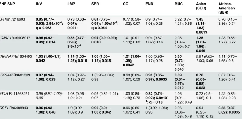
(SER [n = 8,369]; END [n = 2,067]; CC [n = 1,024]; and MUC [n = 943]) were analyzed. The strongest evidence of an association for INV EOC was observed for SNP rs17216603 in
the iron transporter geneHEPH(OR = 0.85, 95%CI = 0.77–0.93, P = 2.55x10⁻⁴; FDR
q-value = 0.065), which was also the most significant SNP associated with SER (P = 1.99x10⁻⁴;
95%CI = 0.73–0.91, FDR q-value = 0.054), and LMP subtypes (P = 0.0206) (Table 1). The most
Table 1. The most significant SNPs in the transport pathway genes and risk of EOC by histology, invasiveness, and race/ethnicity1.
GENE SNP INV LMP SER CC END MUC Asian
(SER)
African-American (SER)
HEPHrs17216603 0.85 (0.77–
0.93); 2.55x10⁻⁴; q = 0.063
0.78 (0.63–
0.97); 0.021;
0.81 (0.73–
0.91); 1.99x10⁻⁴; q = 0.054
0.77 (0.58–
1.02); 0.07
0.9 (0.74–
1.08); 0.26
0.92 (0.7–
1.21); 0.56
1.45 (1.15–
1.83) 0.0019
0.76 (0.15–
3.86); 0.74
SLC39A11rs9908917 0.95 (0.92–
0.99); 0.014
0.85 (0.77–
0.93); 3.9x10⁻⁴
0.94 (0.9–0.99);
0.010
1.01 (0.91–
1.13); 0.82
0.94 (0.87–
1.02); 0.16 0.98 (0.87– 1.00); 0.7 1.25 (1.01– 1.56); 0.049
1.23 (0.85–
1.77); 0.27
SERPINA7Rs1804495 1.05 (1.00–1.1);
0.042
1.14 (1.03–
1.27); 0.016
1.06 (1.00–
1.12); 0.045
1.21 (1.06–
1.39); 0.0042
1.06 (0.96–
1.17); 0.28
0.85 (0.73–
1.00); 0.045
0.81 (0.65–
1.00); 0.05
1.11 (0.75–
1.65); 0.6
SLC25A45Rs681309 0.97 (0.94–
1.00); 0.029
1.04 (0.97–
1.12); 0.27
1 (0.96–1.04); 0.99
0.98 (0.89–
1.07); 0.59
0.91 (0.85–
0.97); 0.0035 0.89 (0.81– 0.97); 0.012 0.78 (0.63– 0.98); 0.033
0.87 (0.6–
1.26); 0.41
UGT1ARs11563251 0.95 (0.91–1.00);
0.05
1.08 (0.96–
1.2); 0.21
0.95 (0.89–1.01); 0.07
1.03 (0.89–
1.18); 0.73
0.82 (0.74–
0.92); 6.8x10
-4; q = 0.18
1.06 (0.91–
1.22); 0.49
0.73 (0.5–
1.06); 0.1
1.22 (0.85–
1.25); 0.28
MGST1Rs6488840 0.96 (0.93–
1.00); 0.048
1.0 (0.92–
1.09); 0.9
0.95 (0.91–
1.00); 0.042
0.96 (0.86–
1.07); 0.41
1 (0.92–1.08); 0.95 0.96 (0.86– 1.08); 0.48 0.54 (0.25– 1.18); 0.12
0.55 (0.37–
0.82); 0.0035
1
INV: all invasive EOC combined; LMP: low malignant potential / borderline tumors; SER: serous; CC: clear cell; End: endometrioid; Muc: mucinous. Statistically significant associations are indicated in bold (P<0.05). Data format is the following: OR (95% CI); p-value; FDR q-value (white-European women). Only significant FDRs (q<0.2) are shown (HEPH: INV and SER; UGT1A: End).
significant association for END EOC was rs11563251 within theUGT1Agene cluster
(OR = 0.82, 95%CI = 0.73–0.92, P = 6.59x10⁻⁴; FDR q-value = 0.177). Only these two SNPs
were associated with q-values<0.20. The most significant association for MUC subtype was
rs681309, near theSLC25A45gene (OR = 0.89, 95%CI = 0.81–0.97, P = 0.012). This SNP was
also associated with the END subtype (P = 0.0035) and INV EOC (P = 0.029). Association with
rs9908917, in the intron ofSLC39A11, was observed for LMP cases (OR = 0.85, 95%CI = 0.77–
0.93, P = 3.9x10⁻⁴). This SNP was also associated with the SER subtype (P = 0.0123) and INV
EOC (P = 0.0144). The SNP rs1804495 inSERPINA7was associated with SER, MUC, CC, INV
and LMP (P<0.05), but not END (P>0.05).
Imputed Variants
In total, 1785 imputed SNPs in six genes (HEPH,MGST1,SERPINA,SLC25A45,SLC39A11
andUGT1A) were examined for association with INV EOC in white-European subjects only.
From these, 274 SNPs were found with p-value<0.05 (S3 Table). Across all six genes, the most
significant imputed SNP was rs117729793 inSLC39A11(per allele, OR = 2.55, 95% CI = 1.5–
4.35, p = 5.66x10⁻⁴). Interestingly, 190 of 274 (~70%) imputed SNPs with p-values<0.05 were
located in or nearSLC39A11.
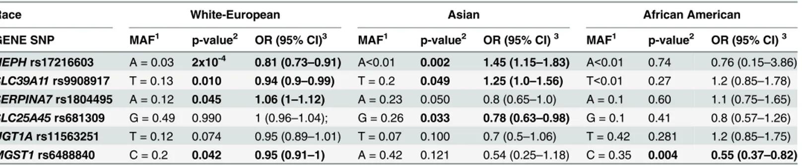
Results in women of African-American (AA) and Asian (AS) ethnicities
We conducted exploratory analyses for other ethnicities and SER EOC. Fourteen SNPs showed significant associations in AS and AA women. Six of the SNPs in the AS women were also sig-nificant in the white-European women, compared to two of the 14 SNPs in the AA women.
The top SNP in women of Asian ancestry (rs17216603 inHEPH) was shared with the
white-European women. TheSLC25A45rs681309 was also shared.SERPINA7rs1804495 was
border-line significant (P = 0.0503), perhaps due to a small sample size. The most significant associa-tion in women of AA ancestry was noted at the SNP rs6488840 near to the microsomal
glutathione S-transferase 1 (MGST1) gene. In our study,MGST1rs6488840 was associated
with statistically significantly reduced SER EOC risk in women of AA ancestry (OR = 0.55; P = 0.0035). This SNP was of borderline significance in women of white-European (OR = 0.95;
P = 0.042), but not Asian (P>0.05), ancestry. In the groups of AS and AA women, no SNPs
had FDR q-value<0.20. Results for the top hits across women of different ancestries are
pre-sented inTable 2.
Table 2. Top SNPs associated with SER EOC across racial groups.
Race White-European Asian African American
GENE SNP MAF1 p-value2 OR (95% CI)3 MAF1 p-value2 OR (95% CI)3 MAF1 p-value2 OR (95% CI)3 HEPHrs17216603 A = 0.03 2x10⁻⁴ 0.81 (0.73
–0.91) A<0.01 0.002 1.45 (1.15–1.83) A<0.01 0.74 0.76 (0.15–3.86)
SLC39A11rs9908917 T = 0.13 0.010 0.94 (0.9–0.99) T = 0.2 0.049 1.25 (1.0–1.56) T<0.01 0.27 1.2 (0.85–1.78)
SERPINA7rs1804495 A = 0.12 0.045 1.06 (1–1.12) A = 0.23 0.050 0.8 (0.65–1.0) A = 0.1 0.60 1.1 (0.75–1.65)
SLC25A45rs681309 G = 0.49 0.990 1 (0.96–1.04); G = 0.26 0.033 0.78 (0.63–0.98) G = 0.1 0.41 0.8 (0.57–1.26)
UGT1Ars11563251 T = 0.12 0.074 0.95 (0.89–1.01) T = 0.07 0.100 0.7 (0.5–1.06) T = 0.42 0.281 1.2 (0.85–1.75)
MGST1rs6488840 C = 0.2 0.042 0.95 (0.91–1) A = 0.42 0.121 0.54 (0.25–1.18) C = 0.35 0.004 0.55 (0.37–0.82)
1MAF, minor allele and its frequency 2p-value<0.05 are in bold
3Odds ratio, 95% con
fidence interval
Discussion
The development and progression of ovarian cancer is accompanied by aberrant cellular
metabolism [33]. Central to cellular metabolic processes are the transport of trace elements
and hormones through cellular and nuclear membranes. In this study, we aimed to elucidate whether germline SNPs in cellular transport genes were associated with EOC risk and
histo-pathologic subtype. We detected nominal associations (P<0.05) with 81 SNPs and EOC risk
in at least one of the histopathologic subtypes (S3 Table). Associations were noted with
rs17216603 inHEPHand SER, INV and LMP subgroups as well as in SER and INV cases in
women of white-European and Asian ancestries. The Hephaestin (HEPH) gene encodes a
transmembrane copper-dependentferroxidase(HEPH protein) responsible for dietary iron
transport from intestinalenterocytesinto the blood stream [34–36].HEPHcatalyzes ferrous
(F2+) iron reoxidation to its ferric (F3+) state [37–38] that can be utilized by the body. The role
of iron homeostasis in cancer progression is yet to be fully understood; however depletion of iron stores in cells induces cell cycle arrest and apoptosis, limits the rate of DNA synthesis, and down-regulates expression of various potentially carcinogenic kinases such as cyclins and
cyclin-dependent kinases [39]. Additionally, iron is known to facilitate generation of
muta-genic reactive oxygen species (ROS) that may drive cancer development and progression [40]
as has been observed in colorectal cancer [41].In silicoanalysis ofHEPHrs17216603
combin-ing results from Snpnexus, SNPinfo and Annovar [42–44] showed that this variant results in
the substitution of Alanine at residue 598 with Threonine and may lead to reduction or loss of
HEPHfunction. Functional analyses of this SNP and gene will be needed to clarify the impact
of this finding. There was no evidence of MAF heterogeneity because the MAF range of
rs17216603 across the studies is 2–5%. This SNP was significantly associated with Invasive
EOC risk in women of white-European descent (P = 0.0003) for the combined results (Fig 1).
The SNP rs9908917 lies within an intron of theSLC39A11gene, and was associated with
SER, all INV and LMP EOC. In addition, of the 1785 SNPs in six genes (HEPH,MGST1,
SER-PINA,SLC25A45,SLC39A11andUGT1A) imputed from the 1000 Genomes Project and
exam-ined for association with INV EOC in white-European subjects, the most significant imputed
SNP was rs117729793 inSLC39A11(per allele, OR = 2.55, 95% CI = 1.5–4.35, p = 5.66x10⁻⁴).
In The Cancer Genome Atlas (TCGA) data [45], expression ofSLC39A11was significantly
higher in ovarian tumors compared to normal tissues (P = 9.99x10⁻⁸). Taken together, these
data highlight a potential role for this gene in EOC pathogenesis. The solute carrier family 39,
member 11 (SLC39A11) is a poorly studied gene belonging to a family of metal ion
transport-ers.SLC39A11may act as a zinc-influx transporter, although the exact functions of the A11
gene have yet to be experimentally established [46]. Other members of theSLC39family
trans-port metal ions, such as iron, copper, cadmium and manganese [47].
SNP rs681309, in the intergenic region nearSLC25A45, showed significant associations
with all INV, MUC, and END and was the most significant SNP among the MUC subtype. The solute carrier family 25 (mitochondrial carrier; adenine nucleotide translocator), member 45 (SLC25A45) belongs to the family of membrane proteins that catalyze the transport of solutes
across the inner mitochondrial membrane [48]. While substrates for theSLC25family carriers
include ADP/ATP, amino acids, malate, ornithine, and citruline [49], the predominant
sub-strate(s) forSLC23A45have not yet been characterized [50], although sequence similarity to
SLC25A29suggests that this protein may be involved in the transport of long-chain fatty acids
such as palmitoylcarnitine and acylcarnitine [51].SLC25A45is expressed in skeletal muscle,
intestine, brain, and testis and is downregulated during ovarian cancer progression [50]. Taken
together, these data suggest that additional studies are warranted on the role ofSLC25A4in
The SNP, rs1804495 inSERPINA7was associated with all INV, SER, MUC, CC and LMP
subtypes, and was the most statistically significant association among the CC subtype. The
ser-pin peptidase inhibitor, clade A (alpha-1 antiproteinase, antitrypsin), member 7 (SERPINA7),
also known as thyroxine-binding globulin (TBG), is a protein that binds thyroid hormones
thyroxin(T4) and 3,5,3’-triiodothyronine (T3) in circulation [52]. Numerous mutations in
SERPINA7have been identified [53–54] leading to partially or completely absent TBG
func-tion. The hallmark of TBG deficiency is abnormally low T3 and T4 combined with normal
thy-roid stimulating hormone (TSH) values [55]. The specific role ofSERPINA7in cancer etiology
has not been established; however, thyroid hormones may support cancer growth [56]. Thus, it
is conceivable that altered TBG production over many years may modulate growth of early-stage ovarian cancer cells. The index SNP, rs1804495, is coding, but the resulting missense change is predicted to have neutral impact on the protein function, suggesting a linked variant may be the causal allele at this locus. We note that in our analyses, rs1804495 confers statisti-cally significantly increased risk for LMP, INV, SER and CC, but decreased risk for mucinous EOC. This observation highlights observations from previously published studies that reveal
differences between MUC and other EOCs [57–59].
Fig 1. Forest plot forHEPHrs17216603 across studies.Squares represent the estimated per-allele odds ratio (OR) for each study. Lines indicate the 95% confidence intervals. Diamond represents the OR estimate and confidence limits. Invasive EOC risk in women of white-European descent only; MAF in controls.
The most significant association among the END subtype was withUGT1A1rs11563251.
UDP glucuronosyltransferase 1 family, polypeptide A cluster (UGT1A) represents a complex
locus which encodes nine human UDP-glucuronosyltransferases.
UDP-glucuronosyltransfer-ase (UGT) enzymes are localized to endoplasmic reticulum (ER) and catalyze glucuronidation,
which is involved in the elimination of bilirubin, steroids, bile acids, toxic dietary components,
and several drugs, including morphine, and irinotecan [60–61]. Genetic variation inUGT1A1
is involved in inherited disorders of bilirubin metabolism such as Crigler-Najjar syndrome,
which is manifested in complete absence (type 1) or diminished (types 2–3) bilirubin
glucuro-nidation and resulting impaired bilirubin excretion. Previously, theUGT1A73allele exhibited
modestly significant association with colorectal cancer (OR = 2.39; P = 0.02) [57], lung cancer
[62], endometrial cancer [63] and pancreatic cancer (OR = 1.98;P= 0.003) [64] with a
particu-larly strong association in smokers with pancreatic carcinoma who were younger than 55 years
(OR = 4.7;P= 0.0009), suggesting the magnitude of the observed associations may be modified
by environmental interactions. Down-regulation ofUGT1Aappears to be an early event in
car-cinogenesis [65]; it is postulated that constitutive expression ofUGT1Afamily genes in normal
mucosa protects organs from carcinogens released in the bladder or absorbed from the diet in
the colon. The rs11563251 variant lies within the 3’UTR of theUGT1A1,-A6and–A10genes,
and so could feasibly impact the RNA stability of these transcripts. Alternatively, since this
SNP also lies within intronic sequences of otherUGT1Agenes, this SNP could possibly be
involved incis-regulation of expression of one or more genes in this cluster.
In this study we conducted exploratory analyses in AS and AA subjects. However, the power to detect associations in women of non-European ancestries was limited due to small sample size and only the SER subtype of EOC was investigated for risk associations. The top
SNPs in the AS ancestry group (rs17216603 inHEPHand rs1552846 inSLC39A11) were also
significant in white-European women. In women of AA ancestry, the most significant SNP
rs6488840 (P = 0.0035) was close to the microsomal glutathione S-transferase 1 (MGST1) gene,
which encodes a protein that catalyzes the conjugation ofglutathioneto electrophiles and the
reduction of lipid hydroperoxides. This protein is localized to theendoplasmic reticulumand
outermitochondrial membranewhere it is thought to protect these membranes fromoxidative
stress. The product of this gene is involved in cellular defenses against toxic, carcinogenic, and
pharmacologically active electrophilic compounds [66].MGST1overexpression has been
dem-onstrated in various cancers (e.g., prostate cancer and lung cancer [67–68]) and has been
asso-ciated with high metastatic potential and chemoresistance [69].MGST1is abundantly
expressed in EOC primary tumors, metastases and effusions [66]. Other significant SNPs in
women of AA ancestry include various members of the SLC39 family:SLC39A11(rs9905659
and rs16977431) andSLC39A8(rs233807).
The main strength of our study is a large sample size of white-European women that afforded sufficient statistical power to detect modest risk differences. Weaknesses, however, include the lack of functional or metabolic studies to establish biological significance of the observed associations. Another weakness is a small sample size in AA and AS women. The con-tribution of genetic and/or biological differences to EOC among different ethnic groups is unclear. However, because ovarian cancer health disparities are observed along the whole
con-tinuum of the disease globally and in the U.S. [70], this topic is without a doubt important and
deserves its own dedicated studies.
In summary, we have found that genetic variation in transmembrane transport genes appear
to be associated with EOC risk across various histologic subtypes (Table 1). These data suggest
Supporting Information
S1 Table. Demographic and clinical characteristics of white-European case subjects with invasive disease (n = 14,525) and controls (n = 23,447) in the COGS study.
(DOCX)
S2 Table. Transport pathway SNPs statistically significantly (p<0.05) associated with at least one histopathologic EOC subtype in white-European women.Significant associations are bolded.
(DOCX)
S3 Table. Imputed SNPs significantly (p<0.05) associated with INV EOC in white-Euro-pean women.SNPs are sorted by p-values.
(DOCX)
Acknowledgments
Individual acknowledgments by study:We thank all the individuals who took part in this study and all the researchers, clinicians and technical and administrative staff who have made possible the many studies contributing to this work. In particular, we thank: D. Bowtell, A. deFazio, D. Gertig, A. Green, P. Parsons, N. Hayward, P. Webb and D. Whiteman (AUS); G. Peuteman, T. Van Brussel and D. Smeets (BEL); the staff of the genotyping unit, S LaBoissiere and F Robidoux (Genome Quebec); U. Eilber and T. Koehler (GER); L. Gacucova (HMO); P. Schurmann, F. Kramer, W. Zheng, T. W. Park, Simon, K. Beer- Grondke and D. Schmidt (HJO); S. Windebank, C. Hilker and J. Vollenweider (MAY); the state cancer registries of AL, AZ, AR, CA, CO, CT, DE, FL, GA, HI, ID, IL, IN, IA, KY, LA, ME, MD, MA, MI, NE, NH, NJ, NY, NC, ND, OH, OK, OR, PA, RI, SC, TN, TX, VA, WA, and WYL (NHS); L. Paddock, M. King, L. Rodriguez-Rodriguez, A. Samoila, and Y. Bensman (NJO); M. Sherman, A.
Hutchin-son,N. Szeszenia—‐Dabrowska, B. Peplonska, W. Zatonski, A. Soni, P. Chao and M. Stagner
(POL); C. Luccarini,P. Harrington the SEARCH team and ECRIC (SEA); R. Royer, S. Zhang (TOR); I. Jacobs, M. Widschwendter, E. Wozniak, N. Balogun, A. Ryan and J. Ford (UKO); Carole Pye (UKR); A. Amin Al Olama, K. Michilaidou, K. Kuchenbaker (COGS). Special thanks to the members of the AOCS management group: Penny Webb (QIMR Berghofer Medical Research Institute, Brisbane, Australia); David Bowtell (Peter MacCallum Cancer Cen-tre, Melbourne, VIC); and Anna deFarzio (Westmead Hospital, Westmead, NSW).
Main funding:The scientific development and funding for this project were funded by the following: NIH R01 CA-1491429 (Phelan PI); the US National Cancer Institute (R01-CA076016); the COGS project is funded through a European Commission's Seventh
Frame-work Programme grant (agreement number 223175 HEALTH F2 2009–223175); the Genetic
Associations and Mechanisms in Oncology (GAME‐ON): a NCI Cancer Post-GWAS Initiative
(U19-CA148112); the Ovarian Cancer Association Consortium is supported by a grant from the Ovarian Cancer Research Fund thanks to donations by the family and friends of Kathryn Sladek Smith (PPD/RPCI.07).
Investigator-specific funding:G.C.-T. is supported by the National Health and Medical
Research Council; B.K. holds an ACS Early Detection Professorship (SIOP—‐06—‐258—‐01
—‐COUN); L.E.K. is supported by a Canadian Institute of Health Research New Investigator
Award (MSH—-87734). AWL is supported by NIEHS T32 training grant (T32ES013678).
Funding of included studies:Funding of the constituent studies was provided by the
Cali-fornia Cancer Research Program (00—‐01389V—‐20170, N01—‐CN25403, 2II0200); the
Victoria; Cancer Council Queensland; Cancer Council New South Wales; Cancer Council South Australia; Cancer Council Tasmania; Cancer Foundation of Western Australia; the Cancer Institute of New Jersey; Cancer Research UK (C490/A6187, C490/A10119, C490/
A10124); the Danish Cancer Society (94—‐222—‐52); the ELAN Program of the University
of Erlangen—‐Nuremberg; the Eve Appeal; the Helsinki University Central Hospital
Research Fund; Helse Vest; the Norwegian Cancer Society; the Norwegian Research Council;
the Ovarian Cancer Research Fund; Nationaal Kankerplan of Belgium; Grant—‐in—‐Aid for
the Third Term Comprehensive 10—‐Year Strategy For Cancer Control from the Ministry of
Health Labour and Welfare of Japan; the L & S Milken Foundation; the Polish Ministry of Science and Higher Education (4 PO5C 028 14, 2 PO5A 068 27); the Roswell Park Cancer
Institute Alliance Foundation; the US National Cancer Institute (K07—‐CA095666, K07—‐
CA143047,K22—‐CA138563, N01—‐CN55424, N01—-PC67001, N01—‐PC067010, N01—‐
PC035137, P01—‐CA017054, P01—‐CA087696, P30—-CA072720, P50—‐CA105009,
P50-CA136393, R01—‐CA014089, R01—‐CA016056, R01—‐CA017054, R01—‐CA049449,
R01—‐CA050385, R01—‐CA054419, R01—‐CA058598, R01—‐CA058860, R01—‐
CA061107, R01—‐CA061132, R01—‐CA067262, R01—‐CA071766, R01—‐CA074850,
R01—‐CA080742, R01—‐CA080978, R01—‐CA083918, R01—‐CA087538, R01—‐
CA092044, R01—‐095023, R01—‐CA122443, R01—‐CA112523, R01—‐CA114343, R01—‐
CA126841, R01—‐CA136924, R03—‐CA113148, R03—‐CA115195, U01—‐CA069417,
U01—‐CA071966 and Intramural research funds); the US Army Medical Research and
Material Command (DAMD17—‐01—‐1—‐0729, DAMD17—‐02—‐1—‐0666, DAMD17—‐
02—‐1—‐0669, W81XWH—-07—-0449, W81XWH—‐10—‐1—‐02802); the US Public
Health Service (PSA—-042205); The National Health and Medical Research Council of
Aus-tralia (199600 and 400281); the German Federal Ministry of Education and Research of
Germany Programme of Clinical Biomedical Research (01GB 9401); the State of Baden—‐
Wurttemberg through Medical Faculty of the University of Ulm (P.685); the Minnesota Ovarian Cancer Alliance; the Mayo Foundation; the Fred C. and Katherine B. Andersen
Foundation; the Lon V. Smith Foundation (LVS—‐39420); the Oak Foundation; the OHSU
Foundation; the Mermaid I project; the Rudolf—‐Bartling Foundation; the UK National
Institute for Health Research Biomedical Research Centres at the University of Cambridge,
Imperial College London, University College Hospital“Womens Health Theme”and the
Royal Marsden Hospital; WorkSafeBC 14.
Author Contributions
JPW KA HAC NA FB HB EVB YTB MWB MB LB NB LAB ABW CHB RB IC KC JCC PJC LSK DWC JMC CC ADM AdB EDicks EDespierre JAD TD MD DFE DME RPE ABE PAF BLF YTG AGM GGG RG MTG JG PHarrington PHA AH FH MATH PHarter CKH EH SH AJakubowska AJensen BTJ BYK LEK MK LAK CK SKK JLAK CK SKKJ JK DLambrechts SLambrechts NDL AWL SLele AL JLubinski DAL DLiang BKL JLissowska KLMatsuo LJ LL LFAGM KM VMG JRM IMN UM RLM FM KBM LN RBN HN SN KO SHO IO SO RPW JP CLP TP LMP MCP EMP HAR BR MAR JHR AR IBR IKR HBS ES IS XOS YBS NS WS HS MCS LS SHT KLT PJT ILT AT SST AMvA RAV IV CSW SWG NW ASW KGW LRW AHW XW YLW HY WZ AZ HNH AB GCT ESI JMS SJR ELG ANAM SAG SAN TAS PDPP CMP.
References
1. Jemal A, Bray F, Center MM, Ferlay J, Ward E, Forman D (2011). Global cancer statistics. CA Cancer J Clin 61: 69–90. doi:10.3322/caac.20107PMID:21296855
2. Buys SS, Partridge E, Black A, Johnson CC, Lamerato L, Isaacs C, et al. (2011) Effect of screening on ovarian cancer mortality: the Prostate, Lung, Colorectal and Ovarian (PLCO) Cancer Screening Ran-domized Controlled Trial. JAMA 305: 2295–2303. doi:10.1001/jama.2011.766PMID:21642681
3. Ries LAG, Young Jr JL, Keel GE, Eisner MP, Lin YD, Horner MJD. (2007) Cancer survival among adults: US SEER program, 1988–2001. Patient and tumor characteristics SEER Survival Monograph Publication: 07–6215.
4. Macheda ML, Rogers S, Best JD (2005) Molecular and cellular regulation of glucose transporter (GLUT) proteins in cancer. J Cell Physiol 202: 654–662. PMID:15389572
5. Guilbert A, Gautier M, Dhennin-Duthille I, Haren N, Sevestre H, Ouadid-Ahidouch H. (2009) Evidence that TRPM7 is required for breast cancer cell proliferation. Am J Physiol Cell Physiol 297: C493–502. doi:10.1152/ajpcell.00624.2008PMID:19515901
6. Pinheiro C, Sousa B, Albergaria A, Paredes J, Dufloth R, Vieira D, et al. (2011) GLUT1 and CAIX expression profiles in breast cancer correlate with adverse prognostic factors and MCT1 overexpres-sion. Histol Histopathol 26: 1279–1286. PMID:21870331
7. Ota I, Sakurai A, Toyoda Y, Morita S, Sasaki T, Chishima T, et al. (2010) Association between Breast Cancer Risk and the Wild-type Allele of Human ABC Transporter ABCC11. Anticancer Research 30 (12): 5189–5194 PMID:21187511
8. Pertega-Gomes N, Vizcaino JR, Miranda-Goncalves V, Pinheiro C, Silva J, Pereira H, et al. (2011) Monocarboxylate transporter 4 (MCT4) and CD147 overexpression is associated with poor prognosis in prostate cancer. BMC Cancer 11: 312. doi:10.1186/1471-2407-11-312PMID:21787388
9. Chen J, Chloupkova M (2009) Abnormal iron uptake and liver cancer. Cancer Biol Ther 8: 1699–1708. PMID:19571663
10. Kurzawski M, Droździk M, Suchy J, Kurzawski G, Białecka M, Górnik W, et al. (2005) Polymorphism in the P-glycoprotein drug transporter MDR1 gene in colon cancer patients. European Journal of Clinical Pharmacology 61(5–6): 389–394
11. Zhang H, Liao L- H, Liu S- M, Lau KW, Lai AK, Zhang JH, et al. (2007) Microsomal glutathione S-trans-ferase gene polymorphisms and colorectal cancer risk in a Han Chinese population. International Jour-nal of Colorectal Disease 22(10): 1185–1194 PMID:17483957
12. Cross AJ, Sinha R, Wood RJ, Xue X, Huang WY, et al. (2011) Iron homeostasis and distal colorectal adenoma risk in the prostate, lung, colorectal, and ovarian cancer screening trial. Cancer Prev Res (Phila) 4: 1465–1475. doi:10.1158/1940-6207.CAPR-11-0103PMID:21685236
13. Filetti S, Bidart JM, Arturi F, Caillou B, Russo D, Schlumberger M. (1999) Sodium/iodide symporter: a key transport system in thyroid cancer cell metabolism. Eur J Endocrinol 141: 443–457. PMID: 10576759
14. Ishihara T, Inoue J, Kozaki K, Imoto I, Inazawa J (2011) HECT-type ubiquitin ligase ITCH targets lyso-somal-associated protein multispanning transmembrane 5 (LAPTM5) and prevents LAPTM5-mediated cell death. J Biol Chem 286: 44086–44094. doi:10.1074/jbc.M111.251694PMID:22009753
15. Nemeth E (2008) Iron regulation and erythropoiesis. Curr Opin Hematol 15: 169–175. doi:10.1097/ MOH.0b013e3282f73335PMID:18391780
16. Lenaz G, Baracca A, Barbero G, Bergamini C, Dalmonte ME, Del Sole M, et al. (2010) Mitochondrial respiratory chain super-complex I-III in physiology and pathology. Biochim Biophys Acta 1797: 633–
17. Foy SP, Labhasetwar V (2011) Oh the irony: Iron as a cancer cause or cure? Biomaterials 32: 9155–
9158. doi:10.1016/j.biomaterials.2011.09.047PMID:21963282
18. Iron Atlas. Available:http://wwwironatlascom.
19. Huang X (2003) Iron overload and its association with cancer risk in humans: evidence for iron as a car-cinogenic metal. Mutation Research/Fundamental and Molecular Mechanisms of Mutagenesis 533(1-2): 153–171
20. Lin SF, Wei H, Maeder D, Franklin RB, Feng P (2009) Profiling of zinc-altered gene expression in human prostate normal vs. cancer cells: a time course study. J Nutr Biochem 20: 1000–1012. doi:10. 1016/j.jnutbio.2008.09.004PMID:19071009
21. Kurian AW, Balise RR, McGuire V, Whittemore AS (2005) Histologic types of epithelial ovarian cancer: have they different risk factors? Gyn Oncol 96: 520–530. PMID:15661246
22. Pharoah PD, Tsai YY, Ramus SJ, Phelan CM, Goode EL, Lawrenson K, et al. (2013) GWAS meta-anal-ysis and replication identifies three new susceptibility loci for ovarian cancer. Nat Genet 45: 362–370, 370e361-362. doi:10.1038/ng.2564PMID:23535730
23. “Gene”database by NCBI:http://www.ncbi.nlm.nih.gov/gene
24. “BioCarta”database:http://www.biocarta.com/
25. “GenomeNet”database:http://www.genome.jp/
26. Howie BN, Donnelly P, Marchini J (2009) A flexible and accurate genotype imputation method for the next generation of genome-wide association studies. PLoS Genetics 5(6): e1000529. doi:10.1371/ journal.pgen.1000529PMID:19543373
27. Howie B, Fuchsberger C, Stephens M, Marchini J, Abecasis GR (2012) Fast and accurate genotype imputation in genome-wide association studies through pre-phasing. Nature Genetics 44(8): 955–959. doi:10.1038/ng.2354PMID:22820512
28. Purcell S, Neale B, Todd-Brown K, Thomas L, Ferreira MA, Bender D, et al. PLINK: a tool set for whole-genome association and population-based linkage analyses. Am. J. Hum. Genet. Sep 2007; 81 (3):559–575. PMID:17701901
29. Petitti D. Meta-analysis decision analysis and cost-effectiveness analysis. New York: Oxford; 1994.
30. Sankararaman S, Sridhar S, Kimmel G, Halperin E. Estimating local ancestry in admixed populations. Am. J. Hum. Genet. Feb 2008; 82(2):290–303. doi:10.1016/j.ajhg.2007.09.022PMID:18252211
31. Price AL, Patterson NJ, Plenge RM, Weinblatt ME, Shadick NA, Reich D. Principal components analy-sis corrects for stratification in genome-wide association studies. Nat. Genet. Aug 2006; 38(8):904–
909. PMID:16862161
32. Storey JD. A direct approach to false discovery rates. Journal of the Royal Statistical Society: Series B 2002; 64:479–98.
33. Anderson AS, Roberts PC, Frisard MI, McMillan RP, Brown TJ, Lawless MH (2013) Metabolic changes during ovarian cancer progression as targets for sphingosine treatment. Exp Cell Res. 10; 319 (10):1431–42. doi:10.1016/j.yexcr.2013.02.017PMID:23518387
34. Fleming RE, Sly WS (2003) The iron gatekeeper: keys to the front and back doors. Blood 102: 1567–
1567.
35. Vulpe CD, Kuo YM, Murphy TL, Cowley L, Askwith C, Libina N, et al. (1999) Hephaestin, a ceruloplas-min homologue implicated in intestinal iron transport, is defective in the sla mouse. Nat Genet 21: 195–
199. PMID:9988272
36. Li L, Vulpe CD, Kaplan J (2003) Functional studies of hephaestin in yeast: evidence for multicopper oxi-dase activity in the endocytic pathway. Biochem J 375: 793–798. PMID:12921533
37. Sebastiani G, Pantopoulos K (2010) Iron Metabolism and Disease. In: Zalups RK, Koropatnick DJ, edi-tors. Cellular and molecular biology of metals: CRC Press.
38. Yeh KY, Yeh M, Glass J (2011) Interactions between ferroportin and hephaestin in rat enterocytes are reduced after iron ingestion. Gastroenterology 141: 292–299. e291. doi:10.1053/j.gastro.2011.03.059 PMID:21473866
39. Merlot AM, Kalinowski DS, Richardson DR (2013) Novel chelators for cancer treatment: where are we now? Antioxid Redox Signal 18: 973–1006. doi:10.1089/ars.2012.4540PMID:22424293
40. Galaris D, Pantopoulos K (2008) Oxidative stress and iron homeostasis: mechanistic and health aspects. Crit Rev Clin Lab Sci 45: 1–23. doi:10.1080/10408360701713104PMID:18293179
42. Zongli Xu, Jack A. Taylor (2009). SNPinfo: Integrating GWAS and Candidate Gene Information into Functional SNP Selection for Genetic Association Studies. Nucleic Acids Research
43. Wang K, Li M, Hakonarson H. ANNOVAR: Functional annotation of genetic variants from next-genera-tion sequencing data<http://nar.oxfordjournals.org/content/38/16/e164>Nucleic Acids Research, 38: e164, 2010
44. Brookes MJ, Hughes S, Turner FE, Reynolds G, Sharma N, Ismail T, et al. (2006) Modulation of iron transport proteins in human colorectal carcinogenesis. Gut 55: 1449–1460. PMID:16641131
45. The Cancer Genome Atlas:http://cancergenome.nih.gov/
46. Eide DJ (2004) The SLC39 family of metal ion transporters. Pflugers Arch 447: 796–800. PMID: 12748861
47. Taylor KM, Morgan HE, Smart K, Zahari NM, Pumford S, Ellis IO, et al. (2007) The emerging role of the LIV-1 subfamily of zinc transporters in breast cancer. Mol Med 13: 396–406. PMID:17673939
48. Palmieri F (2004) The mitochondrial transporter family (SLC25): physiological and pathological implica-tions. Pflugers Arch 447: 689–709. PMID:14598172
49. Haitina T, Lindblom J, Renstrom T, Fredriksson R (2006) Fourteen novel human members of mitochon-drial solute carrier family 25 (SLC25) widely expressed in the central nervous system. Genomics 88: 779–790. PMID:16949250
50. Palmieri F (2013) The mitochondrial transporter family SLC25: identification, properties and physiopa-thology. Mol Aspects Med 34: 465–484. doi:10.1016/j.mam.2012.05.005PMID:23266187
51. Iacobazzi V, Invernizzi F, Baratta S, Pons R, Chung W, Garavaqlia B, et al. (2004) Molecular and func-tional analysis of SLC25A20 mutations causing carnitine-acylcarnitine translocase deficiency. Hum Mutat 24: 312–320. PMID:15365988
52. Law RH, Zhang Q, McGowan S, Buckle AM, Silverman GA, Wong W, et al. (2006) An overview of the serpin superfamily. Genome Biol 7: 216. PMID:16737556
53. Domingues R, Font P, Sobrinho L, Bugalho MJ (2009) A novel variant in Serpina7 gene in a family with thyroxine-binding globulin deficiency. Endocrine 36: 83–86. doi:10.1007/s12020-009-9202-2PMID: 19415532
54. 41. Lacka K, Nizankowska T, Ogrodowicz A, Lacki JK (2007) A Novel Mutation (del 1711 G) in the TBG Gene as a Cause of Complete TBG Deficiency. Thyroid 17(11): 1143–1146. PMID:17887925
55. Kobayashi H, Sakurai A, Katai M, Hashizume K (1999) Autosomally Transmitted Low Concentration of Thyroxine-Binding Globulin Thyroid 9(2): 159–163. PMID:10090316
56. Cristofanilli M, Yamamura Y, Kau SW, Bevers T, Strom S, Patangan M, et al. (2005) Thyroid hormone and breast carcinoma. Primary hypothyroidism is associated with a reduced incidence of primary breast carcinoma. Cancer 103: 1122–1128. PMID:15712375
57. Frumovitz M, Schmeler KM, Malpica A, Sood AK, Gershenson DM (2010) Unmasking the complexities of mucinous ovarian carcinoma. Gynecol Oncol 117: 491–496. doi:10.1016/j.ygyno.2010.02.010 PMID:20332054
58. Shimada M, Kigawa J, Ohishi Y, Yasuda M, Suzuki M, Hiura M, et al. (2009) Clinicopathological charac-teristics of mucinous adenocarcinoma of the ovary. Gynecol Oncol 113: 331–334. doi:10.1016/j. ygyno.2009.02.010PMID:19275957
59. Risch HA, Marrett LD, Jain M, Howe GR. Differences in Risk Factors for Epithelial Ovarian Cancer by Histologic Type. Am J Epidemiol 1996; 144(4):363–72. PMID:8712193
60. Levesque E, Girard H, Journault K, Lepine J, Guillemette C (2007) Regulation of the UGT1A1 bilirubin-conjugating pathway: role of a new splicing event at the UGT1A locus. Hepatology 45: 128–138. PMID:17187418
61. Tukey RH, Strassburg CP (2000) Human UDP-glucuronosyltransferases: metabolism, expression, and disease. Annu Rev Pharmacol Toxicol 40: 581–616. PMID:10836148
62. Araki J, Kobayashi Y, Iwasa M, Urawa N, Gabazza EC, Taquchi O, et al. (2005) Polymorphism of UDP-glucuronosyltransferase 1A7 gene: a possible new risk factor for lung cancer. Eur J Cancer 41: 2360–
2365. PMID:16143514
63. Duguay Y, McGrath M, Lepine J, Gagne JF, Hankinson SE, Colditz GA, et al. (2004) The functional UGT1A1 promoter polymorphism decreases endometrial cancer risk. Cancer Res 64: 1202–1207. PMID:14871858
65. Giuliani L, Ciotti M, Stoppacciaro A, Pasquini A, Silvestri I, De Matteis A, et al. (2005) UDP-glucurono-syltransferases 1A expression in human urinary bladder and colon cancer by immunohistochemistry. Oncol Rep 13: 185–191. PMID:15643497
66. Morgenstern R, Zhang J, Johansson K (2011) Microsomal glutathione transferase 1: mechanism and functional roles. Drug Metab Rev 43: 300–306. doi:10.3109/03602532.2011.558511PMID:21495795
67. Nicolle M. Linnerth, Kelly Sirbovan, Roger A. Moorehead. Use of a transgenic mouse model to identify markers of human lung tumors. International Journal of Cancer Volume 114, Issue 6, pages 977–982, 10 May 2005 PMID:15645424
68. H Chaib EK Cockrell, MA Rubin, JA Macoska. Profiling and verification of gene expression patterns in normal and malignant human prostate tissues by cDNA microarray analysis. Neoplasia 3(1), 2001, p 42–52
69. Thea Eline Hetland Dag Andre Nymoen, Emilsen Elisabeth, Kaern J, et al. (2012) MGST1 expression in serous ovarian carcinoma differs at various anatomic sites, but is unrelated to chemoresistance or survival. Gyn oncol 126 (3):460–5 doi:10.1016/j.ygyno.2012.05.029PMID:22652154