Effect of a 0.5% chlorhexidine gel on dental plaque superinfecting
microorganisms in mentally handicapped patients
Efeito do gel de clorexidina a 0,5% em microrganismos
superinfectantes da placa bacteriana de portadores de
necessidades especiais
Cláudio Mendes Pannuti* Roberto Fraga Moreira Lotufo* Silvana Cai**
Maria da Conceição Saraiva* Nívea Maria de Freitas*** Danilo Falsi****
ABSTRACT:A randomized clinical trial was conducted to investigate the effect of a 0.5% chlorhexidine (CHX) gel on dental plaque superinfecting microorganisms in mentally handicapped patients. Thirty inmates from the institution “Casas André Luiz” were assigned to either test group (CHX gel, n = 15) or control group (placebo gel, n = 15). The gel was administered over a period of 8 weeks. Supragingival plaque samples were collected at baseline, after gel use (8 weeks) and 16 weeks after baseline. The presence of Gram-negative Enterobacteriaceae,Staphylococcusand yeasts was evaluated. No significant growth of any superinfecting microorganism was observed in the CHX group, when com-pared to the placebo group. The results indicated that the 0.5% chlorhexidine gel did not produce an undesirable shift in these bacterial populations.
DESCRIPTORS:Chlorhexidine; Cerebral palsy;Staphylococcus; Enterobacteriaceae; Yeasts.
RESUMO:Foi conduzido um ensaio clínico aleatório com objetivo de investigar o efeito do gel de clorexidina (CHX) a 0,5% sobre microorganismos superinfectantes da placa bacteriana de pacientes especiais. Trinta internos da instituição “Casas André Luiz” foram aleatoriamente divididos em grupo teste (gel de CHX, n = 15) e controle (gel pla-cebo, n = 15). O gel foi utilizado por oito semanas. Amostras de placa supragengival foram coletadas no início do estudo, após o uso do gel (oito semanas) e 16 semanas após o início do estudo. Foi avaliada a presença de bacilos entéricos Gram-negativos,Staphylococcuse leveduras. Não houve diferença entre os grupos quanto à presença desses microorganismos em qualquer momento do estudo. Os resultados indicam que o gel de CHX não provocou mudanças significativas na composição desses microorganismos.
DESCRITORES:Clorexidina; Paralisia cerebral;Staphylococcus; Enterobacteriaceae; Leveduras.
INTRODUCTION
Periodontal disease is the most prevalent oral condition in institutionalized mentally handicap-ped patients4,11. Even though gingivitis and
perio-dontitis can be prevented by the mechanical remo-val of dental plaque, efficient toothbrushing and flossing is a difficult task for mentally handicap-ped subjects because of their physical and mental limitations10
. Consequently, the use of antimicro-bial agents, such as chlorhexidine digluconate (CHX), appears to be particularly suitable for these individuals. Since mentally handicapped patients
are often incapable of rinsing, CHX gel applied with trays is frequently used in institutions12
. In a previous investigation, 43 Brazilian cere-bral palsied institutionalized subjects were studi-ed. The subjects were randomly assigned to a test group (CHX gel 0.5%) and to a control group (pla-cebo gel). The gel was applied over a period of eight weeks. A significant decrease in interdental blee-ding occurred in the test group, while the control group presented a small increase.
Chemotherapeutic products used to control dental plaque should be microbiologically
asses-*PhD in Periodontics; *asses-*PhD in Microbiology, Institute of Biomedical Sciences; ***MSc Student, Discipline of Periodontics, School of Dentistry – University of São Paulo.
sed in order to investigate their effect on the oral microbial flora. Microbiological sampling should demonstrate that pathogenic or opportunistic mi-croorganisms, such as enteric rods, Staphylococ-cusand yeasts do not develop during the use of the agent3,22.Staphylococcus13,17and
Enterobacteriace-ae species15,27
are less susceptible to chlorhexidine than most of the other oral microorganisms. Some clinical studies have demonstrated an overgrowth of enteric rods after the use of chlorhexidine mouthrinses5,8
. Other reports have demonstrated that CHX mouthrinses failed to eradicate yeasts from special patients29,30
. The presence of these or-ganisms is of clinical importance, due to their as-sociation with oral and extra-oral infections9,24,26.
We have reported a high prevalence of enteric rods,Staphylococcusand yeasts on supragingival dental plaque from institutionalized developmen-tally disabled subjects20
. Thus, the aim of this study was to investigate the effect of a chlorhexidi-ne gel on dental plaque enteric rods, Staphylococ-cusand yeasts, in institutionalized cerebral palsi-ed patients.
MATERIAL AND METHODS
A double-blind, randomized clinical trial was conducted at the “Casas André Luiz” Hospital (Gu-arulhos, Brazil). Thirty cerebral palsied inmates, aged 17 to 35 years, were randomly selected from a previous study19
. The institution’s Ethics Commit-tee previously approved the study.
The subjects were randomly assigned to either the test group (n = 15) or the control group (n = 15). The test group used a 0.5% chlorhexidine gel (Fór-mula & Ação, São Paulo, Brazil), and the control group a placebo gel (quinine sulfate, Fórmula & Ação, São Paulo, Brazil). Prior to initiating the study, the Community Periodontal Index for Treat-ment Needs (CPITN) was assessed to verify the pre-sence of periodontal disease1
. Subjects with ad-vanced periodontal disease (CPITN = 4) were excluded. Two weeks before the baseline collecti-on, all subjects in both groups received thorough dental scaling and prophylaxis, and were instruc-ted on how to brush their teeth. Oral hygiene ins-tructions were also given to the caregivers. One day before the beginning of the application of gel, data was collected for the Plaque Control Record18.
All participants received the gel for an 8-week period. Dental hygienists applied the gel to the up-per and lower dental arches by means of flexible
disposable trays (Oral-B, São Paulo, Brazil), twice daily. A volume of 12 ml of the gel was dispensed onto the tray with syringes. During the 1-minute application, saliva and excess gel were removed with a saliva ejector.
Supragingival dental plaque samples were col-lected at baseline, after gel use (8 weeks) and 16 weeks after baseline. A sterile curette of standard volume (0.1 ml) was used for collecting plaque from two first molars, previously isolated and dri-ed. The plaque was pooled into a vial containing 2.0 ml of VMGA III transport medium with glass beads16
. The vials were maintained at room tempe-rature, transferred to the laboratory and processed within 18 h after collection. At the laboratory, the vials were placed in an incubator for 30 min at 37°C. Dental plaque was then dispersed with a vortex mixer at its maximal setting for 60 s. Serial 10-fold dilutions were prepared in peptone water.
100ml aliquots from the VMGA III vial (no diluti-on) and 100 ml aliquots from each dilution were plated on Staphylococcus Medium 110 (Difco La-boratories, Detroit, USA) for the detection of Staph-ylococcus; MacConkey Agar (Probac do Brasil, São Paulo, Brazil) for the detection of Enterobacteria-ceae; Sabouraud Dextrose Agar with 0.1% chlo-ramphenicol (Difco Laboratories, Detroit, USA) for the detection of yeasts; and Brucella Agar (Difco Laboratories, Detroit, USA) containing 5% defibri-nated sheep blood, hemin (10mg/ml) and menadi-one (1 mg/ml), for determining total microbial counts. The MacConkey and Staphylococcus 110 plates were incubated aerobically at 37°C for 24 h. Sabouraud agar plates were maintained at room temperature for 4 days, and Brucella plates were incubated anaerobically (95% N2 and 5% CO2) at
37°C for 5-7 days.
staining, oxidation-fermentation (OF) test and a positive catalase reaction. Staphylococcus aureus was identified based on a positive tube coagulase test and a positive DNase test.
Thec2(chi-square) test was used to verify if the-re was association between experimental groups and qualitative variables, and to assess differences between groups regarding the presence of enteric rods, Staphylococcus and yeasts. Fisher’s exact test was used when the expected frequency of any cell in the table was less than five. A two-way analysis of variance for repeated measures (ANOVA) was used to verify the differences betwe-en groups (indepbetwe-endbetwe-ent variable) and time (repea-ted measure), in relation to the total number of mi-croorganisms (dependent variable). The Tukey’s HSD test was used for multiple comparisons. For all tests, a level of significance ofa< 0.05 was em-ployed.
RESULTS
The effect of the gel on plaque and gingivitis is reported in another publication19
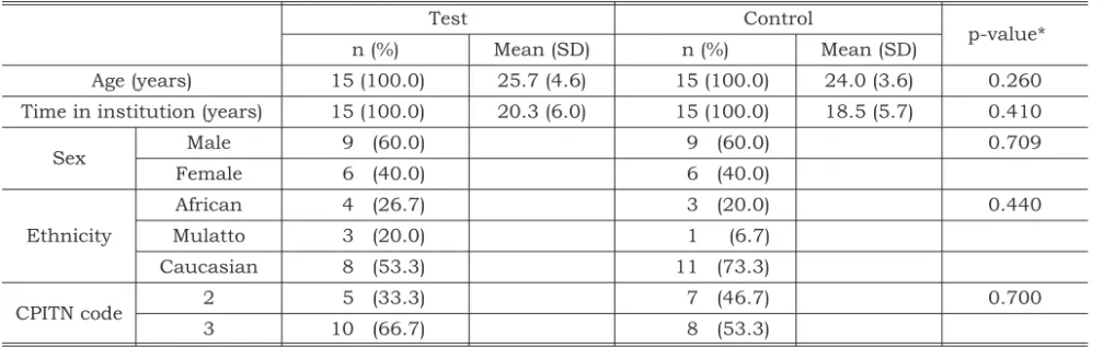
. The patient’s characteristics are presented in Table 1. Most of the inmates had a CPITN code 3 (4 or 5 mm pocket depth) as the most severe periodontal condition, but most of the sites were code 1 (42.1%) or 2 (37.9%). There was no significant association bet-ween group and CPITN code (p = 0.910). Forty-one percent of the subjects presented severe mental deficiency, 37% moderate, 18% mild and 2% pro-found. At baseline, the mean Plaque Control Re-cord was 69.2% in the test group, and 72.7% in the
control group. There was no significant difference between groups in relation to Plaque Control Re-cord (p = 0.500).
There was no difference between groups in rela-tion to the mean total number of microorganisms at baseline and after the use of gel (8 weeks); howe-ver, there was a significant difference (p = 0.03) 16 weeks after baseline collection (Table 2).
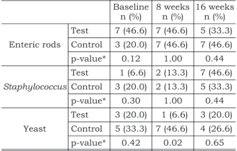
Table 3 shows that there was no difference bet-ween groups in relation to the presence of enteric rods at any time during the investigation. Throug-hout the period of study, the most frequently isola-ted enteric species were Serratia marcescens (13 isolates),Citrobacter freundii(7 isolates) and Kleb-siella pneumoniae(6 isolates). In 13 inmates, ente-ric rods comprised over 1.0% of the total cultivable flora; and in five of these 13 subjects, more than 40% of the total cultivable flora was made up of en-teric rods.
Throughout the investigation there was no dif-ference between groups in relation to the presence of Staphylococcus(Table 3). All isolates were coa-gulase-positive, andStaphylococcusnever compri-sed more than 0.5% of the total number of micro-organisms.
The presence of yeasts was similar in the two groups at the baseline (p = 0.42) and after 16 we-eks (p = 0.65) (Table 3). However, after the use of gel (8 weeks), yeasts were recovered in 7 subjects of the control group and only in one patient in the test group. The difference was significant (p = 0.02).
TABLE 1 -Background and periodontal characteristics of patients.
Test Control
p-value*
n (%) Mean (SD) n (%) Mean (SD)
Age (years) 15 (100.0) 25.7 (4.6) 15 (100.0) 24.0 (3.6) 0.260 Time in institution (years) 15 (100.0) 20.3 (6.0) 15 (100.0) 18.5 (5.7) 0.410
Sex Male 9 (60.0) 9 (60.0) 0.709
Female 6 (40.0) 6 (40.0)
Ethnicity
African 4 (26.7) 3 (20.0) 0.440
Mulatto 3 (20.0) 1 (6.7)
Caucasian 8 (53.3) 11 (73.3)
CPITN code 2 5 (33.3) 7 (46.7) 0.700
3 10 (66.7) 8 (53.3)
DISCUSSION
In this trial, we investigated the effect of a chlor-hexidine gel on dental plaque superinfecting mi-croorganisms, in cerebral palsied patients. Throughout the study there was no statistical dif-ference in relation to the presence of enteric rods and Staphylococcusbetween the two groups. No-netheless, after the gel use, the occurrence of ye-asts was statistically higher in the control group than in the test group. The interpretation of this statistical difference, however, needs to be care-fully done, especially because of the small sample size. The statistical difference arose because of a decrease in yeast occurrence in the test group, and an increase in the control group. Therefore, the dif-ference can be either explained by chance or by a possible inhibitory effect of CHX. This inhibitory effect was reported byin vitrostudies6,7.
The proportion ofStaphylococcusand yeasts in relation to the total cultivable flora was low during the experimental period. In 13 inmates (43.33%), Enterobacteriaceae comprised more than 1% of the cultivable flora. One patient presented high le-vels of enteric rods at baseline (70%), after 8 weeks (74%) and after 16 weeks (76%). According to Loes-che14
, if a microorganism is found to comprise at le-ast 1.0% of the plaque at a given site, it has entered into a stable relationship with the host due to its numerical dominance. A microorganism found at low levels most likely indicates transient presence. The high levels of enteric rods reported in our in-vestigation are not frequent in healthy subjects, but similar results had already been reported in institutionalized elders23, and intensive care
medi-cal patients24
.
There are several factors that may account for the high proportions of enteric rods. Some of the inmates had recently taken antibiotics, which can lead to an overgrowth of these organisms21
. Also, inadequate sanitary conditions may be a source for oral colonization with Enterobacteriaceae spe-cies2,28. In spite of adequate sanitary conditions at
the institution, it is known that mentally handi-capped subjects may have inadequate habits such as drinking inappropriate water, and even taking fecal material to their mouths. Another possibility is that a prolonged transportation time of samples in VMGA III medium may have allowed multiplica-tion of enteric species2. We have tried to minimize
this problem, processing the samples within 18 hours after collection.
The high prevalence of superinfecting orga-nisms found in our study can represent a risk for oral and extra-oral infections. It has been reported that Enterobacteriaceae, Staphylococcus and ye-asts are associated with refractory periodontal di-sease25
. These organisms may also disseminate into the bloodstream and colonize remote sites, le-ading to systemic infections9. Recent
investigati-ons have deminvestigati-onstrated that dental plaque can be a potential reservoir for Enterobacteriaceae and Staphylococcus– which are considered respiratory pathogens. Aspiration of salivary and dental pla-que respiratory pathogens could be a risk factor for pneumonia23,24in these patients.
Based on the findings of these investigations and the Pannuti et al.19
study, we concluded that the 0.5% chlorhexidine gel was efficient and safe. The product was capable of reducing interdental bleeding, and it did not promote any undesirable TABLE 2 -Change in the total number† of
microorga-nisms in the test group (n = 15) and in the control group (n = 15) during the trial.
Trial phases Mean Standarddeviation Range p-value*
Baseline Test 7.2 5.0 0.5-16 0.99 Control 6.5 6.5 0.4-23
8 weeks Test 21.2 22.6 1.0-70 0.99 Control 19.1 29.0 0.2-110
16 weeks Test 6.3 11.7 0.16-36 0.033 Control 142 290 0.26-960
†Total number expressed in 1011.
*p-value for Tukey’s test.
TABLE 3 - Infection with enteric rods, Staphylococcus
and yeast in the test group (n = 15) and in the control group (n = 15).
Baseline n (%)
8 weeks n (%)
16 weeks n (%)
Enteric rods
Test 7 (46.6) 7 (46.6) 5 (33.3)
Control 3 (20.0) 7 (46.6) 7 (46.6)
p-value* 0.12 1.00 0.44
Staphylococcus
Test 1 (6.6) 2 (13.3) 7 (46.6)
Control 3 (20.0) 2 (13.3) 5 (33.3)
p-value* 0.30 1.00 0.44
Yeast
Test 3 (20.0) 1 (6.6) 3 (20.0) Control 5 (33.3) 7 (46.6) 4 (26.6)
p-value* 0.42 0.02 0.65
shift in the colonization patterns of enteric rods, Staphylococcusand yeasts, when compared to the placebo. Based on our results it can be suggested that the application of CHX gel can be safely indi-cated as an adjunct to mechanical plaque removal for cerebral palsied subjects. However, we stress that it cannot completely replace toothbrushing and interdental cleaning; efforts should thus be made to ensure the instruction and motivation of patients and caretakers in relation to the mechani-cal removal of plaque.
CONCLUSIONS
The 0.5% chlorhexidine gel did not promote a shift in the colonization patterns of Gram-negative enteric rods, Staphylococcus and yeasts, when compared to the placebo. In 43.3% of the inmates Enterobacteriaceae comprised more than 1% of the cultivable flora. The most frequent enteric spe-cies isolated were Serratia marcescens (13 isola-tes),Citrobacter freundii(7 isolates) andKlebsiella pneumoniae(6 isolates).
REFERENCES
1. Ainamo J, Barmes DE, Beagrie G, Cutress T, Martins J. Development of the World Health Organization (WHO) com-munity periodontal index of treatment needs. Int J Dent Res 1982;32:281-91.
2. Ali RW, Velcescu C, Jivanescu MC, Lofthus B, Skaug N. Prevalence of 6 putative periodontal pathogens in sub-gingival plaque samples from Romanian adult perio-dontitis patients. J Clin Periodontol 1996;23:133-9. 3. American Dental Association. Guidelines for acceptance of
chemotherapeutic products for the control of supragingival dental plaque and gingivitis. J Am Dent Assoc 1986; 112:529-32.
4. Brown JP. The efficacy and economy of comprehensive dental care for handicapped children. Int Dent J 1980; 30:14-27.
5. Brown AT, Sims RE, Raybould TP, Lillich TT, Henslee TJ, Ferretti GA. Oral Gram-negative bacilli in bone marrow transplant patients given chlorhexidine rinses. J Dent Res 1989;68:1199-204.
6. Candido RC, Azevedo RV, Ito IY. Determinação da concentração inibitória mínima de Cepacol, Malvona e Periogard, ante a Candida albicansisoladas da cavidade bucal. Rev Odontol UNESP 1996;25:79-84.
7. Denton GW. Chlorhexidine. In: Block SS. Disinfection, sterilization and preservation. 3rded. Philadelphia: Lea & Febinger; 1991. p. 438-52.
8. Ferretti GA, Raybould TP, Brown AT, MacDonald JS, Greenwood M, Maruyama Y, et al. Chlorhexidine prophy-laxis for chemotherapy- and radiotherapy-induced stoma-titis: a randomized, double-blind trial. Oral Surg Oral Med Oral Pathol 1990;69:331-8.
9. Fourrier F, Duvivier B, Boutigny H, Roussel-Delvallez M, Chopin C. Colonization of dental plaque: a source of nosocomial infections in intensive care unit patients. Crit Care Med 1998;26:301-8.
10. Francis JR, Hunter B, Addy M. A comparison of three deliv-ery methods of chlorhexidine in handicapped children. J Periodontol 1987;58:451-5.
11. Francis JR, Stevenson DR, Palmer JD. Dental health and dental care requirements for young handicapped adults in Wessex. Community Dent Health 1991;8:131-7.
12. Gabre P, Gahnberg L. Dental health status of mentally re-tarded patients with various living arrangements. Spec Care Dent 1994;14:203-7.
13. Kampf G, Jarosch R, Ruden H. Limited effectiveness of chlorhexidine based hand disinfectant against methicil-lin-resistantStaphylococcus aureus(MRSA). J Hosp Infect 1998;38:297-303.
14. Loesche WJ. Ecology of oral flora In: Nisengard RJ, New-man MG. Oral Microbiology and Immunology. 2nded. To-ronto: W.B. Saunders; 1988. p. 308.
15. Mengistu Y, Erge W, Bellete B. In vitro susceptibility of Gram-negative bacterial isolates to chlorhexidine. East Afr Med J 1999;76:243-6.
16. Möller AJ. Microbiological examination of root canals and periapial tissues of human teeth. Methodological studies. Odontol Tidskr 1966;Dec 20;74:Suppl:1-380.
17. Nicoletti G, Boghossian V, Gurevitch F, Borland R, Mor-genroth P. The antimicrobial activityin vitroof chlorhexidi-ne, a mixture of isothiazolinones (“Kathon” CG) and cetyl trimethyl ammoniun bromide (CTAB). J Hosp Infect 1993;23:87-111.
18. O’Leary TJ, Drake RB, Naylor JE. The plaque control re-cord. J Periodontol 1972;43:38.
19. Pannuti CM, Lotufo RFM, Saraiva MC, Cai S, Ferraro A, Falsi D. Effect of 0.5% chlorhexidine gel on special pati-ents. J Clin Periodontol (in press).
20. Pannuti CM, Lotufo RFM, Cai S, Freitas N, Ferraro AQ. Pre-valência de microrganismos superinfectantes na placa bacteriana de deficientes mentais institucionalizados. RPG Rev Pos Grad 2001;8:35-9.
21. Rams TE, Babalola OO, Slots J. Subgingival occurrence of enteric rods, yeasts and Staphylococci after systemic doxy-cycline therapy. Oral Microbiol Immunol 1990;5:166-8. 22. Ranney RR. Criteria for efficacy of plaque control agents for
periodontal disease: microbiology. J Dent Res 1989; 68:1655-60.
23. Russel SL, Boylan RJ, Kaslick RS, Scannapieco FA, Katz RV. Respiratory pathogen colonization of the dental plaque of institutionalized elders. Spec Care Dent 1999; 19:128-34.
24. Scannapieco FA, Stewart EM, Mylotte JM. Colonization of dental plaque by respiratory pathogens in medical intensi-ve care patients. Crit Care Med 1992;20:740-5.
26. Slots J, Feik D, Rams TE.In vitroantimicrobial sensitivity of enteric rods and pseudomonads from advanced adult periodontitis. Oral Microbiol Immunol 1990;5:298-301. 27. Slots J, Rams TE, Schonfeld SE. In vitro activity of
chlorhexidine against enteric rods, pseudomonads and acinetobacter from human periodontitis. Oral Microbiol Immunol 1991;6:62-4.
28. Slots J, Rams TE, Feik D, Taveras HD, Gillespie GM. Subgingival microflora of advanced periodontitis in the Do-minican Republic. J Periodontol 1991;62:543-7.
29. Spijkervet FKL, van Saene HKF, Panders AK, Vermey A, van Saene JJ, Mehta DM, et al. Effect of chlorhexidine rins-ing on the oropharyngeal ecology in patients with head and neck cancer who have irradiation mucositis. Oral Surg Oral Med Oral Pathol 1989;67:154-61.
30. Thurmond JM, Brown AT, Sims RE, Ferreti GA, Raybould TP, Lillich TT, Henslee PJ. OralCandida albicansin bone marrow transplant patients given chlorhexidine rinses: oc-currence and susceptibilities to the agent. Oral Med 1991;72:291-5.