vol. 39, n. 2, abr./jun., 2003
Botanical (morphological, micrographic), chemical and pharmacological
characteristics of Pfaffia species (Amaranthaceae) native to South Brazil
Grace Gosmann
1, Susana Gattuso
2, Martha Gattuso
2, Raquel Fenner
1, Elyara Fiorin Pacheco
1,
Alexandre Ferraz
1,3, Luciane Anita Savi
4, Célia Regina Monte Barardi
5,
Cláudia Maria Oliveira Simões
4, Maximiliano Sortino
2, Susana Zacchino
2,
Carmela Gnerre
6, Bernard Testa
6, Stela Maris Kuze Rates
1*
1Faculdade de Farmácia, Universidade Federal do Rio Grande do Sul ,UFRGS, 2Facultad de Ciencias Bioquímicas y Farmacéuticas, Universidad Nacional de Rosario, Rosario, Argentina, 3Centro Integrado do Câncer, Universidade Luterana do Brasil, Canoas, RS, 4Laboratório de Virologia Aplicada, Departmento de Ciências Farmacêuticas (CCS),
Universidade Federal de Santa Catarina, 5Departamento de Microbiologia e Parasitologia (CCB), Universidade Federal de Santa Catarina, 6Institute de Chimie Thérapeutique Section de Pharmacie, Université de Lausanne,
Lausanne-Dorigny, Switzerland
Some parameters for the quality control of P. glomerata and P.
paniculata roots using their botanical and chemical characteristics are presented. It was also carried out an in vitro pharmacological screening to evaluate some biological properties of P. glomerata that could be related to its popular use as “tonic”. Relating to biological assays, ethanolic extract from P. glomerata roots did not present antiviral, antiproliferative, antifungal or MAO inhibitory activities. The cytotoxicity evaluation of P. glomerata determined that IC50
is >2,000 μg/mL. The main morphological and micrographic
characteristics of P. glomerata and P. paniculata roots are described in this paper in order to aid in their unequivocal identification.
UNITERMS:
• Pfaffia glomerata
• Pfaffia paniculata
• Brazilian ginseng • Micrography • Quality control
*Correspondence: S. M. K. Rates Faculdade de Farmácia
Universidade Federal do Rio Grande do Sul
Av. Ipiranga 2752, 90610-000 Porto Alegre, RS, Brazil E-mail: [email protected]
INTRODUCTION
Around ninety species of Pfaffia (Amaranthaceae) are known in Central and South America, twenty-seven species have been described in Brazil (Vasconcelos, 1982; Taniguchi et al., 1997). They are popularly known as “corango”, “suma”, “paratudo” and “Brazilian ginseng” since Pfaffia species are commonly used for the same
therapeutic indications as ginseng (Panax spp.
-Araliaceae) (Oliveira, 1986). Although P. paniculata is the most employed in commercial preparations in Brazil, P. glomerata is commonly used in the State of Rio Grande do Sul (South of Brazil) labeled either as P. glomerata or
wrongly as P. paniculata. A study of the differences
between these two latter species is clearly needed in order to avoid adulterations that could result in undesirable or toxic effects.
Regarding chemical constituents from roots of
Pfaffia species, allantoin, ecdysteroids, pfaffic acid and their glycosides (nortriterpene saponins), stigmasterol and sitosterol (Nakai et al., 1984; Nishimoto et al., 1984, 1988; Shiobara et al., 1993 a,b; Takemoto et al., 1983) have been identified.
while crude extracts from P. paniculata displayed anti-inflammatory, sexual stimulant activities and low toxicity in rodents (Mazzanti, Braghirolli, 1994; Arletti et al., 1999). In turn, triterpene saponins from P. paniculata showed to possess antitumoural activity (Nishimoto et al., 1984).
In particular, one of the aims of this work is to provide botanical parameters for the quality control of P. glomerata and P. paniculata. In part due to difficult botanical differentiation among Pfaffia species, it is qui-te common the misidentification of the raw maqui-terial in commercial preparations (Santos et al., 1987; De-Paris et al., 1998a). The major morphological features and the micrographic characters of P. glomerata and P. paniculata
roots are presented here in order to give the necessary background for their unequivocal identification.
In addition, we discuss the thin-layer chromato-graphy (TLC) profile of P. glomerata and P. paniculata
species, and the antifungal, antiproliferative, antiviral, MAO inhibitory activities and cytotoxic properties of P. glomerata roots. These data could be relevant for future assesment of efficacy and safe use of P. glomerata in the traditional medicine of South Brazil.
MATERIALS AND METHODS
Plant Material
Pfaffia glomerata (Spreng.) Pedersen and Pfaffia paniculata (Mart.) O. Kuntze fresh roots were obtained from the cultivated area of the Centro Pluridisciplinar de Pesquisas Químicas, Biológicas e Agrícolas (CPQBA), UNICAMP, Campinas, São Paulo, Brazil. For botanical characterization, the roots were collected on October 1999 by one of the authors (S.J.G.) and voucher specimens (P. glomerata Gattuso 754, P.paniculata Gattuso 755) are kept in both, U.N.R. Herbarium and in the Herbarium of the Vegetal Biological Area, of National University of Rosario, Argentina. For chemical and biological analysis, the roots were collected and identified at CPQBA on
January 2000 by Ílio Montanari (P. glomerata, CPQBA
0238, P. paniculata, CPQBA 0241).
Extraction Procedures
Roots and rhizomes (subterraneous parts) from P. glomerata and P. paniculata were reduced to small pieces, dried in circulating air stove (45 oC) and triturated. The powder was extracted with 60% ethanol under reflux (1:10, plant:solvent, w/v) during 6 hours. The ethanol was removed under “vacuum” and the resulting residue was lyophilized and stored under light protection. The yield of
both lyophilized extracts was 28% in relation to the dried subterraneous parts.
Micrographic Studies
Fresh and fixed (formaldehyde:ethanol:acetic acid:water, 2:10:1:3.5) roots from both species were used. Longitudinal and transverse sections of the roots measuring 10 µm were prepared with microtome and stained with Safranin and Fast Green (Dizeo de Strittmater, 1979). Sections were mounted in synthetic balsam. Roots was macerated by conventional methods (Boodle, 1916). The distributions of calcium oxalate crystals was visualized by a polarizing microscope. Original drawings were made with a drawing device. Symbols proposed by Metcalfe and Chalk (1957) were used in the diagrams. The black and white photomicrographs were performed with the Axiolab Zeiss Automatic System.
TLC Analyses
TLC was carried out on silica gel (Merck, GF254) using either chloroform:ethanol (95:5, v/v) or butanol:ethyl acetate:water:acetic acid (4:1:5:1, v/v). Compounds were visualized by heating (120 oC) using the anisaldehyde-sulfuric acid-sprayed plates. Ecdysterone, oleanolic acid, sitosterol and stigmasterol were used as authentic samples.
Antiviral Activity of P. glomerata
Ethanolic extract was evaluated for cytotoxicity, antiviral and virucidal action according to Simões et al.
(1999), using VERO cells (ATCC:CCL81) and Herpes
simplex virus type 1 (HSV-1)/strains VR733 (ATCC), KOS and 29R/acyclovir resistant (Laboratoire de Pharmacognosie, Faculté de Pharmacie, Université de Rennes I, Rennes, France). To assess the effects of the extract, dilutions ranging from 2,000 to 1.9 mg/mL were prepared in 199 media.
Antiproliferative Activity of P. glomerata
carci-noma, from the American Type Culture Collection (Rockville; MD, USA).
Antifungal Assays of P. glomerata
The fungistatic activity was evaluated by the agar dilution method using Sabouraud-chloramphenicol agar for yeast, filamentous fungi and dermatophytes, according to reported procedures (Mitscher et al., 1972; Zacchino et al., 1998, 1999; Feresin et al., 2001). The followingstrains
were used: Candida albicans ATCC 10231, Candida
tropicalis C 131, Saccharomyces cerevisiae ATCC 9763,
Cryptococcus neoformans ATCC 32264, Aspergillus flavus ATCC 9170, Aspergillus fumigatus ATTC 26934 and Aspergillus niger ATCC 9029. Dermatophytes:
Microsporum canis C 112, Trichophyton rubrum C 110, T. mentagrophytes ATTC 9972,Epidermophyton floccosum
C 114 and Microsporum gypseum C 115 kindly provided
by CEREMIC, Centro de Referencia Micológica, Facul-tad de Ciencias Bioquímicas y Farmacéuticas, Suipacha 531, Rosario, Argentina. The extract was solubilized in DMSO and added to the culture medium to give a final concentration that does not exceed 2% (v/v) to give serial decreasing dilutions ranging from 1000 to 250 µg/mL.
MAO Inhibitory Activity of P. glomerata
The ethanolic extract was assayed for monoamine oxidase A (MAO A) and MAO B inhibitory activity in rat brain mitochondrial preparations at concentrations ranging from 1 to 20 mg/mL, following the methodology
described by Gnerre et al. (2001). The extract was
dissolved in DMSO and added to the cell culture medium to give a final concentration of co-solvent of 5% (v/v).
RESULTS AND DISCUSSION
Morphological and micrographic analyses of roots from Pfaffia glomerata (Spreng) Pedersen and Pfaffia paniculata (Mart.) O. Kuntze
Morphological and micrographic descriptions of the main characteristics of these plants are necessary to provide unique insights into unequivocal recognition of P. glomerata and P. paniculata.
Common characteristics of the roots
Both species consist of underground swollen roots, the outer surface is yellow-brown to brown and transver-sely rugose. The transverse root sections show a narrow
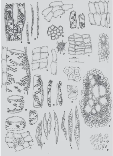
bark composed of almost tabulated phellem cells. The anatomical peculiarity is the anomalous growth in thickness, which takes place by the development of a succession of collateral vascular bundles from rings of secondary meristematic tissue. The secondary bundles are embedded in conjunctive parenchyma. Subsequently, the accessory cambium arises from parenchyma cells on the periphery of the phloem of each vascular bundle. The secondary phloem was composed of sieve tube elements, companion cells, and axial parenchyma. The secondary xylem was composed of vessel, tracheids, xylem fibres and axial parenchyma (Figure 1).
Non-common characteristics of the roots
Comparisons of non-common characteristics of the roots are summarized in Table I. Detailed descriptions are presented below.
Pfaffia glomerata
The root is 2-2,5 cm in diameter. The transverse root sections show a uniform rhytidome, made up of a single
FIGURE 1 - A-D Common characteristcs of Pfaffia
peridermis with 6 to 8 layers of almost tabulet phellem cells (Figure 2, H). The layers were formed by rows of thin-walled cells. The phelloderm was unstratified. Roots show 4-5 rings of collateral vascular bundles embedded in conjunctive parenchyma. Large idioblasts with numerous
minute crystals of calcium oxalateare present in the
conjunctive tissue (Figure 2, F).
In this species the strands of xylem and phloem are quite small, the phloem consisting of 3-4 sieve tubes only. The xylem strands contain 1-2 rows of 2-3 radially placed vessels. The fibres with small simple pits are few and shows a thick-walled (Figure 2, I).
• Macerated roots. The analysis of the material yielded the following constitutive elements:
a) Phellem cells. In cross-sections they were rectangular with thin-walled cells and measuring 50-80 µm in length and 40 µm in wide. In the super-ficial view they are polygonal, attaining a diameter of 50 µm (Figure 2, H, G).
b) Conjunctive parenchyma. It showed polygonal cells. The average dimensions of parenchyma cells are 170 µm in length and 50 µm in wide (Figure 2, E).
c) Vessel. The vessels possessed alternate bordered pits with a simple perforation plate in transverse to slightly oblique end walls and measuring 100 µm in length and 50 µm in diameter (Figure 2, A, D). d) Xileme fibers. They are scanty and measuring
250-300 µm in length and 10-12 µm in diameter (Figu-re 2, A, C).
e) Fibre-tracheid. Measuring 150 µm in length and 10-12 µm in diameter (Figure 2, B).
Pfaffia paniculata
The root is 3-4 cm in diameter. The transverse root sections show a uniform rhytidome, made up of a single peridermis with 12 to 14 layers of almost tabulet phellem cells. The layers were formed by rows of thick-walled cells (Figure 2, Q, R). The phelloderm was unstratified. Roots show 12-14 rings of collateral vascular bundles embedded in conjunctive tissue. Large idioblasts with calcium oxalatecrystals of different shapes are present in the conjunctive tissue; i.e., octahedral prisms, rectangular prisms and octahedrons (Figure 2, O, P).
In this species the strands of xylem and phloem are large, the phloem consisting of 6-8 sieve tubes. The xylem strands contain 1-4 rows of 4-6 radially placed vessels. Fibbers with small simple pits are abundant (Figure 2, S). • Macerated roots. The analysis of the material yielded
the following constitutive elements:
a) Phellem cells. In cross-sections they were rectangular with thick-walled cells and measuring 75 µm in length and 45 µm in wide. In the superfi-cial view they are polygonal, attaining a diameter of 45-50 µm (Figure 2, Q, R).
b) Conjunctive parenchyma. It showed elongated lon-gitudinal cells. The average dimensions of parenchyma cells are 100 µm in length and 50 µm in wide (Figure 2, N).
FIGURE 2 - A-S: Non-common characteristics. A-I
c) Vessel. The vessels possessed alternate bordered pits with a simple perforation plate in transverse to slightly oblique end walls. The mean vessel member
length and diameter are 40 µm and 120 µm
respectively and others vessels measuring 165 µm in length and 65 µm in diameter (Figure 2, J, M). d) Xileme fibres. They are very abundant,
thick-walled, measuring 350 µm in length and 25 µm in diameter (Figure 2, J, L).
e) Fibre-tracheid. Thin-walled, they measured 200 µm in length and 35 µm in diameter (Figure 2, K).
The examined species differ in morphologic and anatomic features, as shown in Table I.
Chemical and biological data
Ethanolic extracts from P. glomerata and P.
paniculata present different TLC profiles at the chroma-tographic conditions used. It was possible to identify the presence of ecdysterone in P. glomerata extract as already described in the literature (Shiobara et al., 1993a), but not
in P. paniculata extract. It seems that this compound could be a good marker for the differentiation of both species. The other substances used in TLC were not good references as both species presented spots with similar Rf and color.
Regarding to biological assays, ethanolic extract from P. glomerata did not present neither antiproliferative, antiviral, antifungal nor MAO inhibitory activities. Nevertheless, it is interesting to note that its cytotoxicity is very low (IC50 > 2,000 µg/mL).
From results reported here, we could observe that roots from P. glomerata and P. paniculata differ in their botanical parameters and TLC profile, being both characteristics useful for avoiding adulterations.
Regarding results obtained in biological assays, P. glomerata showed to be inactive in all the activities tested, such as antifungal, antiviral, antitumoural and MAO inhibition. Interesting enough, our studies demonstrated that this species is not cytotoxic.
Finally, careful evaluation of the available scientific data for Pfaffia species allow us to consider that more studies are necessary to provide chemical,
pharmacolo-TABLE I - Non-common features of Pfaffia glomerata and Pfaffia paniculata
Pfaffia paniculata
Exomorphology
Theroot is 3-4 cm in diameter
Anatomy
· A single peridermis with 12 to 14 layers, thick-walled cells.
· 12-14 rings of large collateral vascular bundles
embedded in conjunctive tissue.
· The xylem strands contain 1-4 rows of 4-6 radially
placed vessels.
Macerate
· Phellem cells: Measuring 75 µm in length and 45 µm
in wide.
· Conjunctive parenchyma: Cell size 100 µm in length
and 50 µm in wide.
Large idioblasts with calcium oxalate crystals, octahedric, rectangular prisms and octahedrons.
· Vessel: 40 µm in length and 120 µm in diameter;
165 µm in length and 65 µm in diameter
· Fibres: thick-walled, 350 µm in length and 25 µm in
diameter.
· Fibre-traqueid: Thin-walled, 200 µm in length and
35 µm in diameter.
Pfaffia glomerata
Exomorphology
Theroot is 2-2.5 cm in diameter
Anatomy
· A single peridermis with 6 to 8 layers, thin-walled cells. · 4-5 rings of small collateral vascular bundles embedded
in conjunctive tissue.
· The xylem strands contain 1-2 rows of 2-4 radially
placed vessels.
Macerate
· Phellem cells: Measuring 50-80 µm in length and
40 µm in wide.
· Conjunctive parenchyma: Cell size 100 µm in length
and 50 µm in wide.
Large idioblasts with numerous minute crystals of calcium oxalate.
· Vessel: 100 µm in length and 100 µm in diameter.
· Fibres: 250-300 µm in length and 10-12 µm in
diameter.
gical and technological basis for their therapeutic use. This is really important taking into account that several
phytopharmaceutical preparations containing P.
glomerata or P. paniculata are available in Brazilian market without definitive proofs of their efficacy and safety. Even if eventual claim for traditional use of these preparations is considered, there are not consistent ethnobotanic reports on P. glomerata and P. paniculata. Furthermore, it is necessary to point out that the quality criteria for both - raw materials and phytopharmaceutical preparations - are still not well established. This study intends to have contributed specially toward the latest question.
RESUMO
Características botânicas (morfológicas, micrográficas), químicas e farmacológicas de espécies de Pfaffia (Amaranthaceae) nativas do Sul
do Brasil
Alguns parâmetros são apresentados para o controle de qualidade de raízes de Pfaffia glomerata e Pfaffia paniculata, utilizando suas características botânicas e químicas. Realizou-se, também, um screening farmaco-lógico de P. glomerata direcionado às atividades relacio-nadas com seu uso como “tônico”. O extrato etanólico das raízes de P. glomerata não apresentou atividade antiviral, antiproliferativa, antifúngica ou atividade ini-bitória de monoaminoxidases. A avaliação da citoto-xicidade determinou que a CI50 é > 2.000 μg/mL. As prin-cipais características morfológicas e micrográficas das raízes de P. glomerata e P. paniculata descritas permitem auxiliar na sua identificação segura.
UNITERMOS:Pfaffia glomerata. Pfaffia paniculata. Ginseng brasileiro. Micrografia. Controle de qualidade.
ACKNOWLEDGMENTS
We are grateful to Ílio Montanari from CPQBA (Universidade de Campinas, UNICAMP, São Paulo) for furnishing cultivated material. Micrographic study was supported by U.N.R. (Universidad Nacional de Rosario). G.G. and S.M.K.R. thank CNPq (Brazil) for fellowships and grant. Part of this work was supported by grants from SAZ (Agencia de Promociones Científicas y Tecnológicas de la Argentina PICT99 # 06-06454) and OEA, Proyecto Aprovechamiento de la Flora Regional; and it is part of the collaborative research within the “Bioactive Natural Products and their Applications” nucleus from the
Association of Universities of the Montevideo Group (AUGM). Collaboration from the Iberoamerican Program of Science and Technology for the Development (CYTED) (Project X.7) is gratefully acknowledged.
REFERENCES
ARLETTI, R.; BENELLI, A.; CAVAZZUTI, E.; SCARPETTA, G.; BERTOLINI, A. Stimulating property of Turnera diffusa and Pfaffia paniculata
extracts on the sexual behavior of male rats.
Psychopharmacology, v. 143, p. 15-19, 1999.
BOODLE, I. A. A method of macerating fibres. Royal
Botanical Garden, Kew Bulletin Miscellanous Informative, v. 4, p. 108-110, 1916.
DE-PARIS, F.; SALGUEIRO, J.; MOREIRA ROSA, R.; GOSMANN, G.; IZQUIERDO, I.; RATES, S. M. K. Análise cromatográfica e atividade depressora central de
produtos vegetais comercializados como Pfaffia
paniculatta Mart. Kuntze (Amaranthaceae) no RS (Brasil). JORNADA DE JÓVENES INVESTIGADORES GRUPO DE MONTEVIDEO, 6., Santa Fe, 1998.
Resúmenes. Santa Fé: Universidade Nacional del Litoral, 1998a. p. 119.
DE-PARIS, F.; QUEVEDO, J., SALGUEIRO, J.; RATES, S. M. K.; IZQUIERDO, I. Potencial atividade depressora do extrato de Pfaffia glomerata sobre o sistema nervoso central. Rev. Med. ATM 1998, v. 18, p. 12-17, 1998b.
DE-PARIS, F., NEVES, G.; SALGUEIRO, J. B.; QUEVEDO, J.; IZQUIERDO, I.; RATES, S. M. K. Psychopharmalogical screening of Pfaffia glomerata
Spreng. (Amaranthaceae) in rodents. J.
Ethnopharmacol., v. 73, p. 261–269, 2000.
DIZEO DE STRITTMATER, C. Modificación de una coloración Safranina Fast green. Bol. Soc. Arg. Botánica, v. 18, p. 121-122, 1979.
FERESIN, G. E.; TAPIA, A.; LÓPEZ, S. N.; ZACCHINO, S. A. Antimicrobial activity of plants used in traditional
medicine of San Juan province, Argentina. J.
Ethopharmacol., v. 78, p. 103-107, 2001.
LIKHITWITAYAWUID, K.; ANGERHOFER, C. K.; CORDELL, G. A.; PEZZUTO, J. M.; RUANGRUNGSI, N. Cytotoxic and antimalarial bisbenzylisoquinoline alkaloids from Stephania erecta.
J.Nat. Prod., v. 56, p. 30-38, 1993.
MAZZANTI, G.; BRAGHIROLLI, L. Analgesic and anti-inflammatory action of Pfaffia paniculata (Martius) Kuntze. Phytother. Res., v. 8, p. 413-416, 1994.
METCALFE, C.; CHALK, L. Anatomy of the Dicotyledons. Oxford: Clarendon, 1957. v. 1, p. 1067-1074.
MITSCHER, L.; LEU, R.; BATHALA, M.; WU, W.; BEAL, J. Antimicrobial agents from higher plants. I. Introduction, rationale and methodology. Lloydia, v. 35, p. 157-166, 1972.
NAKAI, S.; TAKAGI, N.; MIICHI, H.; HAYASHI, S.; NISHIMOTO, N.; TAKEMOTO, T.; KIZU, H. Pfaffosides and nortriterpenoid saponins from Pfaffia paniculata. Phytochemistry, v. 23, p. 1703-1705, 1984.
NISHIMOTO, N.; NAKAI, S.; TAKAGI, N.; HAYASHI, S.; TAKEMOTO, T.; ODASHIMA, S.; KIZU, H.; WADA, Y. Pfaffosides and nortriterpenoid saponins from Pfaffia paniculata. Phytochemistry, v. 23, p. 139-142, 1984.
NISHIMOTO, N.; SHIOBARA, Y.; INOUE, S-S.; FUJINO, M.; TAKEMOTO, T.; YEOH, C. L.; OLIVEIRA, F.; AKISUE, G.; AKISUE, M. K.; HASHIMOTO, G. Three
ecdysteroid glycosides from Pfaffia iresinoides.
Phytochemistry, v. 27, p. 1665-1668, 1988.
OLIVEIRA, F. Pfaffia paniculata (Martius) Kuntze–
Brazilian Ginseng. Rev. Bras.Farmacognosia, v. 1, p. 86-92, 1986.
SANTOS, R. I.; SANTOS, M. A.; SCHENKEL, E. P. Análise cromatográfica de amostras comercializadas como Panax ginseng e Pfaffia paniculata. Cad. Farm.,
Porto Alegre, v. 3, p. 59-65, 1987.
SHIOBARA, Y.; INOUE, S-S.; KATO, K.; NISHIGUCHI, Y.; OISHI, Y.; NISHIMOTO, N.; OLIVEIRA, F.; AKISUE, G.; AKISUE, M. K.; HASHIMOTO, G. A nortriterpenoid, triterpenoids and ecdysteroids from
Pfaffia glomerata.Phytochemistry, v. 32, p. 1527-1530, 1993a.
SHIOBARA, Y.; INOUE, S-S.; KATO, K.; NISHIGUCHI, Y.; NISHIMOTO, N.; OLIVEIRA, F.; AKISUE, G.; AKISUE, M. K.; HASHIMOTO, G. Pfaffane-type
nortriterpenoids from Pfaffia pulverulenta.
Phytochemistry, v. 33, p. 897-899, 1993b.
SIMÕES, C. M. O.; FALKENBERGER, M.; AULER MENTZ, L.; SCHENKEL, E. P.; AMOROS, M.; GIRRE, L. Antiviral activity of South Brazilian medicinal plant extracts. Phytomedicine, v.6, p. 205-214, 1999.
TAKEMOTO, T.; NISHIMOTO, N.; NAKAI, S.; TAKAGI, N.; HAYASHI, S.; ODASHIMA, S.; WADA, Y. Pfaffic acid, a novel nortriterpene from Pfaffia paniculata
Kuntze. Tetrahedron Lett., v. 24, p. 1057-60, 1983.
TANIGUCHI, S. F.; BERSANI-AMADO, C. A.; SUDO, L.
S.; ASSEF, S. M. C.; OGA, S. Effect of Pfaffia
iresinoides on the experimental inflammatory process in rats. Phytother. Res., v. 11, p. 568–571, 1997.
VASCONCELOS, J. M. O. Estudo taxonômico sobre
Amaranthaceae no RS, Brasil. Porto Alegre, 1982. 151 p. [Dissertação de Mestrado. Curso de Botânica, Universidade Federal do Rio Grande do Sul].
ZACCHINO, S.; SANTECCHIA, C.; LÓPEZ, S.; GATTUSO, S.; MUÑOZ, J.; CRUAÑES, A.; VIVOT, E.; CRUAÑES, J.; SALINAS, A.; RUIZ, R.; RUIZ, S. In vitro antifungal evaluation and studies on mode of action of eight selected species from the Argentina flora. Phytomedicine, v. 5, p. 389-395, 1998.
ZACCHINO, S.; LÓPEZ, S.; PEZZENATI, G.; FURLAN, R.; SANTECCHIA, C.; MUÑOZ, L.; GIANNINI, F.;
RODRÍGUEZ, A.; ENRIZ, R. In vitro evaluation of
antifungal properties of phenylpropanoids and related compounds acting against dermatophytes. J. Nat. Prod., v. 62, p. 1353-1357, 1999.