Clinical Diagnosis versus Autopsy1
444
Records from 910 autopsies performed at a university hospital in Salvador, Bahia, Brazil, were examined in order to assess the accuracy of clinical diagnoses of the patients’ underlying causes of death. This study found inaccurate clinical diagnoses in 31% of the cases.
7he overall rate of diagnostic error appeared to remain fairly stable from 1970 to 1982, being highest for older patients. Thirty-six percent of the 263 cancer deaths were incorrectly diagnosed, and a number of pathologies considered relatively easy to diagnose were not always correctly identified. Quite aside from their direct medical implications, diagnostic errors of the magnitude observed in this and other studies seriously jeopardize the quality of vital statistics and such statistics’ useful- ness for improving public health.
T
he accuracy of clinical diagnosis is traditionally assessed by comparing it with autopsy results. On the basis of postmortem diagnoses, doctors can in- crease their clinical knowledge and diag- nostic ability so as to improve the quality of their work with other patients. The au- topsy is thus an important learning tool, especially in university hospitals (Z-3).Pathologists indicate that, on the aver- age, significant diagnostic errors are re- vealed by about 40% of all autopsies (l--4), the range varying from 6% to 68% depending on a variety of circumstances (5). Curiously, this high proportion of er- rors has remained stable in recent dec- ades despite major technologic advances in diagnostic methods (6).
The aim of the work reported here was to determine the accuracy of clinical diag- noses of underlying causes of death through autopsies carried out at a univer- sity hospital in Salvador, Brazil. (It is im-
This article will also be published in Spanish in the
Bolefin de la Oficina Sanitaria Panamericana, vol. 110.
*Department of Preventive Medicine, Federal Uni- versity of Bahia. Mailing address: Rua Padre Feij6, 29 Canela, 40140 Salvador, Bahia, Brazil.
portant that the underlying cause of death not be confused with the terminal or immediate “causa mortis”-7). Vari- ables considered included the decedent’s age, sex, and year of death, as well as the kind of pathology involved.
MATERLALS AND METHODS
Most hospital instruction at the Federal University of Bahia’s Medical School takes place at the Professor Edgard Santos University Hospital. The patients admitted to this referral hospital are gen- erally suffering from chronic diseases; emergency, obstetric, and tuberculosis patients are not normally admitted.
Autopsies are carried out by resident physicians under the supervision of pro- fessors and physicians in the hospital’s Department of Pathological Anatomy. Ba- sic data on each patient (age, sex, clinical diagnosis, date of death, and underlying cause of death at-autopsy) were routinely placed in the files of the Department of Pathological Anatomy. Since 1970, this ma- terial has been organized in a standardized manner by resident physicians.
The study reported here was based on 1,222 autopsies carried out in all the even-numbered years from 1970 through 1982. During these seven years 1,852 deaths occurred (proportion autopsied: 66%). From 1982 onwards, with the re- emergence of a crisis in Brazil’s univer- sity hospitals, the number of admissions and autopsies fell sharply.
Of the 1,222 cases, 312 were excluded from the study. In some instances (about two-thirds) this was because the available information was incomplete, while in the remainder it was because the diagnosis could have given rise to mistaken inter- pretations (the anatomopathologic diag- nosis being twofold, imprecise, unusual, or hard to reconcile with clinical and anatomopathologic terminology). The bulk of the cases excluded came from the hospital’s surgical clinics.
The clinical diagnosis made by the clin- ical teams in the hospital was compared with the anatomopathologic diagnosis of the underlying cause of death at autopsy. On this basis, the clinical diagnosis was placed in one of three categories: com- pletely correct-total agreement between the clinical and anatomopathologic diag- noses; purfiaIZy correct-partial agreement between the two diagnoses; incorrecf- total disagreement between the diag- noses. In general, the partially correct di- agnoses reflected situations where the underlying disease identified by the pa- thologist was mentioned in the clinical diagnosis among other suspected dis- eases, without having been indicated as the principal cause of death. In contrast, where the diagnosis was classified as in- correct the underlying cause of death found by the pathologist was not even mentioned in the clinical diagnosis.
Care was taken to ensure that differ- ences between clinical and anatomopatho- logic terminology did not lead to discrepan- cies between the two kinds of diagnosis. For example, heart failure, myocardial in-
sufficiency, and atheromatosis of the coro- nary arteries were regarded as the same diagnosis, irrespective of distinctions cited in the International Classification of Dis- eases. Clinical diagnoses that could not be associated with morphologic diagnostic findings at autopsy (such as hydroelec- trolytic disturbances, cardiac arrhythmias, etc.) were fully accepted, thereby justifying the clinical diagnosis.
RESULTS AND DISCUSSION
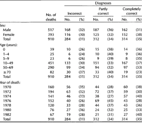
Of the 910 study cases, the clinical di- agnosis of the underlying cause of death was found to be completely correct in 314 cases (35%), partially correct in 312 (34%), and incorrect in 284 (31%). Though the latter 31% figure may seem high for incorrect diagnoses, in fact it is comparable to levels observed at hospi- tals in developed countries (2, 5, 6, 8).
As Table 1 indicates, the accuracy of the clinical diagnoses was approximately the same for both sexes. However, like previous studies, the data show a ten- dency for the rate of incorrect clinical diagnoses to increase with patient age
(1, 5, 8).
It is also noteworthy that the percent- age of incorrect clinical diagnoses did not show any pronounced overall tendency to increase or diminish over the course of the study period. This relative stability could conceivably be associated with a steady decline in the autopsy percentage at the study hospital (from 81% to 58%) during the same period. However, Gold- man and others (6), who analyzed 100 randomly selected autopsies performed at a university hospital in Boston in 1960, 1970, and 1980, found that introduction of advanced diagnostic procedures such as scanning, ultrasound, and computer- ized tomography did not lead to a reduc- tion in the proportion of diagnostic er- rors. It was also observed that, over the period in question, kidney diseases and
Table 1. Accuracy of clinical diagnoses of the underlying cause of death of the 910 study subjects grouped according to sex, age, and year of death.
Diagnoses
Partly Completely
No. of Incorrect correct correct
deaths No. (%I No. w No. PM
Sex:
Male 517 168 (32) 187 (36) 162 (31)
Female 393 116 (30) 125 (32) 152 (38)
Total 910 284 (31) 312 (34) 314 (35)
Age CyearsJ:
0 39 10 (26) 15 (38) 14 (36) l-4 25 6 (24) 10 (40) 9 (36) 5-9 23 6 (26) 9 (39) 8 (35) 1 o-49 451 133 (30) 151 (33) 167 (37) 50-69 290 99 (34) 94 (32) 97 (33) 270 82 30 (37) 33 (40) 19 (23) Total 910 284 (31) 312 (34) 314 (35) Year of death:
1970 160 56 (35) 44 (28) 60 WY 1972 194 63 (32) 72 (37) 59 (30) 1974 141 46 (33) 38 (27) 57 (40) 1976 152 40 (26) 69 (45) 43 (28) 1978 120 33 (28) 44 (37) 43 F-6) 1980 76 27 (36) 24 (32) 25 (33) 1982 67 19 (28) 21 (31) 27 (40) Total 910 284 (31) 312 (34) 314 (35)
pulmonary embolisms became less fre- quent causes of death. On the other hand, frequencies of systemic infections with fungal, viral, and bacterial origins increased significantly-and in 1980 clini- cal diagnoses failed to identify these in- fections in 24% of the cases studied.
In our 910 study cases, 263 deaths were found at autopsy to have been due to cancer, while 647 were assigned to other causes. The proportions of incorrect diag- nosis in these two groups of deaths were 36% and 29%, respectively. The differ- ence between these figures was found to be statistically significant at the 5% prob- ability level (chi-square with continuity correction = 4.22).
Table 2 shows that among the deaths from cancer, the proportion of incorrect diagnosis varied considerably-from 17% (for cancer of the pancreas) to 56% (for
esophageal cancer). Interestingly, com- parison of the proportions of incorrect cancer diagnosis found by us with those found by Cameron and McGoogan (8, 9) in studying 320 deaths from cancer at Scottish hospitals (percentage autopsied: 25%) indicates that their observed pro- portions were higher than ours for can- cers of the liver (66%), gall bladder and bile ducts (43%), and pancreas (43%), but were lower than ours for lung cancer (28%) and similar to ours for cancer of the stomach (33%) and lymphoma (32%).
The 45% proportion of incorrect diag- nosis shown for the category “other can- cers” may seem very high. The explana- tion should be sought in the “bias” caused by referrals (20). Specifically, many patients with relatively rare forms of cancer and other hard to diagnose dis- eases tend to be referred to the Professor
Table 2. The accuracy with which cancer deaths were clinically diagnosed in the 910 study subjects, as indicated by autopsy data.
Diagnoses
No. of % % partly % completely Cancer deaths incorrect correct correct
Esophagus 9 56 33 11
Gall bladder/bile ducts 22 32 64 4
LeukemiaIlymphoma 50 28 20 52
Liver 11 27 46 27
Lung 21 43 33 24
Pancreas 17 17 65 18
Stomach 35 31 29 40
Other cancers 98 45 35 20
Total 263 36 36 28
Edgard Santos University Hospital for di- agnostic clarification. It would therefore be wrong to consider the cases studied as constituting a representative sample of fatal cancer cases occurring in the Bahia population.
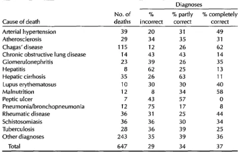
Table 3 shows the accuracy of clinical diagnoses for 14 selected causes of death. These data draw attention to certain causes of death that are being incorrectly diagnosed in our hospitals, including
such
relatively easy to diagnose diseases as arterial hypertension (20% incorrect), Chagas’ disease (12% incorrect),3 chronic obstructive lung disease (43% incorrect), pneumonialbronchopneumonia (75% in- correct), and schistosomiasis (36% incor- rect) .It could be argued that the data shown demonstrate nothing more than that cli- nicians are not filling out their patients’ death certificates properly. However, even this “minor” oversight would seri- ously jeopardize the quality of data pro- vided to our vital statistics services; and
Thagas’ disease is very common in the state of Bahia. Important research is conducted at the uni- versity hospital, which usually attracts patients from endemic areas of the state. Serology for this disease is routinely performed for all cardiac pa- tients as well as for gastrointestinal patients sus- pected of having “mega” forms of the disease.
inaccuracies in the information derived from death certificates could make such information unsuitable for use in re- search and planning. Data from the inter- American investigations of mortality (1 I) and childhood mortality (22) in the city of
SO Paulo indicate that the underlying causes of death among study subjects were correctly identified on their death certificates in 67% of the cases among in- dividuals between the ages of 15 and 74 years and in 52% of the cases among chil- dren below the age of five. (It should be noted, however, that the proportion of autopsies among the study subjects was 11.8% among those aged 15-74 and 20.1% among the children under age 5.)
As regards our 910 cases, we are more inclined to believe the bulk of the dis- crepancies arose from actual errors in clinical diagnosis of the underlying cause of death, in line with results obtained by studies made in other parts of the world (I, 2, 4-6, 8, 9).
These various findings, as well as ours, make it clear that the medical profession is not taking full advantage of its oppor- tunities to increase knowledge through analysis of its own errors. In the devel- oped countries, autopsies are becoming less and less frequent, stated reasons being a low cost-benefit ratio and fear of
Table 3. The accuracy with which selected causes of death (and other causes of
death not specified here) were clinically diagnosed in the 910 study subjects, as indicated by autopsy data.
Diagnoses
No. of % % partly % completely Cause of death deaths incorrect correct correct Arterial hypertension 39 20 31 49 Atherosclerosis 29 34 35 31 Chagas’ disease 115 12 26 62 Chronic obstructive lung disease 14 43 43 14 Clomerulonephritis 23 39 26 35
Hepatitis 8 62 25 13
Hepatic cirrhosis 35 26 63 11 Lupus erythematosus 10 30 30 40
Malnutrition 12 8 34 58
Peptic ulcer 7 43 57 0
Pneumonitironchopneumonia 12 75 17 8 Rheumatic disease 36 31 25 44 Schistosomiasis 36 36 30 34 Tuberculosis 28 36 39 25 Other diagnoses 243 35 39 36
Total 647 29 34 37
inaccuracies in the information derived from death certificates could make such
information unsuitable for use in re-
search and planning. Data from the inter- American investigations of mortality (1 I) and childhood mortality (12) in the city of Sso Paulo indicate that the underlying
causes of death among study subjects
were correctly identified on their death certificates in 67% of the cases among in- dividuals between the ages of 15 and 74 years and in 52% of the cases among chil- dren below the age of five. (It shbuld be
noted, however, that the proportion of
autopsies among the study subjects was
11.8% among those aged 15-74 and
20.1% among the children under age 5.)
AntGnio Almeida of the Department of
Pathological Anatomy at the Professor
Edgard Santos University Hospital for
their support and encouragement, and
also the medical students of the Federal
University of Bahia in Epidemiology
Course 85.2, Class P02, for their assis- tance with data collection.
REFERENCES
1.
2.
McGoogan E. The autopsy and clinical di- agnosis. JR Co11 Pathol. 1984;18:240-43. Pounder DJ, Roland R, Horowitz M, et al. The value of the autopsy in medical audit: a combined clinical and pathological as- sessment of 100 cases. Aust N Z J Med. 1983;13:478-82.
3. Cameron HM, McGoogan E, Watson H.
Necropsy: a yardstick for clinical diag- noses. Br Med J. 1980;281:985-88.
4.
5.
6.
7.
8.
Scottolini AG, Weinstein SR. The autopsy in
1983 clli&~~92-~aIity control. JAMA.
Britt6n M. Diagnostic errors discovered at autopsy. Acta Med Stand. 1974;196:203-10. Goldman L, Sayson R, Robbins S et al. The value of the autopsy in three medical eras. N Erg1 J Med. 1983;308:1000-5. Laurenti R, Mello Jorge MHI? 0 atestado de
o’bito. Sgo Paulo: WHO Center for Classi-
fication of Diseases in Portuguese; 1981.
Cameron HM, McGoogan E. A prospec- tive study of 1,152 hospital autopsies: I, inaccuracies in death certification. J Pa- thol. 1981;133:273-83.
9. Cameron, HM, McGoogan E. A prospec-
tive study of 1,152 hospital autopsies: II, analysis of inaccuracies in clinical diag- noses and their significance. J Puthol. 1981;133:285-300.
10. Sackett DL, Haynes RB, Tugwell I? Clini- cal epidemiology: a basic science
for
clinical medicine. Boston: Little Brown; 1985:162. 11. Puffer RR, Griffith GW. Patterns of urbanmortality. Washington, DC: Pan American Health Organization; 1967. (PAHO scien- tific publication 151).
12. Puffer RR, Serrano CV. Patterns
of
mortal- ity in childhood. Washington, DC: PanAmerican Health Organization; 1973.
(PAHO scientific publication 262).
13. Hasson J, Gross H. The autopsy and qual- ity assessment of medicaI care. Am J Med. 1974;56:137-40.
14. Roberts WC. The autopsy: its decline and suggestions for its revival. N Engl J Med. 1978;299:332-38.
Meeting on Hunger Research
The Fourth Annual Hunger Research Briefing and Exchange will be held at Brown University in Providence, Rhode Island (USA), from 3 to 5 April 1991. Cosponsored by Brown University’s Alan Shawn Feinstein World Hunger Program and InterAction-The American Council for Voluntary International Action, the briefing is designed to encourage dialogue among researchers and practitioners concerned with alleviating hunger.
The topic of this year’s briefing will be the Bellagio Declaration, a docu- ment created at an international meeting held in Bellagio, Italy, in 1989, that identifies four goals for the 1990s: 1) eliminating deaths from famine, 2) ending hunger in half of the poorest households, 3) reducing by half the rate of malnutrition among mothers and small children, and 4) eradicating iodine and vitamin A deficiencies. The briefing will focus on implementa- tion strategies for these goals including identification of resources, devel- opment of ongoing programs, and linkage of grassroots efforts with those from higher levels. The program will be supplemented by an extensive ex- hibit of hunger-related publications and a notebook of abstracts of recently completed or ongoing research and project reports.
For further information, contact Briefing Coordinator, World Hunger Program, Box 1831, Brown University, Providence, RI 02912, USA; tele- phone (401) 863-2700; telex 952095; fax (401) 863-2192.