Expression of inducible nitric oxide
synthase is increased in patients with
heart failure due to ischemic disease
1Unidade de Aterosclerose, Instituto do Coração (Incor), Hospital das Clínicas,
Faculdade de Medicina, Universidade de São Paulo, São Paulo, SP, Brasil
2Departamento de Farmacologia, Instituto de Ciências Biomédicas,
Universidade de São Paulo, São Paulo, SP, Brasil
3Hospital do Coração, Associação do Sanatório Sírio,São Paulo, SP, Brasil
C.R. Ferreiro3, A.C.P. Chagas1,
M.H.C. Carvalho2,
A.P. Dantas2, C. Scavone2,
L.C.B. Souza3, E. Buffolo3
and P.L. da Luz1
Abstract
The objective of the present study was to determine the relationship between nitric oxide synthases (NOS) and heart failure in cardiac tissue from patients with and without cardiac decompensation. Right atrial tissue was excised from patients with coronary artery disease (CAD) and left ventricular ejection fraction (LVEF) <35% (N = 10), and from patients with CAD and LVEF >60% (N = 10) during cardiac surgery. NOS activity was measured by the conversion of L-[H3
]-arginine to L-[H3]-citrulline. Gene expression was quantified by the
competitive reverse transcription-polymerase chain reaction. Both endothelial NOS (eNOS) activity and expression were significantly reduced in failing hearts compared to non-failing hearts: 0.36 ± 0.18 vs 1.51 ± 0.31 pmol mg-1 min-1 (P < 0.0001) and 0.37 ± 0.08 vs 0.78 ±
0.09 relative cDNA absorbance at 320 nm (P < 0.0001), respectively. In contrast, inducible NOS (iNOS) activity and expression were significantly higher in failing hearts than in non-failing hearts: 4.00 ± 0.90 vs 1.54 ± 0.65 pmol mg-1 min-1 (P < 0.0001) and 2.19 ± 0.27 vs 1.43 ± 0.13 cDNA absorbance at 320 nm (P < 0.0001), respectively. We conclude that heart failure down-regulates both eNOS activity and expression in cardiac tissue from patients with LVEF <35%. In contrast, iNOS activity and expression are increased in failing hearts and may represent an alternative mechanism for nitric oxide produc-tion in heart failure due to ischemic disease.
Correspondence
P.L. da Luz
Unidade de Ateroscleroses Incor, HC, FM, USP Av. Dr. Enéas C. Aguiar, 44 0540-3000 São Paulo, SP Brasil
Fax: +55-11-3069-5547 E-mail: [email protected]
Publication supported by FAPESP.
Received February 24, 2003 Accepted April 7, 2004
Key words
•Nitric oxide synthase •Left ventricle ejection
fraction •Heart failure
Introduction
Nitric oxide (NO) is an important cellular signaling molecule (1-5) synthesized by three different isoforms of the enzyme nitric oxide synthase (NOS). Two isoforms are constitu-tively expressed while one is induced in response to cytokines and endotoxins among other stimuli (6-10).
More-over, exposure of cardiac muscle to cyto-kines impairs cardiac contractility, an effect that seems to be mediated by NO (12). In patients with heart failure, the functional significance of modified myocardial expres-sion of NOS for left ventricular performance remains unclear (13). Studies carried out to investigate the relationship between NOS activity, gene expression and heart failure using myocardial biopsies are controversial. de Belder et al. (14) reported increased myo-cardial activity of the inducible NOS isoform (iNOS) in patients with dilated cardiomyop-athy, but not in ischemic or valvular disease. Thoenes et al. (15) found immunoreactive iNOS protein only in septic hearts, but not in other forms of cardiomyopathy. Others dem-onstrated iNOS mRNA and immunoreactiv-ity in hearts from patients with dilated car-diomyopathy (16-18), but also in ischemic and valvular disease (16). Conflicting re-sults have also been reported regarding the endothelial isoform (eNOS). Some studies have suggested that eNOS expression and activity are reduced in the failing human heart, while others have observed increased levels (19,20).
The purpose of the present investigation was to examine the activity and gene expres-sion of eNOS and iNOS in atrial tissue from patients with coronary artery disease (CAD), with and without heart failure, who were subjected to cardiac surgery. The specific goal was to assess the relationship between enzymatic activity and left ventricular ejec-tion fracejec-tion (LVEF).
Patients and Methods
Patients
Twenty consecutive patients with CAD submitted to coronary angiography before cardiac surgery were divided into two groups: A, 10 patients with LVEF >60%, and B, 10 patients with LVEF <35%.
In group A there were 8 smokers, 8
pa-tients were whites and 2 blacks. In this group, surgery consisted of isolated by-pass grafts in all patients. Exclusion criteria were: pre-vious myocardial infarction, prepre-vious car-diac surgery, diabetes mellitus, use of angio-tensin-converting enzyme (ACE) inhibitors and/or ß-blockers. In group B all patients had previous myocardial infarction, 6 pa-tients had diabetes mellitus and 8 were smok-ers, and there were 7 whites and 3 blacks. Surgeries in this group were: by-pass grafts plus left ventricular aneurysmectomy in 4 patients, left ventricular remodeling (modi-fied Baptista’s surgery) in 4, and isolated by-pass in 2. In this group atrial fibrillation was the only exclusion criterion. For ethical rea-sons, heart failure therapy was maintained and consisted of ACE inhibitors (N = 9), diuretics (N = 8), digitalis (N = 8), and ß-blockers (N = 4). All patients were operated upon under extracorporeal circulation.
Methods
All patients gave written informed con-sent to participate in the study before the procedure. The protocol was approved by the Human Subjects Review Committee of Hospital do Coração, São Paulo. The inves-tigation was carried out according to the principles outlined in the Declaration of Helsinki.
Left ventricular angiograms and Doppler echocardiography were used to measure LVEF by the area-length method and Simpson’s method (21), respectively. He-modynamic data were obtained 1-4 weeks before cardiopulmonary by-pass. Heart rate was recorded by direct monitoring with a Hewlett Packard device (Handover, MS, USA) and arterial blood pressure was ob-tained by a direct invasive method using a pressure transducer (HP-104).
subse-quent biochemical analysis.
Measurement of NOS activity
NOS activity was measured in superna-tants from right atrial tissue as described by McKee et al. (22). The NOS assay is based on the biochemical conversion of L-arginine to L-citrulline by NOS. The tissue was ho-mogenized in ice-cold Tris-HCl buffer (20 mM Tris-HCl, 10 mM EDTA, and 10 mM EGTA, pH 7.4) using a Teflon homogenizer. Each homogenate was centrifuged at 12,000
g for 5 min at 4ºC. Supernatants were
re-moved and the NOS assay was performed by incubation (37ºC for 60 min) of 100 µg (20 µl) of protein in a final volume of 60 µl of assay mixture containing 50 mM Tris-HCl, 6 µM tetrahydrobiopterin, 2 µM FAD, 2 µM FMN + 10 mM NADPH, 100 mM L-argi-nine/L-[H3]-arginine (5 µCi/ml), 6 mM
CaCl2, and 0.1 µM calmodulin. For iNOs
activity (calcium/calmodulin-free activity) EDTA/EGTA were added and CaCl2 and
calmodulin were omitted. Calcium-depend-ent (eNOS) activity was calculated as the difference between the calcium-calmodulim sample and the EDTA/EGTA sample. The reaction was stopped with 400 µl of ice-cold stop buffer (50 mM HEPES and 5 mM EDTA, pH 5.5) and 100 µl of cation-exchange resin (Dowex, Na+ form, equilibrated with 50 mM
HEPES, pH 5.5) was added to each reaction mixture to remove excess L-[H3]-arginine.
The aliquots were placed in spin cups and centrifuged for 1 min at 12,000 g. The
super-natants were collected into vials, scintilla-tion liquid (4 ml) was added and radioactiv-ity was quantified. Samples of rat cerebel-lum were analyzed simultaneously as a posi-tive control. Protein concentrations in samples of human right atrium homogenates were determined by the Bradford Coomassie brilliant blue method (23) with bovine serum albumin as the standard and homogenization buffer as the blank. This method does not allow separation of neuronal and endothelial
components of NOS. Because the neuronal component is probably small and mRNA assessment is specific for eNOS (see ahead), we refer to the activity here as eNOS.
Determination of NOS expression by RT-PCR
Total cellular RNA was isolated from human right atrium using TRizol Reagent (Gibco-BRL, Life Technologies, Rockville, MD, USA). After DNA digestion (RQ1 DNAse RNAse-free; Promega Corporation, Madison, WI, USA), 1 µg total RNA from each preparation was reverse transcribed in the presence of an RNAse inhibitor (RNasIn®,
Promega Corporation) in a 20-µl reaction volume containing 50 mM Tris-HCl, pH 8.3, 75 mM KCl, 3.0 mM MgCl2, 10 mM
dithio-threitol, 2.0 mM deoxynucleotidetriphos-phates (dNTP), 200 U of Moloney murine leukemia virus reverse transcriptase (Gibco-BRL) and 1 µg of oligo (dT)12-18 primer. The reaction was carried out at room temper-ature for 10 min and at 37ºC for 60 min and stopped by heating at 100ºC for 5 min. The reverse-transcribed cDNA (2 µl) was ampli-fied in a final volume of 50 µl by PCR under standard conditions (1.5 mM MgCl2, 450
µM dNTP, 2.5 U Taq polymerase) with spe-cific primers for human eNOS, iNOS and rat glyceraldehyde-3-dehydrogenase (GAPDH) designed on the basis of published cDNA sequences (24). GAPDH was used as an internal control for co-amplification.
agarose gel containing 0.5 µg/ml ethidium bromide. The gel was subjected to ultravio-let light and photographed. Band intensities were measured using a software package (Kodak Digital Science, Eastman Kodak Company, New Haven, CT, USA) and the signals were expressed relative to the inten-sity of the GAPDH amplicon in each co-amplified sample.
Statistical analysis
Multivariate analysis of variance was used to compare eNOS and iNOS activities and their gene expressions between groups A and B. Age and weight distributions be-tween patients with and without heart failure were compared by the Mann-Whitney test. Sex differences were compared by the Fisher test. Pearson’s correlation coefficients were calculated to determine the relation of eNOS and iNOS activity and expression with LVEF and functional class. A P value <0.05 was considered significant.
Results
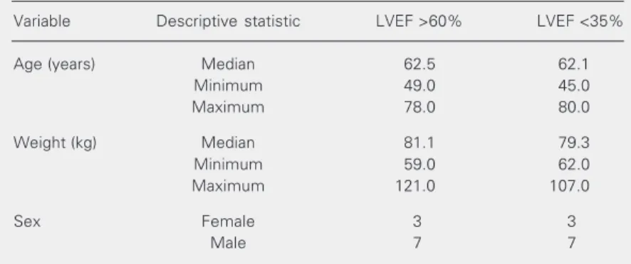
The A group consisted of 10 patients, 7 males and 3 females, without heart failure and LVEF >60%. All patients were in func-tional class 0 of the New York Heart Asso-ciation (NYHA). The B group consisted of 10 patients, 7 males and 3 females, with heart failure and LVEF <35%. In this group 3 patients were in functional class II, 4 in class III and 3 in class IV of NYHA. Patient characteristics are shown in Tables 2, 3, and 4.
eNOS and iNOS activities
eNOS activity was significantly lower in failing hearts than in non-failing hearts: 0.36 ± 0.18 vs 1.51 ± 0.31 pmol mg-1 min-1
(P < 0.0001; Figure 1A). In contrast, iNOS activity was significantly increased in failing hearts compared with non-failing hearts: 4.00 ± 0.90 vs 1.54 ± 0.65 pmol mg-1 min-1 Table 2. Comparisons of age, weight and sex variables between the groups with left
ventricular ejection fraction (LVEF) >60% and LVEF <35%.
Variable Descriptive statistic LVEF >60% LVEF <35%
Age (years) Median 62.5 62.1
Minimum 49.0 45.0
Maximum 78.0 80.0
Weight (kg) Median 81.1 79.3
Minimum 59.0 62.0
Maximum 121.0 107.0
Sex Female 3 3
Male 7 7
There were no statistical differences between groups for age (Mann-Whitney test), weight (Mann-Whitney test) or sex (Fisher test).
2.0
1.5
pmol mg
-1 min
-1
1.0
0.5
0.0
P < 0.0001 P < 0.0001
6.0
5.0
pmol mg
-1
min
-1
4.0
3.0 2.0
1.0 0.0
LVEF >60% LVEF <35% LVEF >60% LVEF <35%
A B
Figure 1. Individual values of (A) endothelial and (B) inducible nitric oxide synthase (NOS) activity in the right atrial appendage of patients in the groups with left ventricular ejection fraction (LVEF) >60% and LVEF <35%. The symbols to the right of individual values are the mean ± SEM. N = 10 patients in each group.
Table 1. Primers and experimental conditions used for the determination of NOS expression by RT-PCR.
Target gene Sequences (5'→3' ) Annealing Number
temperature (ºC) of cycles
eNOS CCAGCTAGCCAAAGTCACCAT (S) 55 35
GTCTCGGAGCCATACAGGATT (AS)
iNOS GAGGAAGTGGGCAGGAGAATG (S) 50 35
GTAGTAGAAAGGGGACAGGAC (AS)
GAPDH GTGAAGGTCGGTGTGAACGGATTT (S) 60 20
CACAGTCTTCTGAGTGGCAGTGAT (AS)
eNOS = endothelial nitric oxide synthase; GAPDH = glyceraldehyde-3-dehydrogen-ase; iNOS = inducible nitric oxide synthglyceraldehyde-3-dehydrogen-ase; S = sense; AS = antisense.
(P < 0.0001; Figure 1B).
eNOS and iNOS expression
eNOS expression was significantly lower in failing hearts than in non-failing hearts: 0.37 ± 0.08 vs 0.78 ± 0.09 absorbance
c-DNA (P < 0.0001; Figure 2A). In contrast, iNOS expression was significantly increased in hearts with LVEF <35% compared with hearts with LVEF >60%: 2.19 ± 0.27 vs 1.43
± 0.13 absorbance cDNA (P < 0.0001; Fig-ure 2B).
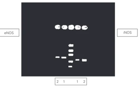
The results of RT-PCR assays for the detection of eNOS and iNOS expression
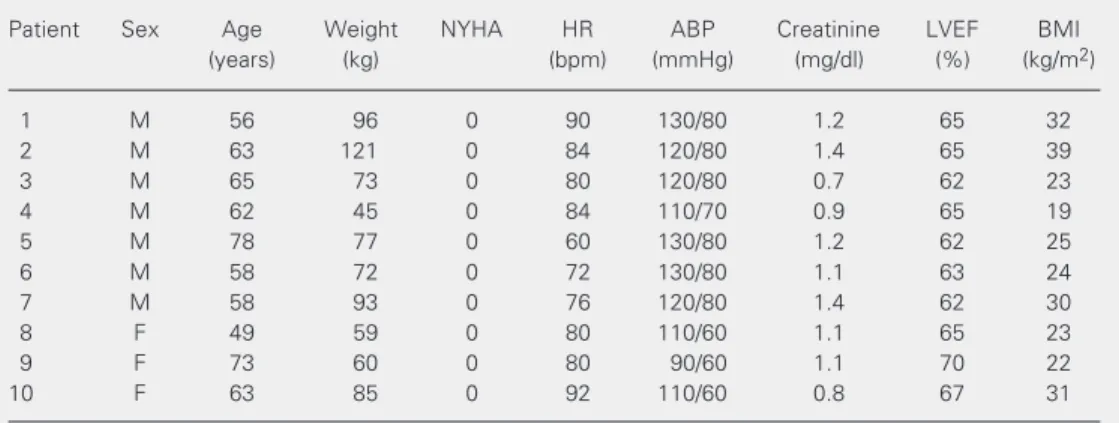
Table 4. Clinical and hemodynamic characteristics of patients with left ventricular ejection fraction <35%.
Patient Sex Age Weight NYHA HR ABP Creatinine LVEF BMI
(years) (kg) (bpm) (mmHg) (mg/dl) (%) (kg/m2)
1 M 66 81 III 100 83/67 2.0 21 28
2 M 77 97 II 88 90/60 1.2 35 34
3 M 62 70 III 90 110/70 1.3 17 25
4 M 80 55 II 92 130/80 1.4 35 23
5 M 45 88 III 98 110/70 1.8 35 30
6 M 55 83 II 94 90/55 1.3 35 28
7 F 46 65 IV 98 90/60 1.4 23 22
8 F 57 68 IV 90 90/60 1.6 20 24
9 M 78 107 III 88 90/50 1.9 35 34
10 F 47 62 IV 104 130/80 3.0 26 26
ABP = arterial blood pressure; BMI = body mass index; HR = heart rate; LVEF = left ventricular ejection fraction; NYHA = New York Heart Association Classification of Cardiac Heart Failure.
measured in one patient of each group are shown in Figure 3 (see legend).
There were no statistically significant cor-relations between functional class and NOS activities or gene expression in either group (P > 0.05).
The present study shows that activity and gene expression of eNOS in atrial tissue from patients with CAD were significantly lower in those with heart failure and LVEF <35% than in those without heart failure and LVEF >60%. In contrast, iNOS activity and expression were significantly higher in patients with heart failure and LVEF <35% compared with non-failing hearts with LVEF >60%.
Table 3. Clinical and hemodynamic characteristics of patients with left ventricular ejection fraction >60%.
Patient Sex Age Weight NYHA HR ABP Creatinine LVEF BMI
(years) (kg) (bpm) (mmHg) (mg/dl) (%) (kg/m2)
1 M 56 96 0 90 130/80 1.2 65 32
2 M 63 121 0 84 120/80 1.4 65 39
3 M 65 73 0 80 120/80 0.7 62 23
4 M 62 45 0 84 110/70 0.9 65 19
5 M 78 77 0 60 130/80 1.2 62 25
6 M 58 72 0 72 130/80 1.1 63 24
7 M 58 93 0 76 120/80 1.4 62 30
8 F 49 59 0 80 110/60 1.1 65 23
9 F 73 60 0 80 90/60 1.1 70 22
10 F 63 85 0 92 110/60 0.8 67 31
Discussion
Although the importance of NO for the regulation of vasomotor tone has been estab-lished, the physiological role of NO in car-diac function and structure remains incom-pletely understood. Removal of the endocar-dium or endothelium modulates cardiac con-traction, suggesting that the NO released from endothelial and endocardial cells modu-lates cardiac contraction (25).
Ischemia elicits a variety of adaptive re-sponses at the tissue, cellular and molecular
levels. A physiological response to ischemia requires the existence of a signal transduc-tion system which should be linked or coupled to an O2 sensor (26). The results of the
present study provide evidence that heart failure probably induces iNOS gene expres-sion in cardiac tissue from patients with ischemic cardiomyopathy. Unlike eNOS, iNOS is not usually expressed in healthy tissues (27). One of the difficulties in study-ing diseased human myocardium is the ab-sence of a readily avaliable source of normal control tissue. Our knowledge about iNOS levels in normal cardiac tissue derives from studies that investigated iNOS mRNA ex-pression in heart donors (28) and that re-vealed no expression or activity of iNOS. In the present study iNOS activity and expres-sion in patients with non-failing hearts were surprisingly high and may have been related to CAD or to cardiothoracic surgery itself.
In addition, the present study provides evidence that eNOs and iNOS activity and gene expression are related to ventricular function. de Belder et al. (19) obtained simi-lar results, but only for nonischemic dilated cardiomyopathy. Heymes et al. (27) found a linear correlation between LVEF and eNOS expression, also in patients with nonischemic dilated cardiomyopathy. On the other hand, Drexler et al. (28) reported a higher activity and expression of iNOS in patients with heart failure due to several etiologies. These investigators used explanted hearts from do-nors as matched controls, and did not find any iNOS activity in such hearts. A possible explanation for this result may be the abnor-mal conditions associated with brain death, ventilation, and explantation of the donor’s heart. Our results about iNOS activity and expression agree with previous studies (18,27,28) although our samples were taken from the right atrium.
We also investigated a possible correla-tion between funccorrela-tional class and eNOS and iNOS activity levels or gene expression, but found no correlations. In a previous study
eNOS iNOS
2 1 1 2
Figure 3. Representative RT-PCR assay for the detection of endothelial nitric oxide syn-thase (eNOS) and inducible nitric oxide synsyn-thase (iNOS) expression. The lower expression of eNOS in a patient with heart failure (number 2) can be seen on the left side; on the right, iNOS expression is higher in the patient with heart failure (number 2) than in the patient without heart failure (number 1). The line in the middle represents the standard values.
1.2 1.0
eNOS cDNA absorbance
0.8
0.6
0.4
P < 0.0001 3.0 P < 0.0001
2.5
2.0
1.5 1.0 0.5
0.0
LVEF >60% LVEF <35% LVEF >60% LVEF <35%
A B
0.2
0.0 iNOS cDNA absorbance
(16) it was demonstrated that patients with NYHA II presented higher iNOS expression than those with normal functional class. On the other hand, Satoh et al. (18) showed that iNOS expression was related predominantly to LVEF and not to functional class.
Comparison between eNOS activity and gene expression in both groups supports the idea that eNOS is down-regulated as LVEF decreases, in agreement with data reported by Heymes et al. (27) who investigated heart failure patients with LVEF lower than 40%. Furthermore, the right atrium may not be the ideal sampling site for biopsies, because its cells are not well localized to sense shear stress induced by pulsatile flow. On the other hand, right atrium cells are well localized to act as O2 sensors, especially cells of the
endocardium, which may be particularly suited to sensing changes in preload.
NO and cyclic guanosine monophosphate (cGPM) induce a concentration-dependent biphasic contractile response: low NO doses cause a positive inotropic response, while higher doses cause a negative inotropic re-sponse (28). Increased iNOS activity prob-ably represents the pathway for increased NO production in heart failure, which would be an attempt to counterbalance the vaso-constrictor state found in patients with re-duced LVEF. In a previous study (29) we analyzed the relation between hypoxia and NOS in children with congenital heart de-fects and showed that iNOS activity and gene expression are up-regulated in right atrial hypoxic tissue compared with non-hypoxic tissue. Thus, it is possible that both hypoxic and ischemic hearts share a
com-mon adaptive mechanism, namely increased NO production via iNOS. In addition, NO acts as a bifunctional regulator of apoptosis. Physiologically relevant NO levels seem to suppress the apoptotic pathway, while NO levels may overwhelm cell protective mechanisms and exert proapoptotic and cy-totoxic effects in patients with heart failure (30-32). Indeed, excessive NO production secondary to the induction of iNOS in failing cardiac tissue would be expected to depress cardiac contraction, as observed in septic shock (33,34). This idea is further supported by the observation of high plasma and tissue levels of various cytokines, such as TNF-α, known to induce iNOS (35-40). Since NO was not assessed in the present study, its possible relation to other phenomena occur-ring duoccur-ring heart failure, including apoptosis and cytokine release, is speculative.
Study limitations
The biochemical conversion of L-argi-nine to L-citrulline assay as a measure of NOS activity has its limitations, and our data should be interpreted carefully; however, the simultaneous indications of the direction of the change of NOS gene expression and chemical activity of both NOS forms add confidence to the measurements.
We conclude that in the present study iNOS gene expression and activity were in-creased in cardiac tissue from patients with heart failure and presumably this phenome-non led to increased NO bioavailability. This may represent an important adaptive mech-anism in heart failure.
References
1. Moncada S & Higgs A (1993). The L-arginine-nitric oxide pathway.
New England Journal of Medicine, 329: 2002-2012.
2. Furchgott RF & Zawadzki JV (1980). The obligatory role of endothe-lial cells in the relaxation of arterial smooth muscle by acetylcholine.
Nature, 288: 373-376.
3. Palmer RMJ, Ferrige AG & Moncada S (1987). Nitric oxide release
accounts for the biological activity of endothelium-derived relaxing factor. Nature, 327: 415-423.
4. Moncada S, Higgs A & Furchgott R (1997). XIV International union of pharmacology nomenclature in nitric oxide research. Pharmacologi-cal Reviews, 49: 137-142.
PL, Laurindo FRM & Chagas ACP (Editors), Endotélio e Doenças Cardiovasculares. Vol. 4. Editora Atheneu, São Paulo, SP, Brazil, 43-51.
6. Papapetropoulos A, Rudic RD & Sessa WC (1999). Molecular con-trol of nitric oxide synthases in the cardiovascular system. Cardio-vascular Research, 43: 509-520.
7. Zweier JL, Wang P & Samouilov A (1995). Enzyme-independent formation of nitric oxide in biological tissues. Nature Medicine, 8: 804-809.
8. Wilcox JN, Subramanian RRCL & Sundell CL (1997). Expression of multiple isoforms of nitric oxide synthase in normal and atheroscle-rotic vessels. Arteriosclerosis, Thrombosis, and Vascular Biology, 17: 2479-2488.
9. Nadaud S & Sobrier F (1996). Molecular biology and molecular genetics of nitric oxide synthase genes. Clinical and Experimental Hypertension, 18: 113-143.
10. Forstemann U, Closs EI & Pollock JS (1994). Nitric oxyde synthase isozymes: characterization, purification, molecular cloning and func-tions. Hypertension, 23 (Part 2): 1121-1131.
11. Brutsaert DL, Meulemans AL, Sipido KR & Sys SU (1988). Effects of damaging the endocardial surface on the mechanical performance of isolated cardiac muscle. Circulation Research, 62: 357-366. 12. Finkel MS, Oddis CV, Jacob TD, Watkins SC, Hattler BG & Simmons
RL (1992). Negative inotropic effects of cytokines on the heart mediated by nitric oxide. Science, 257: 387-389.
13. Schulz R, Nava E & Moncada S (1992). Induction and potential biological relevance of a Ca2+-independent nitric oxide synthase in the myocardium. British Journal ofPharmacology, 105: 575-580. 14. de Belder AJ, Radomski MW, Why HJ, Richardson PJ & Martin JF
(1995). Myocardial calcium-independent nitric oxide synthase activ-ity is present in dilated cardiomyopathy, myocarditis, and portpartum cardiomyopathy but not in ischaemic or valvar heart disease. British Heart Journal, 74: 426-429.
15. Thoenes M, Forstermann U & Tracey WR (1996). Expression of inducible nitric oxide synthase in failing and non-failing human heart.
Journal of Molecular and Cellular Cardiology, 28: 165-169. 16. Haywood GA, Tsao PS & Von Der Leyen HE (1996). Expression of
inducible nitric oxide synthase in human failing heart. Circulation, 93: 1087-1094.
17. Habib FM, Springal DR & Davies GJ (1996). Tumour necrosis factor and inducible nitric oxide synthase in dilated cardiomyopathy. Lan-cet, 347: 1151-1155.
18. Satoh M, Nakamura M & Tamura G (1997). Inducible nitric oxide synthase and tumor necrosis factor-alpha in myocardium in human dilated cardiomyopathy. Journal of the American College of Cardiol-ogy, 29: 716-724.
19. de Belder AJ, Radomski M, Why H, Richardson PJ & Moncada S (1993). Nitric oxide synthase activities in human myocardium. Lan-cet, 341: 84-85.
20. Stein B, Eschenhagen T & Rudiger J (1998). Increased expression of constitutive nitric oxide synthase III, but not inducible nitric oxide synthase II, in human heart failure. Journal of the American College of Cardiology, 32: 1179-1186.
21. Nissen SE, Elion JL, Grayburn P, Booth DC, Wisenbaugh TW & DeMaria AN (1987). Determination of left ventricular ejection frac-tion by computer densitometric analysis of digital subtracfrac-tion an-giography: experimental validation and correlation with area-length methods. American Journal of Cardiology. 59: 675-680.
22. McKee M, Scavone C & Nathanson JA (1994). Nitric oxide, cGMP, and hormone regulation of active sodium transport. Proceedings of the National Academy of Sciences, USA, 91: 12056-12060. 23. Bradford M (1976). A rapid and sensitive method for the
quantita-tion of microgram quantities of protein utilizing the principle of protein-dye binding. Annals of Biochemistry, 72: 248-254. 24. Teng KS, Murthy JF & Kuemmerle JR (1998). Expression of
endo-thelial nitric oxide synthase in human and rabbit gastrointestinal smooth muscle cells. American Journal of Physiology, 275: G342-G351.
25. Winegrad S (1997). Endothelial cell regulation of contractility of the heart. Annual Review of Physiology, 59: 505-525.
26. Michel T & Feron O (1997). Nitric oxide synthases: which, where, how, and why? Journal of Clinical Investigation, 100: 2146-2152. 27. Heymes C, Vanderheyden M & Bronzwaer JGF (1999).
Endomyo-cardial nitric oxide synthase and left ventricular preload reserve in dilated cardiomyopathy. Circulation, 99: 3009-3016.
28. Drexler H, Kastner S & Strobel A (1998). Expression and functional significance of inducible nitric oxide synthase in the failing human heart. Journal of the American College of Cardiology, 32: 955-963. 29. Ferreiro CR, Chagas ACP, Carvalho MHC, Dantas AP, Jatene MB,
Souza LCB & da Luz PL (2001). Influence of hypoxia on nitric oxide synthase activity and gene expression in children with congenital heart disease. Circulation, 103: 2272-2276.
30. Kim YM, Bombeck CA & Billiar TR (1999). Nitric oxide as a bifunc-tional regulator of apoptosis. Circulation Research, 84: 253-256. 31. Ing DJ, Zang J & Dzau VJ (1999). Modulation of cytokine-induced
cardiac myocyte apoptosis by nitric oxide, Bak, and Bcl-x. Circula-tion Research, 84: 21-33.
32. Pinsky D, Cai B & Yang X (1994). Nitric oxide-dependent killing of cardiac myocytes by adjacent macrophages. Circulation, 90 (Suppl I): I-192.
33. Price S, Anning PB, Mitchell JA & Evans TW (1999). Myocardial dysfunction in sepsis: mechanisms and therapeutic implications.
European Heart Journal, 20: 715-724.
34. Ullrich R, Scherrer CM & Bloch KD (2000). Congenital deficiency of nitric oxide synthase 2 protects against endotoxin-induced myocar-dial dysfunction in mice. Circulation, 102: 1440-1446.
35. Kasai K, Hattori Y & Banba N (1997). Induction of tetrahydrobiop-terin synthesis in rat cardiac myocytes: impact on cytokine-induced NO generation. American Journal of Physiology, 273: H665-H672. 36. Sasayama S, Matsumori A & Kihara Y (1999). New insights into the
pathophysiological role for cytokines in heart failure. Cardiovascular Research, 42: 557-564.
37. Torre AG, Kapadia S & Lee J (1996). Tumor necrosis factor-alpha and tumor necrosis factor receptors in the failing human heart.
Circulation, 93: 704-711.
38. Oral H, Dorn 2nd GW & Mann DL (1997). Sphingosine mediates the immediate negative inotropic effects of tumor necrosis factor-alpha in the adult mammalian cardiac myocyte. Journal of Biological Chemistry, 272: 4836-4842.
39. Kubota T, McTiernan CF & Frye CS (1997). Dilated cardiomyopathy in transgenic mice with cardiac-specific overexpression of tumor necrosis factor-alpha. Circulation Research, 81: 627-635.