Faculdade de Medicina de Ribeirão Preto – USP, Faculdade de Medicina da UNAERP e CECORP – Centro Especializado do Coração e Pulmão de Ribeirão Preto Correspondência: Paulo Roberto B. Evora - Rua Rui Barbosa, 367/7 - 14015-120 - Ribeirão Preto, SP – Brazil - E-mail: [email protected]
Fernanda Viaro, Fernando Nobre, Paulo Roberto B. Evora
Ribeirão Preto, SP - Brazil
Expression of Nitric Oxide Synthases in the Pathophysiology
of Cardiovascular Diseases
Nitric oxide (NO) is not only a potent vasodilator, it also inhibits platelet adherence and aggregation, reduces adhe-rence of leukocytes to the endothelium, and suppresses proliferation of vascular smooth muscle cells. The knowledge of nitric oxide synthases (NOSs) is of extreme scientific impor-tance, not only for understanding new pathophysiological mechanisms but, also as a target for therapeutic intervention 1.
Therefore, the intention of this overview is to review the role of nitric oxide synthases in the physiological and patho-logical mechanisms of the cardiovascular system.
Basic concepts
NOSs, from the biochemical point of view, are a family of complex enzymes that catalyze the oxidation of L-argini-ne to form NO and L-citrulliL-argini-ne. The three human NOS isofor-ms have been identified to date, ecNOS(endothelial consti-tutive NOS), nNOS (neuronal NOS), and iNOS (inducible NOS), their genes are found on human chromosomes 7, 12, and 17, respectively, and so were named for the tissue in which they were first cloned and characterized 1,2 .
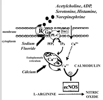
Endothelial constitutive nitric oxide synthase (ec-NOS) - The role of nitric oxide in regulating vascular tone and mediating platelet function is attributable to the ongo-ing activity of the endothelial constitutive form of NOS. Inactivation of the ecNOS pathway limits the contribution of NO to vessel homeostasis and results in increased vascular tone and platelet adhesion and aggregation. The complete signal transduction pathway of ecNOS activation is repre-sented in figure 1, where the activities of ecNOS are regula-ted by the intracellular free calcium concentration and calci-um-calmodulin complexes. ecNOS is a constitutively expres-sed protein predominantly associated with the particulate subcellular fraction, suggesting that the native enzyme is a membrane-bound protein. A recent detailed analysis of the membrane association of ecNOS showed that this enzyme is localized to the Golgi apparatus as well as to specific
structures in the plasmalemmal membrane called caveolae. The association of ecNOS with a region of the plasma membrane in which several key signal transducing comple– xes are concentrated (such as G-proteins) is likely to have profound repercussions on enzyme activity as well as on its accessibility to intracellular mechanisms of the pathway release, including mechanisms independents of intracellu-lar calcium release 3-5.
Neuronal nitric oxide synthase (nNOS) - This isoform is present in central and peripheral neuronal cells and cer-tain epithelial cells. Its activity is also regulated by Ca2+ and
calmodulin. Its functions include long-term regulation of sy-naptic transmission in the central nervous system, central re-gulation of blood pressure, smooth muscle relaxation, and vasodilation via peripheral nitrergic nerves. It has also been implicated in neuronal death in cerebrovascular stroke 6.
Inducible nitric oxide synthase (iNOS) - The expres-sion in this enzyme is induced in a multitude of different cells, including macrophages, endothelial cells, vascular smooth muscle cells and cardiac myocytes after stimulation with lipopolysaccharide (LPS), cytokines (such as IL-1β, TFN-α, IFN-γ, IL-6), and others; thus it has an important role in antimicrobial, antiparasitic and antineoplastic activity 7.
This isoform is not regulated by Ca2+. It produces large
amo-unts of NO that have cytostatic effects on parasitic target cells by inhibiting iron-containing enzymes and causing DNA fragmentation. The induction of iNOS is involved in the pathophysiology of autoimmune diseases and septic shock 6.
Based on the difficulties the authors of this review had in relation to the inconsistent NOS isoform nomenclature we decided to create a table of the various names used for each isoform to help futures research and reading on the subject (table I).
Methods for studying NOS expression
immunoche-mistry or messenger ribonucleic acid (mRNA) by in situ hy-bridization, bioassays, inhibition of nitric oxide synthase ac-tivity, iron responsive element-binding protein acac-tivity, and production of nitrate/nitrite, L-citrulline, or cyclic guanosine monophosphate (cGMP). Careful evaluation of potential pitfalls associated with these indirect methods of detecting nitric oxide effects prior to their use will prevent misinterpre-tation of results 8.
Unspecific inhibitors - Inhibitors of both ecNOS and iNOS are metilates and nitrics forms of L-arginine that act by competition. Among them the most popular are L-NMMA, o L-NAME, e and o L-NOARG. Another example is ADMA (asymmetric dimethylarginine), a circulating endogenous NOS inhibitor. Studies have shown that plasma ADMA level is positively correlated with risk factors for atheroscle-rosis thus suggesting that this endogenous antagonist of NO synthase may be a marker of atherosclerosis 9.
Specific inhibitors - Specifically inhibit the isoform INOS. They include the glucocorticoids (such as dexame-thasone), aminoguanidine, L-canavanine, N6- (1-imioetyl) lisine (L-NIL) and 2,4-diamino 6-hydroxy-pyrimidine 10.
Knowledge about the specific NOSs inhibitors is of pi-votal importance for experimental and therapeutical pharma-cologic assays (figure 2).
Some comparisons of the differents NOS isoforms ex-pressions would be helpful in a review of basic concepts.
As already mentioned ecNOS is an enzyme that is mem-brane associated whereas iNOS and nNOS are largely cyto-solic 1. Another difference between the NOS isoforms is
amount and duration of NO produced. Molecule NO is syn-thesized for short periods of time (seconds to minutes) fol-lowing enzyme activation of ecNOS or nNOS. In contrast, the iNOS is expressed after cell activation only and then produces NO for comparatively long periods of time (hours to days).
Anatomic distribution of NOS in the normal
heart
Most of the constitutive nitric oxide synthase activity in the normal heart is present in endothelium along the ex-tensive network of arteries, veins and capillaries within the myocardium. This endothelial isoform of nitric oxide syn-thase also exists in the endocardium lining the cardiac cavi-ties. Neuronal nitric oxide synthase appears much less pro-minent, although the exact amount of this isoform in the heart is uncertain. Although no inducible nitric oxide syn-thase occurs in the normal heart, macrophages associated with repair of various types of cardiac damage contain this isoform. For all nitric oxide synthases, however, species va-riation and variability among models underscore the impor-tance of correlative studies of structure and function 11.
Autocrine and paracrine function of NOS in
the normal heart
From the point of view of autocrine and paracrine func-tion, the different cell types comprising cardiac muscle ex-press one or more of the three isoforms (neuronal NOS, or nNOS; inducible NOS, or iNOS; and endothelial NOS, or ecNOS) of nitric oxide synthase (NOS). The nNOS is Table I - Nomenclature of oxide nitric synthases (NOSs)
Enzyme Nomenclature
Endothelial NOS eNOS, ecNOS, cNOS, NOS III ou NOS3 Neuronal NOS bNOS(brain),NOS I ou NOS1 Inducible NOS iNOS, NOS II ou NOS2 cytoplasm
Fig. 1 - Pathway of release of nitric oxide (NO); PIP2= phosphatidylinisitol 4,5-bi-phosphate; IP3= inositol tri4,5-bi-phosphate; ecNOS=endothelial constitutive nitric oxide synthase.
membrane
Acetylcholine, ADP, Serotonine, Histamine, Norepinephrine
Sodium Fluoride
Endoplasmatic reticulum
Cálcium
L-ARGININE
CALMODULIN
NITRIC OXIDE
blood
Fig. 2 - Inhibitors of the oxide nitric synthases (NOS).
L-ARGININE
AMINOGUANIDINE
DEXAMETASONE
endothelium cell
expressed in sympathetic nerve terminals and regulates the release of catecholamines in the heart. The ecNOS cons-titutively expressed in endothelial cells inhibits contractile tone and the proliferation of underlying vascular smooth muscle cells, inhibits platelet aggregation and monocyte adhesion, promotes diastolic relaxation, and decreases O2 consumption in cardiac muscle through paracrinally pro-duced NO. The ecNOS is also constitutively expressed in cardiac myocytes from rodent and human species, where it autocrinally opposes the inotropic action of catecholamines after muscarinic cholinergic and beta-adrenergic receptor stimulation. The iNOS gene transcription and protein ex-pression are induced in all cell types after exposure to a vari-ety of inflammatory cytokines. Aside from participating in the immune defense against intracellular microorganisms and viruses, the large amounts of NO produced autocrinal-ly or paracrinalautocrinal-ly mediate the vasoplegia and myocardial de-pression characteristic of systemic immune stimulation and promote cell death through apoptosis. In cardiac myocytes, NO may regulate L-type calcium current and contraction through activation of cGMP-dependent protein kinase and cGMP-modulated phosphodiesterases. Other mechanisms independent of cGMP elevations may operate through inte-raction of NO with heme proteins, nonheme iron, or free thiol residues on target signaling proteins, enzymes, or ion chan-nels. Given the multiplicity of NOS isoforms expressed in cardiac muscle and of the potential molecular targets for the NO produced, tight molecular regulation of NOS expression and activity at the transcriptional and posttranscriptional level is necessary to coordinate the many roles of NO in heart function in health and disease 12.
NOS and cardiovascular diseases
Myocardial ischemia - This part of the text will include general aspects of myocardial ischemia: ischemia reperfusi-on injury, precreperfusi-onditireperfusi-oning and acute myocardial infarctireperfusi-on (AMI).
Ischemia reperfusion injury - In vivo findings de-monstrate a cardioprotective role for ecNOS-derived NO in ischemic-reperfused hearts. Myocardial ischemia-reperfu-sion injury is exacerbated in the absence of ecNOS 13.
Al-though endothelial dysfunction occurs following ischemia and has been attributed to altered NO formation, the bio-chemical basis for this dysfunction is unknown. Therefore, studies were performed to determine the effects of myocar-dial ischemia and reperfusion on ecNOS expression in isolated rat hearts subjected to periods of global ischemia or ischemia followed by reperfusion. While activity was preserved after 30 min of ischemia, it decreased by 77% after 60 min and became nearly undetectable after 120 min. Reper-fusion resulted in only a partial restoration of activity. The decline in activity with ischemia was due, in part, to a loss of ecNOS protein. Hemodynamic studies have shown that the onset of impaired vascular reactivity paralleled the loss of functional ecNOS. Thus, the loss of endothelial function following ischemia parallels a loss of ecNOS activity. Itis
due perhaps to a combination of pH-dependent denatura-tion and proteolysis 14.
Preconditioning (PC) - Brief periods of myocardial is-chemia preceding a subsequent more prolonged ischemic period 24-72 h later confer protection against myocardial in-farction (‘delayed preconditioning’ or the ‘second window’ of preconditioning). Three structurally different NOS inhibitors: 1) N-omega-nitro-L-arginine (L-NA - nonselec-tive NOS inhibitor); 2) Aminoguanidine (AMG - selecnonselec-tive iNOS inhibitors) e; 3) Smethylisothiourea sulfate (SMT -selective iNOS inhibitors) given 24 hours after the precon-ditioning (PC) ischemia, consistently abolish late PC against myocardial stunning in conscious rabbits, indicating that this cardioprotective effect is mediated by the activity of NOS. The results obtained with AMG and SMT specifically implicate iNOS isoform as the mediator of the protection on day 2. Previous studies have shown that NO triggers the development of late PC. The present results indicate that NO plays a dual role in late PC against stunning, acting ini-tially as the trigger and subsequently as the mediator of the protection 15,16.
In the same line of research, one nice recent study exa-mined the effects of specific iNOS blockers on delayed pro-tection conferred by ischemic preconditioning 48 h later in an anaesthetized rabbit model of myocardial infarction. In rabbits receiving no pharmacological intervention, the per-centage of myocardium infarcted within the risk zone was 43.9+5.0%. This percentage was significantly reduced to 18.5+5.6%, 48 h after ischemic preconditioning with four 5-min coronary occlusions. Ad5-ministration of the iNOS ex-pression inhibitor dexamethasone (4 mg.Kg-1iv) 60 min
before ischemic preconditioning completely blocked the infarct-limiting effect of ischemic preconditioning. Further-more, administration of aminoguanidine (300 mg.Kg-1 sc), a
relatively selective inhibitor of iNOS activity, 60 min before sustained ischemia also abolished the delayed protection afforded by ischemic preconditioning. Neither amino-guanidine (AMG) nor dexamethasone per se had a signifi-cant effect on myocardial infarct size. These data provide pharmacological evidence that the induction of iNOS, follo-wing brief periods of coronary occlusion, is associated with increased myocardial tolerance to infarction 48 h later 17.
Pretreatment with monophosphoryl lipid A (MLA) can pharmacologically mimic the second window of ischemic preconditioning, significantly reduce infarct size and neu-trophil infiltration. Inhibition of iNOS activity by AMG abolishes the infarct size reductive effect of MLA. Amino-guanidine also blocks the ability of MLA to significantly re-duce neutrophil infiltration. These results suggest that MLA pretreatment may enhance iNOS enzyme activity du-ring ischemia, which may be responsible for the observed cardioprotection 18.
localized in endothelial and endocardial cells in normal and infarcted tissues. The presence of iNOS activity in macro-phages in infarcted myocardium was identified immunohis-tochemically, but cardiomyocytes and neutrophils did not label with the antibodies to ecNOS and iNOS. In summary: 1) infiltrating macrophages are the main site of increased iNOS activity in infarcted rabbit myocardium; 2) ecNOS activity is not significantly increased in infarcted tissues as compared with normal myocardium; 3) neutrophils and cardiomyocy-tes do not express NOS immunoreactivity in infarcted and normal rabbit myocardium 19.
Experimental data suggest that NO derived from the iNOS isoform contributes to some of the myocardial injury following AMI, possibly by causing myocardial cell death in lining areas of ischemic region of the heart. So far, induc-tion of myocardial iNOS after 72 h of AMI contributes to the development of left ventricular dysfunction, and modu-lation of iNOS activity by SMT improves left ventricular performance and may be beneficial after AMI. These fin-dings suggest that selective inhibition of iNOS activity may provide a therapeutic strategy in cardiac disorders such as AMI, by improving the left ventricular dysfunction and re-duction of myocardial infarct size 20- 22.
Hypertension - The available data on the role of the L-arginine/nitric oxide (NO) pathway in the genesis of hyper-tension in spontaneously hypertensive rats (SHR) are limi-ted and contradictory. The expression of nNOS was revea-led in the media of SHR arteries but not in healthy rats. The ecNOS expression was observed in the endothelium, but no detectable levels of iNOS were found in these tissues. These results demonstrate the expression of nNOS in rat vascular smooth muscle cells and its activation on stimula-tion by Ang II in spontaneously hypertensive, but not nor-motensive, animals 23. Male SHR were studied during the
early phase of evolution of hypertension (age 8 to 12 weeks) to distinguish the primary changes of NO metabolism from those caused by advanced hypertension (vasculopathy and aging) late in the course of the disease. The SHR exhibited a marked rise in arterial blood pressure and a signi-ficant increase in urinary excretion and plasma concentra-tion of NO metabolites (nitrite/nitrate [NOx]). Likewise, the SHR showed a significant elevation of thoracic aorta NOS activity coupled with significant increases of kidney, aorta, iNOS, and ecNOS proteins. In an attempt to determine was whether the enhanced L-arginine/NO pathway was a con-sequence of hypertension, studies were repeated using 3-week-old animals before the onset of hypertension. The study revealed significant increases in urinary NOx excre-tion as well as vascular ecNOS and renal iNOS proteins. In conclusion, the L-arginine/NO pathway is up-regulated in young SHR both before and after the onset of hyperten-sion. Thus, development of hypertension is not due to a primary impairment of NO production in SHR. On the contra-ry, NO production is increased in young SHR both before and after the onset of hypertension 24.
Considering the factors age and hypertension, the ac-tivity and protein expression of ecNOS and iNOS were
investigated during the development of hypertension in spontaneously hypertensive rats (SHR). SHR and Wistar-Kyoto rats (WKY) were studied at three different ages: 4, 14 to 17, and 63 weeks of age. After treatment with saline or li-popolysaccharide (LPS, 10 mg/kg IV) for 3 hours, the aortas were removed for measurement of NOS activity and protein expression. Plasma levels of nitrite/nitrate (NO2-/NO3-) and tumor necrosis factor-alpha (TNF-alpha) were also determi-ned. At 14 to 17 weeks and 63 weeks, the basal activity and protein expression of ecNOS in the aortas were significantly lower in SHR than in WKY. In addition, the aged WKY exhi-bited lower ecNOS activity than that of adult WKY, but this change was not seen in SHR. By comparison, the basal acti-vity and protein expression of iNOS were only observed in SHR of the 14-to-17-week group and in the 63-week group. The SHR still exhibited higher activities of iNOS, and these differences were further exaggerated by treatment with LPS. These results demonstrated that alterations of activity and protein expression of ecNOS and iNOS occurred in SHR. In addition, aging may reduce the activity of ecNOS in WKY but not in SHR. The decline of either ecNOS activity, or ex-pression or both may contribute to the development of hy-pertension whereas the increase of iNOS expression may be a consequence of the pathological state of vessels associa-ted with hypertension in SHR. However, the augmenassocia-ted expression of iNOS in SHR was attenuated by antihyper-tensive therapy, suggesting that the abnormal expression of iNOS, which needs stimulation by citokines, is associa-ted with hypertension, but this association is relatively rare and considered controversial 25.
Another study about the role of iNOS in hypertension using a different model of experimental hypertension deser-ves to be mentioned. Salt-sensitive hypertension in the Dahl/Rapp rat (S strain) is prevented by L-arginine. Based on the observations that dexamethasone prevented the an-tihypertensive effect of L-arginine in these animals and the suggestion that a locus in or near an iNOS gene on chromo-some 10, one study explored the hypothesis that the vascu-lar smooth muscle isoform of iNOS was abnormal in S rats. Primary cultures of aortic smooth muscle cells from S rats demonstrated impaired iNOS production, which improved with increased L-arginine in the medium. A possible mutation of iNOS and the role of this enzyme in the pathoge-nesis of salt-sensitive hypertension in the Dahl/Rapp rat re-quire further investigation 26.
capacity to synthesize NO and suggest that the impaired renal medullary control of arterial pressure of genetic hypertension is not due to a reduced NO production by the kidney 27.
Current renal studies, in conjunction with studies in the heart and aorta, strongly suggest that in hypertension, increased ecNOS activity may provide a protective home-ostatic role in all the end-organs that are targets of hyper-tensive injury 28. The mRNA levels of NOS in rat kidneys
du-ring states of stimulated and reduced renin gene expression were determined, to find out whether renal mRNA levels of NOS were correlated with the activity of the renin system. Stimulation of the renin system was achieved by unilateral renal artery clipping (2-kidney/1-clip rats), treatment with the angiotensin II (ANG II) antagonist losartan (40 mg/kg), application of furosemide (12 mg/Kg x day) and a low-sodium diet (0.02% w/w Na+). Inhibition of the renin system
was achieved in the nonclipped (contralateral) kidneys of 2-kidney/1-clip rats and in the kidneys of rats that were fed a high-sodium diet. In both cases renin mRNA levels decrea-sed to about 50% of the control values. The first screening of the gene expression of NOS (nNOS, ecNOS and iNOS) during all these alterations of the renin system indicated that only nNOS mRNA levels change concordantly with the levels of renin. These changes in nNOS mRNA levels were checked by further studies, which proved that the renal levels of nNOS mRNA were significantly increased by about 50% after a low-sodium diet and hypoperfusion of the kidney. Given a stimulatory role of NO on the renin system these findings may provide the first evidence that increases of in renal levels of nNOS mRNA and, as a consequence, of renal NO formation could be important mediators of the well-known effect of salt intake and hypoperfusion on the renin system 29.
The role of nNOS in arterial pressure, renal hemodyna-mics, and renal excretory changes that occur in Dahl salt-re-sistant (DR) and salt-sensitive (DS) rats during changes in Na intake is another interesting aspect of the expression of this NOS isoform. The expression of nNOS in these two types of animals was evaluated by its inhibition with 7-nitro-indazole (7NI). After 7 days of 7NI, DS-high Na rats, which had a control arterial pressure 31 mmHg higher than the comparable DR rats, increased their arterial pressure to 114+/-3% control, which was not significantly different from the DS-high Na alone pressure. No significant changes oc-curred in the glomerular filtration rate, effective renal plasma flow, urinary Na excretion, or urine volume because of 7NI. However, plasma renin activity decreased significantly in DR and DS rats on low Na intake with 7NI infusion. The data demonstrate that the highly salt-resistant DR rat became salt-sensitive during nNOS inhibition with 7NI. However, the arterial pressure of the DS rat was not affected by 7NI. This suggests that nitric oxide produced by nNOS in the DR rat normally helps to prevent salt-sensitive hypertension and that low functional levels of nNOS in the DS rat may contribute to its salt-sensitivity 30.
The mechanism underlying the central hypertensino-genic effects of mineralocorticoids remains unclear. Given
that NO is thought to act at autonomic sites in the brain to re-gulate arterial blood pressure, the effects of the potent mine-ralocorticoid aldosterone and 19-noraldosterone on the abundance of nNOS mRNA in the brain were investigated. Compared with controls, rats treated with aldosterone or 19-noraldosterone for 4 weeks showed significant decrea-ses in the amount of nNOS mRNA in the hypothalamus and rostral and caudal ventrolateral medulla. These data sug-gest that reduced nNOS activity may contribute to the in-crease in blood pressure in rats with central mineralocorti-coid-induced hypertension 31.
One criterious review must include data about special situations involving arterial hypertension: pre-eclampsia, aortic coarctation and cyclosporin A induced arterial hyper-tension. The syncytiotrophoblast (ST) cell layer of the hu-man villous placenta expresses NOS. Because NO is a potent relaxant of vascular smooth muscle and inhibitor of platelet activity, it is possible to postulate that exaggerated intervillous aggregation of platelets and reduced fetopla-cental blood flow in pre-eclampsia result from reduced ex-pression of NOS (and production of NO) by the ST. But, contrary to any expectations, the NOS expression was not significantly different between villous placenta obtained from normal first pregnant and pre-eclamptic women.
The placental NOS were also comparable among multi-parous normal and pre-eclamptic women, as well as women with gestational hypertension. When compared with the enzyme activity of the villous, that of the basal plate was reduced by approximately one-half in all placentae. The calcium-independent activity was consistently fortyfold less than the calcium-dependent activity, and it was similar between villous and basal plate, and between placentae from normal and hypertensive women. These data suggest that expression of NOS is not different in placentae obtai-ned from normal and pre-eclamptic women 32. The
corre-lation nNOS activity with renal hypertension has been in-vestigated in a coarctation rat model. Significant elevation of blood pressure occurred along with left ventricular hyper-trophy 8 weeks after coarctation. The nNOS activity was also significantly reduced in coarctated animals. The resul-ts suggest that an inverse correlation exisresul-ts between nNOS activity and blood pressure level in aorta coarctation 33.
related to impaired NO production stimulated by the NOS isoform. If true, strategies designed to restore NO availability may mitigate HTN and other vascular complications of CsA therapy 34.
The adaptive changes that occur in the left ventricle (LV) and vessels in response to hypertension, namely, mus-cle hypertrophy/hyperplasia, endothelial dysfunction, and extracellular matrix increase, do not depend solely on blood pressure elevation. These changes are, in fact, maladaptive because they are forerunners of cardiac failure, stroke, and renal failure. Investigations were done about the relation-ships among LV and aortic ecNOS activity, with LV hyper-trophy and aortic hyperhyper-trophy in spontaneously hyperten-sive rats (SHR) and Dahl salt-sensitive (DS) rats matched for blood pressure and age. Compared with their normotensive counterparts, aortic ecNOS activity was increased in SHR but reduced in DS rats. The correlation between blood pres-sure and aortic ecNOS activity was positive in SHR and ne-gative in DS rats. LV ecNOS activity was increased in SHR compared with that in normotensive Wistar-Kyoto rats . On the other hand, LV ecNOS activity was not increased in hy-pertensive DS rats compared with normotensive DS rats. In SHR, aortic hypertrophy did not increase significantly and LV hypertrophy increased only 15% whereas in hyperten-sive DS rats the aorta and LV hypertrophied 36% and 88%, respectively. Moreover, in DS rats a negative correlation occurred between ecNOS activity and aortic hypertrophy. In DS rats, antihypertensive therapy consisting of an angio-tensin-converting enzyme inhibitor, perindopril, and a diure-tic, indapamide, normalized blood pressure, aortic ecNOS activity, and LV hypertrophy and reduced aortic hypertro-phy. These studies imply that up-regulation of vascular ecNOS activity has a protective cardiovascular homeostatic role in hypertension. Clinically, the variable end-organ disease observed in individuals with similar severity of hypertension may be explained, at least in part, by geneti-cally conditioned differences of vascular ecNOS activity in response to hypertension 35.
Diabetes mellitus - Investigations of platelet NOS ac-tivity in insulin-dependent (IDDM) and non-insulin-dependent diabetes mellitus (NIDDM), which are characte-rized by enhanced platelet activation and platelet membrane Na+/K+ ATPase activity were determined in 19 IDDM patients, 21 NIDDM patients and 31 healthy control subjec-ts. Both NOS and Na+/K+ ATPase activity were significan-tly reduced in diabetic subjects compared with that in con-trol subjects. NOS showed a significant positive relation wi-th Na+/K+ ATPase activity in diabetic patients. It is hypo-thesized that the decreased NOS activity might play a role in the pathogenesis of diabetic vascular complications 36 .
The radical NO is a possible mediator of pancreatic be-ta-cell damage in early insulin-dependent diabetes mellitus (IDDM). Different NOS isoforms exist, but in the context of immune mediated beta-cell damage the iNOS is the most re-levant. The beta-cell iNOS is similar and encoded by the same gene on chromosome 17 as the iNOS expressed in macrophages and other nucleated cells. The iNOS
activa-tion depends on gene transcripactiva-tion and de novo enzyme synthesis, and NO seems to induce a negative feedback on iNOS expression. Although iNOS mRNA is induced by interleukin-1 beta (IL-1 beta) alone in rodent insulinprodu-cing cells, a combination of two (IL-1 beta + interferon gam-ma) (IFN-gamgam-ma) or three (IL-1 beta + IFN gamma + tumour necrosis factor alpha), cytokines is required for iNOS acti-vation in human pancreatic islets. Regulation of iNOS and other related genes in beta cells is complex and differs in se-veral aspects from that observed in macrophages. Impor-tant differences also exist in iNOS regulation between ro-dent and human pancreatic islets. A detailed knowledge of the molecular regulation of these genes in beta cells may be instrumental in the development of new approaches to pre-venting beta-cell destruction in early IDDM 37.
Evidence indicates that insulin can down-regulate the iNOS pathway in vivo. The iNOS pathway is up-regulated in diabetes-prone rats and mice and is associated with an au-toimmune process. However, some experiments indicate that macrophage NO production and iNOS mRNA expression are also elevated in rats or mice made diabetic by streptozotocin injection in which no primary autoimmune component exists. Insulin administration reduces NO production in autoim-mune-prone and streptozotocin-induced diabetic rodents. Finally, insulin decreases macrophage NO production in nor-mal hosts. These results indicate that the autoimmune para-digm is inadequate for explaining increased NO in diabetes. As a potential mechanism to for explaining insulin-mediated regulation of NO production, TGF beta 1 may be involved be-cause 1) macrophages from diabetic mice produce less TGF-beta1 than macrophages from healthy hosts; 2) the circula-ting TGF-beta1 level is lower in diabetic mice; and 3) insulin administration increases circulating TGF-beta1 in normal mice. Together, these results provide evidence that increased NO in diabetes is not only a cause but also an effect of beta-cell destruction and results in part from a heretofore unre-cognized immunomodulatory activity of insulin 38.
The overproduction of NO is reported in the diabetic kidney and is considered to be involved in glomerular hyper-filtration. The precise mechanism of NO production in the diabetic kidney is, however, not known. One recent report compares the localization ecNOS isoform expression in the kidney tissue of streptozotocin (STZ)-induced diabetic rats and 5/6 nephrectomized rats and clarifies the pivotal role of ecNOS for the glomerular hyperfiltration in the early stages of diabetic nephropathy. In diabetic rats, the diameters of afferent arterioles, the glomerular volume, creatinine clearance, and urinary NO
-2/NO
-3were increased after the
in-duction of diabetes. Efferent arterioles were, however, not altered. Insulin or L-NAME treatment returned the diame-ters of afferent arterioles, glomerular volume, creatinine clearance, and urinary NO
-2/NO
-3 to normal. The expression
expressi-on was up-regulated in both afferent and efferent arterioles and in the glomeruli of 5/6 nephrectomized rats, where the dilatation of afferent and efferent arterioles and glomerular enlargement were observed. Treatment with L-NAME ameliorated the ecNOS expression and dilatation of arterio-les. It was concluded that enhanced NO synthesis by ecNOS in afferent arterioles and glomerular endothelial cells in response to the hyperglycemic state could cause prefe-rential dilatation of afferent arterioles, which ultimately induces glomerular enlargement and glomerular hyperfiltra-tion 39. Another study was aimed at investigating this role of
NO in the pathogenesis of glomerular hyperfiltration and hyperperfusion in streptozotocin-induced diabetic rats. To evaluate the role of NO in diabetic hyperfiltration, plasma and urine concentrations of NO2-/NO3-, stable metabolic products of NO and protein expressions of three isoforms of NOS in streptozotocin-induced diabetic rats were measured. Also, renal hemodynamic changes, such as glo-merular filtration rate (GFR) and renal plasma flow (RPF), in responses to acute and chronic administration of NO syn-thesis inhibitor, nitro-L-arginine methyl ester (L-NAME) were investigated in diabetic and control rats. Diabetic rats exhibited significantly elevated plasma and urinary NO
-2/
NO
-3 levels at 28 days after streptozotocin injection, and
to-tal excretion of NO -2/NO
-3 was approximately fivefold
hi-gher in diabetic rats than controls. The three isoforms of NOS (nNOS, iNOS, and ecNOS) were all increased in the re-nal cortex, whereas they remained ure-naltered in the rere-nal me-dulla at day 28. GFR and RPF were significantly elevated in diabetic rats, and acute and chronic inhibition of NO syn-thesis by L-NAME attenuated the renal hemodynamic changes. These studies concluded that NO synthesis was increased due to enhanced NOS expression in diabetic rats, and chronic NO blockade attenuated renal hyperfiltration and hyperperfusion in diabetic rats. In addition, diabetic rats exhibited enhanced renal hemodynamic responses to acute NO inhibition and excreted increased urinary NO
-2/
NO
-3. These results suggest that excessive NO production
may contribute to renal hyperfiltration and hyperperfusion in early diabetes 40.
To determine the possible role of nNOS expression in the pathogenesis of diabetic neuropathy, nociception and nNOS expression in dorsal root ganglion (DRG) of rats with streptozocin-induced diabetes were evaluated. Paw with-drawal threshold to noxious mechanical stimuli was decrea-sed in both L-NAME-treated and diabetic rats. The number of positive neurons to nNOS (by histochemistry) was signi-ficantly decreased in untreated diabetic rats compared with controls. Decreased expression of nNOS protein was confir-med by molecular biology techniques. Insulin treatment completely prevented decreases in withdrawal threshold and nNOS expression. Cyclic GMP content paralleled nNOS expression in experimental animals. These results suggest that decreased nNOS-cGMP system in DRG may play a role in the pathogenesis of diabetic sensory neuropathy 41.
With regard to diabetic retinopathy: 1. NOS activity was studied in the retinas from normal rats and in the retinas
from two groups of streptozotocin-induced (8 days and 4 months) diabetic rats. In each animal group, the NOS activi-ty correlated with the concentration of amino acids related to L-arginine metabolism and to L-arginine uptake. 2. Reti-nas from both groups of streptozotocin-induced diabetes (8 days and 4 months) showed an increased NOS activity compared with the NOS activity in retinas from healthy rats. In retinas lysate from healthy rats, the NOS activity was most potently inhibited by NO-Arg (1 mM) whereas in both groups of streptozotocin-induced diabetes, the NOS activi-ty was most potently inhibited by the iNOS inhibitor amino-guanidine (0.5 mM). 3. The basal levels of the amino acids related to L-arginine metabolism-namely, L-arginine, L-cit-rulline, L-ornithine and L-glutamine in retinas from both groups of rats with streptozotocin-induced diabetes were decreased compared with the amino acid levels in retinas from normal rats. 4. The uptake of L-[3H]arginine in retinas from both groups of rats with streptozotocin-induced dia-betes was increased compared with the uptake of of L-[3H]arginine in retinas from healthy rats. A close associa-tion of neuronal nitric oxide synthase-immunoreactive (nNOS-IR) neurons with the retinal vasculature has been re-ported, and it is proposed that activation of these neurons could be the mechanism by which retinal blood flow and me-tabolism are linked. These studies suggest that the action of Aminoguanidine in restoring the number of nNOS-contai-ning retinal neurons is mediated by the inhibition of AGE formation. The depletion of nNOS-containing neurons may contribute to alterations in the autoregulation of blood flow that occurs in diabetes 42.
Nitric oxide is an important inhibitory neurotransmitter in the gut. Alterations in NO mediated responses have been described in diabetic animals. The presence of NOS reflects the potential for NO synthesis and is found in neurons in the myenteric plexus. A study was designed to determine changes in NOS expression in the myenteric plexus of the gastrointestinal tract of diabetic rats at three months of streptozotocin-induced diabetes. Diabetic animals showed a decrease in NOS expression in the antrum, compared with that in controls. The NOS expression in the duodenum, ile-um, and colon of diabetic animals was not statistically diffe-rent from that in controls. Decreased expression of NOS in the antrum may contribute to altered gastric emptying ob-served in diabetes 43.
water), an unspecific inhibitor of NOS. Saline solution was injected subcutaneously in the control groups. During the experimental period, body weight gain was greater in the in-sulin-treated groups than in the control groups whereas water intake was considerably decreased in the insulin-treated groups. Insulin treatment resulted in a decrease in plasma glucose and blood pressure and an increase in both NO metabolites (NOx) in the plasma and NOS activity in the aorta tissue. L-NAME treatment blunted not only the anti-hypertensive effect of insulin but also the changes in NOx and NOS activity. These findings suggest that insulin redu-ces blood pressure in the ZDF rat by stimulating NOS acti-vation and NO production 44.
Hypercholesterolemia - Hypercholesterolemia is as-sociated with impairments in endothelium-dependent vas-cular relaxation. Paradoxically, endothelial production of ni-trogen oxide is increased in early stages of hypercholestero-lemia. Oxidized low-density lipoproteins (ox-LDL) inhibit vascular relaxation by decreasing the synthesis or rapid de-gradation of NO. Human neutrophils, which are also stimu-lated to generate NO, by lipoproteins, were incubated with native-LDL, ox-LDL, HDL or HDL+ox-LDL, and NO syn-thase activity was measured as the conversion of [3H]L-ar-ginine to [3H]L-citrulline. Ox-LDL, but not native-LDL or HDL, significantly decreased NOS expression. This effect of ox-LDL was incubation time and concentration depen-dent. The incubation of cells with HDL or L-arginine dimi-nished the effects of ox-LDL on NOS. Thus, ox-LDL de-creases the activity of NOS, and this effect of ox-LDL can be modified by HDL and L-arginine 45. Oxidized low-density
lipoprotein has both stimulatory and inhibitory effects on ecNOS expression and has focused on lysophosphatidyl choline (LPC) as a component of oxidized LDL that may modulate this effect. Another biologically active com-ponent of oxidized LDL is 13-hydroperoxyoctadecadienoic acid (13-HPODE), an oxidized form of linoleic acid. Twenty-four-hour treatment of bovine aortic endothelial cells with HPODE caused a dose-dependent increase in ecNOS mRNA levels. The time response studies show that HPODE treatment significantly increased ecNOS mRNA levels at 12 and 24 h, inducing an up-regulation of ecNOS expression. These observations suggest that endothelial cells may attempt to compensate for oxidative injury by increasing ex-pression of ecNOS in early stages of hypercholesterolemia 46.
Hypercholesterolemia is a central pathogenic factor of endothelial dysfunction caused in part by an impairment of endothelial NO production through mechanisms that re-main poorly characterized. The activity of the ecNOS was recently shown to be modulated by its reciprocal interacti-ons with the stimulatory Ca2+-calmodulin complex and the
inhibitory protein caveolin. Hypercholesterolemia may re-duce NO production through alteration of this regulatory equilibrium. Bovine aortic endothelial cells were cultured in the presence of serum obtained from normocholesterolemic (NC) or hypercholesterolemic (HC) human volunteers. Expo-sure of endothelial cells to the HC serum up-regulated ca-veolin availability without any measurable effect on ecNOS
protein levels. This effect of HC serum was associated with an impairment of basal NO release paralleled by an increase in inhibitory caveolin-ecNOS complex formation. Similar treatment with HC serum significantly attenuated the NO production stimulated by the calcium ionophore A23187. Accordingly, higher calmodulin levels were required to dis-rupt the enhanced caveolin-ecNOS heterocomplex from HC serum-treated cells. Finally, cell exposure to the low-density li-poprotein (LDL) fraction alone reproduced dose-dependent inhibition of basal and stimulated NO release, as well as the up-regulation of caveolin expression and its heterocomplex formation with ecNOS, which were unaffected by cotreat-ment with antioxidants. Together, these data establish a new mechanism for the cholesterol-induced impairment of NO production through the modulation of caveolin abun-dance in endothelial cells, a mechanism that may participate in the pathogenesis of endothelial dysfunction and the proatherogenic effects of hypercholesterolemia. The ecNOS function is rapidly regulated by agonists and blood flow and chronically by factors that regulate mRNA stability and gene transcription. Recently, localization of ecNOS to specialized plasma membrane invaginations called caveolae has been proposed to be required for ma-ximal ecNOS activity. Caveolae are highly enriched in cho-lesterol, and hypercholesterolemia is associated with in-creased NO production. Reactive oxygen species (ROS) contribute to endothelial dysfunction in hypercholesterole-mia. Experimental cholesterol treatment increases ecNOS expression whereas ROS treatment decreases ecNOS ex-pression, suggesting that oxidative stress modulates endo-thelial function by regulating caveolae formation, ecNOS expression and ecNOS-caveolin interactions 47,48.
Atherosclerosis - Normal and atherosclerotic human vessels were studied by in situ hybridization and immuno-cytochemistry by using probe-specific ecNOS, iNOS, and nNOS isoforms. The ecNOS was detected in endothelial cells overlying normal human aortas, fatty streaks, and ad-vanced atherosclerotic lesions. A comparison of the relative expression of ecNOS to von Willebrand factor on serial sec-tions of normal and atherosclerotic vessels indicated that a decrease occurs in the number of endothelial cells expres-sing ecNOS in advanced lesions. The iNOS and nNOS were not detected in normal vessels, but widespread production of these isoforms was found in early and advanced lesions associated with macrophages, endothelial cells, and mesen-chymal-like intimal cells. These data suggest that (1) a loss of ecNOS expression by endothelial cells over advanced atherosclerotic lesions and (2) a significant increase in overall NO synthesis by other cell types in advanced lesi-ons composed of the ecNOS, nNOS, and iNOS isoforms oc-cur. The increased expression of NOS and presumably NO in atherosclerotic plaques may be related to cell death and necrosis in these tissues 49.
released from the endothelium, act synergistically to inhibit platelet aggregation and adhesion. These autacoids also inhibit the adhesion and migration of leukocytes and, in some arteries, they synergize in terms of vasodilation. 2) The development of atherosclerosis and hyperlipemia per se is accompanied by impairment of endothelium-depen-dent vasodilation. 3) Atherosclerosis is associated with marked changes in the activity of isoforms of NOS in the artery wall, including increased expression of iNOS isoform in complex human lesions as well as in the neointima of expe-rimental animal models. 4) Failure of NO release from the endothelium with normal physiological stimuli, which has been attributed to a defect in the operation of the ecNOS, provides conditions propitious for leukocyte adhesion, va-sospasm, thrombosis and, in addition, may promote increased proliferation of intimal cells. 5) NO and superoxide anions generated by inflammatory cells in atherosclerosis react to form cytodestructive peroxynitrite radicals, poten-tially causing injury to the endothelium and myocytes, and this may be a factor in apoptosis of cells leading to plaque rupture 50.
Regarding ecNOS expression in atherosclerosis, studies in normal human mammary arteries and atheroscle-rotic carotid arteries showed reduced NO release in atheros-clerotic segments accompanied by marked reduction of immunoreactive ecNOS in luminal endothelial cells. Endo-thelial cells of vasa vasorum of atherosclerotic segments, however, remained positive for ecNOS, as was the endothe-lium of normal arteries. These studies show that in clinically relevant human atherosclerosis ecNOS protein expression and NO release are markedly reduced. This may be involved in the progression of atherosclerosis 51.
Concerning the effects of lipoproteins on NOS expres-sion, molecular biology analyses indicate that both endo-thelial NO-synthase mRNA and protein are down-regulated by atherogenic concentrations of nLDL (180 and 240 mg cholesterol/dl) after 48 h of incubation, perhaps at a trans-criptional level. Additionally, treatment of the cells with high-density lipoproteins, at human physiological concen-trations (45 mg cholesterol/dl), does not appear to alter the expression of endothelial NO synthase, which seems to indicate that nLDL affect the gene transcription rate by a specific and concentration-dependent mechanism. These findings may have important implications because they provide a novel mechanism by which hypercholesterolemia induces early changes on endothelial cells that could have pathophysiological significance in the atherosclerotic pro-cess 52.
The expression of iNOS as well as its functional activi-ty has recently been reported in atherosclerotic lesions. The iNOS expression was not revealed in arteries from control rabbits and in fatty streaks found in carotid and femoral arte-ries from hypercholesterolemic rabbits. In transitional lesio-ns from the thoracic and abdominal aortas, the coronary and pulmonary arteries, a punctiform iNOS staining was detec-ted in the intima. When lesions were more advanced, iNOS expression was found more intense and diffuse and
locali-zed in the subendothelial layer as well as in the media. Smo-oth muscle cell accumulation in intimal layers of the arteries is a marker of the degree of evolution of the atherosclerotic lesion. Based on these observations, it is possible to assu-me a correlation between the smooth muscle cell infiltration in the intima and the iNOS expression in the intima and the subendothelial layer, suggesting a link between the severity of the lesion and the iNOS expression 53. Inflammatory
cyto-kines associated with atherosclerosis may be capable of sti-mulating the synthesis and activity of iNOS, which could further influence the pathologic features associated with the disease. Studies assessing the localization of iNOS within healthy human and atherosclerotic vessels confirmed the presence of iNOS in atherosclerotic vessels, in which it was specifically localized to macrophages, foam cells, and the vascular smooth muscle. The distribution of immunostai-ning for nitrotyrosine, which means peroxynitrite formation, was virtually identical to that seen for iNOS and was pre-sent in macrophages, foam cells, and the vascular smooth muscle. In conclusion, these studies have demonstrated that stimulated expression of iNOS is associated with athe-rosclerosis and that the activity of this enzyme under such conditions preferentially promotes the formation and activi-ty of peroxynitrite. This may be important in the pathology of atherosclerosis, which contributes to lipid peroxidation and to vascular damage 54.
Recently, a circulating endogenous NOS inhibitor, asymmetric dimethylarginine (ADMA), has been detected in human plasma. One study was planned to examine the rela-tionship between plasma ADMA and atherosclerosis in humans who underwent a complete history and physical examination, determination of serum chemistries and AD-MA levels, and duplex scanning of the carotid arteries. These individuals had no symptoms of coronary or periphe-ral artery disease and were taking no medications. Univariate and multivariate analyses revealed that plasma levels of ADMA were positively correlated with age, mean arterial pressure, and Sigma glucose (an index of glucose tole-rance). Most intriguingly, stepwise regression analysis re-vealed that plasma ADMA levels were significantly corre-lated to intima-media thickness of the carotid artery (as measured by high-resolution ultrasonography). This study reveals that plasma ADMA levels are positively correlated with risk factors for atherosclerosis. Furthermore, plasma ADMA level is significantly correlated with carotid intima-media thickness. These results suggest that this endogeno-us antagonist of NOS may be a marker of atherosclerosis 55.
obvious differences in the staining of the endothelium of cardiac blood vessels from nonfailing and failing human hearts. However, NOS III-immunoreactivity in cardiomyo-cytes was significantly more intense in failing compared with to nonfailing hearts. Low expression of iNOS (NOS II) mRNA was detected in only 2 of 30 failing human hearts and was not found in nonfailing hearts. These studies conclu-ded that the increased NOS III expression in the ventricular myocardium of failing human hearts may contribute to the contractile dysfunction observed in heart failure or it may play a role in morphologic alterations such as hypertrophy and apoptosis of cardiomyocytes, or it may do both56.
A number of studies have shown altered NO produc-tion by the ecNOS isoform, but very little informaproduc-tion is available on the role of the inducible isoform. Inducible ni-tric oxide synthase (iNOS or NOS II) generates a prolonged release of large amounts of NO that may be cytotoxic or inhibit myocyte contractility, or both. It has been suggested that this mechanism specifically contributes to heart failure caused by dilated cardiomyopathy. To test this hypothesis an interesting study compared the myocardial amount and localization of iNOS in myocardial biopsies from patients with dilated cardiomyopathy or ischemic heart disease, collected during heart transplantation. Twenty-two patients included in this study were in NYHA class III-IV and iNOS was detected in all biopsies. Intriguingly, the amount of iNOS mRNA did not differ significantly between the two groups. Similarly, no intergroup differences in the amount of iNOS protein were observed. iNOS was invariably located in vascular endothelial and smooth muscle cells. In conclusion, on the basis of these studies it appears that iNOS is expres-sed in the myocardium of all patients with heart failure cau-sed by either dilated or ischemic cardiomyopathy. iNOS is located prima–rily and invariably in the endothelium and vascular smooth muscle cells of the myocardial vasculature, and its expression appears to be associated with the condi-tion of heart failure per se rather than related to the heart fai-lure etiology. Also, this is true for valvular heart disease 57,58.
Heart failure is associated with activation of cytokines and expression of iNOS (NOS II), which generates NO from L-arginine. Nitric oxide has been shown to modulate myo-cardial performance, raising the possibility that cardiac ge-neration of NO by NOS II modulates cardiac contraction in the failing human heart. Cardiac production of NO by NOS II attenuates the positive inotropic effects of beta-adrenergic stimulation and hastens relaxation in failing human hearts. A significant activity of iNOS has been reported in biopsies from failing hearts due to idiopathic dilated cardiomyo-pathy. Thus, a potential pathophysiological role of iNOS in this type of heart disease has been stated. By measuring iNOS protein expression and cGMP content in left ventricu-lar myocardium from nonfailing and failing human hearts it is possible to conclude that the induction of iNOS may play a role in contractile dysfunction observed in septic shock but is unlikely to be of major pathophysiological importance in end-stage heart failure of any cause 57 iNOS is capable of
producing large amounts of NO once induced by mediators
such as interleukin (IL-1, IL-2, IL-6), tumor necrosis factor (TNF)-alpha, and interferon-gamma. Endothelial NO synthase is present in the endocardium, cardiac myocytes, and cardiac conduction tissue. Inducible NO synthase is present in cardiac myocytes, endocardium, vascular smooth muscle cells, and infiltrating inflammatory cells. Evidence from both animal models and patients suggests that NO exerts a negative inotropic effect. Increased iNOs, TNF-alpha, and IL-6 have been found in patients with heart failure in several studies. In other studies, decreased ecNOS was found in patients with heart failure. TNF-alpha and IL-6 may be produced in heart failure and may induce iNOS, re-sulting in NO production, which acts as a negative inotrope. eNOS may be decreased as a result of down-regulation by TNF-alpha or inducible NO synthase. The possible role of these mediators in heart failure deserves further evaluation because these findings could have novel therapeutic impli-cations 59.
Patients with heart failure exhibit high plasma levels of nitrite/nitrate (NOx), a stable metabolite of NO, and of cyto-kines. The increased iNOS activity in cardiac tissue from pa-tients with dilated cardiomyopathy, raises the possibility of local or systemic overproduction of NO induced by cytoki-nes. This exerts a chronic negative inotropic effect on the myocardium and may have detrimental effects on systemic hemodynamics in patients with heart failure. Plasma levels of NG-dimethylarginine (asymmetric dimethylarginine; ADMA), a circulating endogenous NO synthase inhibitor, were measured in control subjects and patients with valvu-lar, hypertensive, ischemic heart diseases or idiopathic cardiomyopathy. The plasma levels of NOx and ADMA, as-sessed by high performance liquid chromatography, were significantly elevated in patients with heart failure. Both NOx and ADMA were positively correlated with New York Heart Association functional class. A significant inverse correlation occurred between plasma NOx and ejection fraction, as estimated by echocardiography. A significant relationship between plasma NOx and ADMA was found only in patients with moderate to severe heart failure. These findings suggest a compensatory role of a circulating endo-genous NOS inhibitor against induced NOS activity in patients with heart failure 60.
con-tractile performance of the skeletal muscle suggest that the expression of iNOS may be responsible for the exercise into-lerance seen in patients with chronic heart failure 61.
Evidence exists indicating that NO availability is redu-ced in the peripheral vasculature of patients with congesti-ve heart failure (CHF). In CHF animals, the ecNOS location in the aorta is altered: the endothelial protein expression is substantially reduced, whereas the expression of ecNOS in the smooth muscle is increased. The total aortic ecNOS is di-minished in CHF compared with that in control animals. On the contrary, no difference in ecNOS protein expression was observed in the extensor digitorum longus and soleus muscles. Furthermore, iNOS was not detected in any of the tissues considered. In conclusion, experimental CHF cau-ses a resetting of the ecNOS protein expression in the des-cending aorta but not in skeletal muscles. The marked re-duction of ecNOS in the aortic endothelium is consistent with the impairment of the vasodilating function reported in patients with CHF 62.
The molecular mechanisms underlying exercise intole-rance in CHF are still unclear. Expression of inducible nitric oxide synthase (iNOS) and reduced phosphocreatine re-synthesis have been described in the skeletal muscle of pa-tients with CHF. Increased expression of iNOS in skeletal muscle of patients with CHF was inversely correlated with mi-CK expression and exercise capacity. These findings ex-tend our knowledge of the pathophysiology of exercise in-tolerance in CHF 63.
Flow-mediated dilatation (FMD) of the peripheral arte-ries may be impaired in chronic heart failure (CHF), and this may contribute to the increased peripheral resistance and exercise intolerance that occur with this disease. Physical exercise improves the FMD of large conduit arteries in CHF, but whether a similar impairment also occurs in smaller arte-ries is unknown. CHF abolishes the FMD of small artearte-ries by impairing the nitric oxide pathway, increasing oxidant stress, and releasing a prostanoid-contracting factor. Exercise partially restores FMD by increasing expression of endothelial nitric oxide synthase and preventing the pro-duction of vasoconstrictor prostanoids and free radicals. Such restoration of FMD might contribute to the increase in exercise capacity after physical exercise in CHF 64.
Investigations of the cellular expression and activity of ecNOS and iNOS in failing human hearts with special refe-rence to the underlying lesion and drug therapy have been tried. The expression of ecNOS but not iNOS in the myocy-tes was intimately associated with beta-adrenergic therapy, being more abundant in patients on beta-blockers compared with a diminished presence in patients on beta-agonists 65.
Some reports have identified iNOS within the myo-cyte component of the failing human heart, and NO is kno-wn to decrease the contraction amplitude of isolated ventri-cular myocytes. But treatment of myocytes from a failing human ventricle with a NOS inhibitor, NG-monomethyl-L-ar-ginine (L-NMMA), in an attempt to restore contractile function, did not increase the isoprenaline/Ca2+ ratio in
myocytes from failing hearts, which suggests no functional
role for tonic NO production in this model of heart failure. Also, the beta-adrenoceptor desensitisation in myocytes from the failing human ventricle is improbable 66.
Whether cytokine inhibitors, which decrease iNOS expression, will offer a new therapeutic insight into heart failure is a matter of speculation. As we learn more about the pathophysiological and pathogenetic role of cytokines in heart failure, it should be possible to design better and more targeted pharmacological agents. Furthermore, the investi-gation of inotropic agents that are effective against the pro-duction of cytokines may help in the classification of these agents 67.
Conclusion
In concluding this review, it is important to add some information about gene therapy and try to analyse particu-larities of NOS expression in cardiovascular disease.
Gene therapy involves the transfer of a functional gene into host cells to correct the malfunction of a specific gene or to alleviate the symptoms of a disease. For gene transfer to the cardiovascular system, adenoviral vectors are the most efficient means of transfer. Recently, transfer and func-tional expression of recombinant NOS genes to cerebral and cardiovascular beds have been demonstrated both ex vivo and in vivo. Studies have demonstrated successful transfer of ecNOS into porcine coronary arteries as verified by histo-chemical localization of recombinant protein with an increa-se of NO releaincrea-se as demonstrated by enhanced nitrite pro-duction and an alteration in vasomotor function. Although the feasibility of the NOS gene transfer approach has been demonstrated in animal models, currently available vectors have a number of technical and safety limitations that have to be solved before human NOS gene therapy for cardio-vascular disease can be attempted, and represent a poten-tial therapeutic strategy in the treatment of vascular prolife-rative disorders. More than 100 protocols have been propo-sed for human gene therapy in the United States, but no ef-fective results have been reported in the gene therapy field. This failure in gene therapy mainly results from the lack of ef-fective gene transfer vectors. As far as somatic gene therapy is concerned, it will be very hard to control systemic disor-ders even after a much more powerful vector system is de-veloped. However, local disorders will be better regulated by gene therapy. In this regard, cardiovascular diseases will be suitable and promising targets for future gene therapy 68,69.
antiproliferative strategies. To date, adenoviral vectors are, by far, the most efficient vectors to perform in vivo arterial gene transfer. These vectors, as well as others, have been recently used to demonstrate that therapeutic genes enco-ding cytotoxic (herpes virus thymidine kinase) or cytostatic (hypophosphorylatable Rb, Gax, endothelial nitric oxide synthase) products successfully inhibit smooth muscle cell proliferation and related intimal hyperplasia. Despite subs-tantial progress, major technical issues, including the toxi-city of first-generation adenoviral vectors, inefficient trans-duction of atherosclerotic arteries, and the risk of extra-arte-rial transfection remain to be addressed before gene therapy is applied to clinical restenosis 70,71.
Conventional antithrombotic treatments with antipla-telet, anticoagulant or fibrinolytic drugs are not uniformly successful and are associated with hemorrhagic side effec-ts. Thus, new approaches to the prevention and treatment of arterial thrombosis are desirable. The gene transfer ap-proach is particularly attractive because of its unique ability to express an antithrombotic gene at selected sites of the vessel wall (where thrombosis is threatened) while avoiding systemic anticoagulation. Clinical conditions potentially amenable to antithrombotic gene therapy include coronary artery bypass grafting, percutaneous transluminal corona-ry angioplasty, peripheral artecorona-ry angioplasty or thrombecto-my, intravascular stenting, and vascular graft prostheses. Gene therapy may prove effective in preventing subacute thrombosis in these settings and, eventually, may play an adjuvant role to systemic thrombolysis in the treatment of acute arterial occlusion. The introduction of an antithrom-botic gene into the arterial wall can be achieved either by direct in vivo gene transfer (e.g., by luminal administration of a viral vector) or by in vitro genetic manipulation of cells before their seeding onto vascular grafts, stents, or denu-ded arteries. The direct gene transfer approach has been used to deliver antithrombotic genes to animal arteries in vivo. Antithrombotic genes used to date include those en-coding enzymes of the prostacyclin synthetic pathway, nitric oxide synthase, the thrombin inhibitor hirudin, and thrombomodulin. The in vitro gene transfer approach has been used to enhance the fibrinolytic activity of vascular grafts by overexpressing plasminogen activators. If the ini-tial successes of gene therapy for thrombotic disease in ani-mal models are confirmed by longer-term experiments, and if new vectors are developed that permit prolonged transgene expression without inflammation, human studies can be ini-tiated 72.
Finally, to understand the NOS isoform’s real role in car-diovascular diseases is a giant task. The simplest idea is consider the constitutive isoforms (ecNOS and iNOS) as “hero” and the inducible isoform (iNOS) as “villain”. But this single idea many times is not true, because the isoform’s NOS expressions have a dual effect. In other words, the NOS expression can act as either hero or villain, or both in certain situations related to the same cardiovascular disease.
Considering myocardial ischemia, in vivo findings de-monstrate a cardioprotective role for ecNOS-derived NO in
the ischemic-reperfused mouse heart. Myocardial ische-mia-reperfusion injury is exacerbated in the absence of ecNOS (“hero”). Otherwise, studies indicate that NO plays a dual role in late preconditioning against stunning, acting initially as the trigger (“villain”) and subsequently as the mediator of the protection (“hero”). As reviewed in this text administration of the iNOS expression inhibitor dexametha-sone before ischemic preconditioning completely blocked the infarct-limiting effect of ischemic preconditioning (“hero”). Furthermore, administration of aminoguanidine, a relatively selective inhibitor of iNOS activity, 60 min before sustained ischemia also abolished the delayed protection afforded by ischemic preconditioning (“hero”). Neither aminoguanidine nor dexamethasone per se had a signifi-cant effect on myocardial infarct size (neither “hero” nor “villain”). These data provide pharmacological evidence that the induction of iNOS, following brief periods of coro-nary occlusion, is associated with increased myocardial tolerance to infarction 48 h later, showing iNOS’s cardiopro-tective role (“hero”). Induction of myocardial iNOS after 72 hours of AMI contributes to the development of left ventri-cular dysfunction (“villain”), and modulation of iNOS activi-ty by SMT improves left ventricular performance and may be beneficial after AMI. These findings suggest that selec-tive inhibition of iNOS activity may provide a therapeutic strategy in cardiac disorders such as AMI by improving the left ventricular dysfunction (“hero”) and reduction of myo-cardial infarct size (not clear, because the same authors did not prove that the effect of iNOS inhibition demonstrated a significant effect on myocardial infarct size).
In hypertension the role of ecNOS is clearly that of the “hero”, because its activity declines and its expression may contribute to the development of hypertension. The in-crease of iNOS expression may be a consequence of the pa-thological state of vessels associated with hypertension in SHR, because the augmented expression of iNOS in SHR was attenuated by antihypertensive therapy, suggesting that the abnormal expression of iNOS is associated with hypertension (“villain”). But, by reviewing papers concer-ning hypertension and NOS, it is possible to observe that the great majority of the investigations in clinical and expe-rimental hypertension show a pivotal “villain” nNOS role. The curious experimental data concerning the nNOS ex-pression in hypertension and its apparent controversial effect in mice genetically lacking ecNOS, suggesting that NO released by non-ecNOS isoforms increases blood pres-sure. If this is true, nNOS can be elected as the “great villain”. To release a kind of “vasoconstrictive NO” is too much, even for all great villains.
In diabetes, in the context of immune mediated beta-cell damage, the iNOS is the most relevant “villain” and evidences indicates that insulin can down-regulate the iNOS pathway in vivo.
that (1) a loss of ecNOS expression by endothelial cells occurs over advanced atherosclerotic lesions (“hero”) and (2) a significant increase occurs in overall NO synthesis by other cell types in advanced lesions composed of the ecNOS, nNOS, and iNOS isoforms. The increased expres-sion of NOS and presumably NO in atherosclerotic plaques may be related to cell death and necrosis in these tissues. Studies have demonstrated that stimulated expression of iNOS is associated with atherosclerosis and that the activity of this enzyme under such conditions preferentially promo-tes the formation and activity of peroxynitrite. According to these data, all three NOS isoforms could be “villains” consi-dering the existence of atherosclerotic plaques.
Molecular biology studies have shown that ecNOS expression increases about twofold in failing compared to with nonfailing hearts. Studies concluded that the increased ecNOS expression in the ventricular myocardium of failing human hearts may either contribute to the contractile dys-function observed in heart failure or may play a role in mor-phologic alterations such as hypertrophy and apoptosis of cardiomyocytes (here ecNOS as a “villain”), or may do both.The reduction of ecNOS in the aortic endothelium is consistent with the impairment of the vasodilating function reported in patients with CHF (“hero” again). TheiNOS is expressed in the myocardium of all patients with heart
failure caused by either dilated or ischemic cardiomyopathy (“villain”). The increased iNOS activity in cardiac tissue from patients with dilated cardiomyopathy, raises the possibility that local or systemic overproduction of NO induced by cytokines exerts a chronic negative inotropic effect on the myocardium and may have detrimental effects on systemic hemodynamics in patients with heart failure (“villain”).
The above considerations may be consequences of our difficulties to in understanding some of concepts con-cerning the NOS expressions. The main idea of this review, is to call attention to these difficulties and to express feelings that the “end of the question” is very far from here. Finally, the idea remains of thinking about NOS expression and NO released (set free for the expression of each one of them) always as “heroes”, coming back to the classic representa-tion of the two plates balanced in equilibrium with relaxing factors on one side and contracting factors on the other side. Thus, the NOS isoform expressions would be interpreted as “heroes” who have lost the war with “villains”. Unhappily, this war many times is, definitively, lost as in real life. For this idea, experiments would have to be delineated in parallel studies of genetic expressions of relaxing, antithrombotic and antiproliferative factors and genetic expressions of factors with opposing effects to these.
References
1. Cooke JP, Dzau VJ. Nitric oxide synthase: role in the genesis of vascular disease. Annu Rev Med 1997; 48: 489-509.
2. Rees DD, Palmer RMJ, Moncada S. Role of endothelium-derived nitric oxide in the regulation of blood pressure. Proc Natl Acad Sci USA 1989; 86: 3375-8. 3. Wang Y, Marsden PA. Nitric oxide synthases: gene structure and regulation.
Adv Pharmacol 1995; 34: 71-90.
4. Schmidt HH, Pollock JS, Nakane M, Forstermann U, Murad F. Ca2+/calmodulin-regulated nitric oxide synthases. Cell Calcium 1992; 13: 427-34.
5. Fleming I, Busse R. NO:the primary EDRF. J Mol Cell Cardiol 1999; 31: 5-14. 6. Forstermann U, Closs EI, Pollock JS, Nakane M, Schwarz P, Gath I, Kleinert H.
Nitric oxide synthase isozymes. Characterization, purification, molecular clo-ning, and functions. Hypertension 1994; 23 : 1121-31.
7. Forstermann U, Kleinert H. Nitric oxide synthase: expression and expressional control of the three isoforms.Department of Pharmacology, Johannes Gutenberg University, Mainz, Germany. Naunyn Schmiedebergs Arch Pharmacol 1995; 352 :351-64.
8. Kiechle FL, Malinski T. Indirect detection of nitric oxide effects: a review. Ann Clin Lab Sci 1996; 26: 501-11.
9. Miyazaki H, Matsuoka H, Cooke JP, et al. Endogenous nitric oxide synthase inhibitor: a novel marker of atherosclerosis. Circulation 1999; 99: 1141-6. 10. Kroncke KD, Fehsel K, Kolb-Bachofen V. Inducible nitric oxide synthase in
hu-man diseases. Clin Exp Immunol 1998; 113: 147-56.
11. Ursell PC, Mayes M. Anatomic distribution of nitric oxide synthase in the heart. Int J Cardiol 1995; 50: 217-23.
12. Balligand JL, Cannon PJ. Nitric oxide synthases and cardiac muscle. Autocrine and paracrine influences. Arterioscler Thromb Vasc Biol 1997; 17: 1846-58. 13. Jones SP, Girod WG, Palazzo AJ, et al. Myocardial ischemia-reperfusion injury
is exacerbated in absence of endothelial cell nitric oxide synthase. Am J Physiol 1999; 276 : H 1567-73.
14. Giraldez RR, Panda A, Xia Y, Sanders SP, Zweier JL. Decreased nitric-oxide syn-thase activity causes impaired endothelium-dependent relaxation in the postis-chemic heart. J Biol Chem 1997; 272: 21420-6.
15. Bolli R, Manchikalapudi S, Tang XL, et al. The protective effect of late preconditi-oning against myocardial stunning in conscious rabbits is mediated by NOS.
Evidence that NO acts both as a trigger and as a mediator of the late phase of ische-mic preconditioning. Circ Res 1997; 81: 1094-107.
16. Takano H, Manchikalapudi S, Tang XL, et al. Nitric oxide synthase is the mediator of late preconditioning against myocardial infarction in conscious rabbits. Cir-culation 1998; 98 : 441-9.
17. Imagawa J, Yellon DM, Baxter GF. Pharmacological evidence that nducible nitric oxide synthase is a mediator of delayed preconditioning.Br J Pharmacol 1999 ; 126 : 701-8.
18. Zhao L, Weber PA, Smith JR, Comerford ML, Elliott GT. Role of inducible nitric oxide synthase in pharmacological “preconditioning” with monophosphoryl lipid A. J Mol Cell Cardiol 1997; 29: 1567-76.
19. Wildhirt SM, Dudek RR, Suzuki H, Pinto V, Narayan KS, Bing RJ. Immunohisto-chemistry in the identification of nitric oxide synthase isoenzymes in myocardial infarction. Cardiovasc Res 1995; 29: 526-31.
20. Suzuki H, Wolf WP, Akiyama K, et al. Effect of inhibitors of inducible form of nitric oxide synthase in infarcted heart muscle. Proc Assoc Am Physicians 1996 108: 173-8.
21. Wildhirt SM, Suzuki H, Horstman D, et al. Selective modulation of inducible ni-tric oxide synthase isozyme in myocardial infarction. Circulation 1997; 96: 1616-23.
22. Wang D, Yang XP, Liu YH, Carretero OA, LaPointe MC. Reduction of myocar-dial infarct size by inhibition of inducible nitric oxide synthase. Am J Hypertens 1999; 12: 174-82.
23. Bologna CM, Heymes C, Benessiano J, Geske RS, Lévy BI, Vanhoutte PM. Neu-ronal nitric oxide synthase is expressed in rat vascular smooth muscle cells: acti-vation by angiotensin II in hypertension. Circ Res 1998; 83: 1271-8. 24. Vaziri ND, Ni Z, Oveisi F. Upregulation of renal and vascular nitric oxide
syntha-se in young spontaneously hypertensive rats. Hypertension 1998; 31: 1248-54. 25. Chou TC, Yen MH, Li CY,Ding YA. Alterations of nitric oxide synthase
expres-sion with aging and hypertenexpres-sion in rats. Hypertenexpres-sion 1998; 31: 643-8. 26. Chen PY, Gladish RD, Sanders PW. Vascular smooth muscle nitric oxide
synthase anomalies in Dahl/Rapp salt-sensitive rats. Hypertension 1998; 31: 918-24