Aspects of the blood chemistry of kidney
transplant patients
Aspectos da bioquímica sanguínea de pacientes transplantados renais
Monnic M. L. Rocha; Adagmar Andriolo
Escola Paulista de Medicina da Universidade Federal de São Paulo (EPM/Unifesp).
First submission on 13/11/14; last submission on 25/06/15; accepted for publication on 27/06/15; published on 20/10/15
ABSTRACT
Introduction: Normal renal function is a prerequisite for maintaining the normal balance of calcium and phosphorus. The incidence of 1,25-dihydroxyvitamin D (1,25(OH)2D, or calcitriol) deiciency in renal transplant patients is 50%. The causes are multifactorial, including low availability of 25-hydroxyvitamin D (25(OH)D). Although kidney transplant is the treatment of choice, some patients maintain bone changes resulting from the interplay of several factors, such as persistence of mineral disorders due to graft dysfunction and effect of immunosuppressive drugs. Objective: To evaluate the dynamics of some biochemical parameters after kidney transplant. Material and methods: Thirteen patients followed from pre-transplant to three months post-transplant, with measurements of creatinine, calcium, phosphate, parathyroid hormone (PTH), 25(OH)D, and calcitriol. Results: Normalization of calcium was observed in 10 of the 13 patients (77%), and only one did not have phosphate levels reduced after transplant; all patients presented elevated PTH in pre-transplant, four of which (31%) maintained this change; nine of the 13 patients (69%) had low levels of 25(OH)D, which remained essentially unchanged after transplantation. Low pre-transplant levels of calcitriol were observed in 46% of the patients, and remained in only one. This patient had a very low pre-transplant level of 25(OH)D (8 ng/ml) that did not change after surgery. A possible explanation for this is the reduced substrate availability for 1-alpha-hydroxylase. Conclusion: Our data, in agreement with the literature, conirm that even patients with good outcomes after renal transplantation can still present important biochemical changes associated with deterioration of bone quality.
Key words: renal transplantation; 25-hydroxyvitamin D; calcitriol; bone metabolism.
INTRODUCTION
Normal kidney function is a prerequisite for maintaining normal bone metabolism, since the kidney is the site of conversion of 25-hydroxyvitamin D (25(OH)D) to 1,25-dihydroxyvitamin D (1,25(OH )2D) – also known as calcitriol, the biologically active form of vitamin D – by the action of the enzyme
1-alpha-hydroxylase(1). Reduction in the glomerular iltration rate (GFR)
is associated with hyperphosphatemia and hypocalcemia, which play a crucial pathophysiological role in the bone disease and the vascular calciication observed in patients with chronic renal failure. Calcitriol deiciency results in the development of secondary hyperparathyroidism, as low levels of this vitamin promote growth of parathyroid glands, the consequent increased synthesis of parathyroid hormone (PTH), and loss of ability to regulate the expression of vitamin D receptor in parathyroid cells(2). Although the changes in vitamin D metabolism and
calcium-phosphorus balance are observed early in the course of chronic kidney disease, severe disorders usually occur only when the patient reaches advanced stages, with GFRs below 45 ml/minute/1,73m2(3-5).
Increased concentration of PTH and hyperphosphatemia have been reported as risk factors for mortality in dialysis patients(6), and
there is evidence that maintaining adequate levels of vitamin D can signiicantly reduce the effects of hyperparathyroidism and hyperphosphatemia on cardiovascular mortality(7). This
knowledge may be relevant not only to dialysis patients, but to the broader population of individuals with kidney disease, including renal transplant recipients.
The incidence of vitamin D deiciency in patients with kidney transplantation has been reported to be about 50%, and its cause is multifactorial, including low sun exposure, continued use of sunscreen, and increased catabolism of the vitamin by immunosuppressants(8, 9). Reduced supply of substrate 25(OH)D for
1-alpha-hydroxylase may be an important cause for the enduring low levels of calcitriol in chronic renal disease(10).
Renal transplantation is the treatment of choice for patients with end-stage renal disease, as these patients have a prolonged life expectancy when compared to those maintained on dialysis and/or
hemodialysis(11). In general, successful transplantation corrects
or signiicantly improves disorders of mineral metabolism, and hence, bone disease during the irst year post-transplant. Some patients, however, still present bone changes as a result of the complex interplay of several factors, including the persistence of mineral disorders due to impaired function of the graft and the action of immunosuppressive drugs(12). The incidence of bone fractures in
renal transplant patients has been reported to be 7%-24%, with an estimated ive times higher risk than in the general population, reaching up to 23 times higher considering the spine(13-16).
There is some evidence of association between the levels of 25-vitamin D and renal graft function, and that vitamin D deiciency may be a worse predictor of graft function(17). In a
large prospective observational study of patients receiving renal transplantation, the low concentration of 25(OH)D at three months post-transplantation was independently associated with a reduction in glomerular iltration nine months later. Although the reduction was only minor, it was consistent with adverse outcomes of chronic interstitial and tubular injury(18).
From this piece of information, we consider it relevant to assess the dynamics of some of the biochemical parameters involved in bone metabolism three months after successful kidney transplantation.
OBJECTIVE
Assessing the concentrations of total calcium, phosphate, PTH, 25(OH)D and 1,25(OH )2D in a group of patients immediately before transplantation and three months after successful renal transplantation.
MATERIAL AND METHODS
Patients treated at Hospital do Rim e Hipertensão of Fundação Oswaldo Ramos-Escola Paulista de Medicina, Universidade Federal de São Paulo (EPM/Unifesp), from January to April 2012, were included as long as they met the following criteria: be enrolled in the institution kidney transplant program; agree to participate in the study; have blood samples collected at set times; not be making use of vitamin D analogues, and have clinical and laboratory
evolution after transplant considered compatible with successful kidney transplant. The concept of successful kidney transplant encompasses the patient’s perception of normality, without clinical complaints; tolerance to the prescribed drugs; physical examination without exceptions; blood pressure, serum creatinine and creatinine clearance within normal ranges. The study group was composed of thirteen adult patients (six men), aged between 19 and 52 years, who were followed from immediate pre-transplantation up to three months post-transplantation. Their serum creatinine, calcium, phosphorus, PTH, 25(OH)D and 1,25(OH)2D were measured in the pre-transplantation period and three months after pre-transplantation. Blood samples were collected in the morning by venipuncture of the supericial forearm vein, using a vacuum tube or syringe and needle according to the technique recommended by the Clinical and Laboratory Standards Institute (CLSI)(19). All analyses were
performed in serum. Creatinine was measured by a colorimetric method based on the alkaline picrate reaction; calcium, by the colorimetric reaction with 5-nitro-5’-methyl-BAPTA; phosphate, by reaction with ammonium molybdate; 25(OH)D and PTH were determined by electrochemiluminescence immunoassay (Cobas E - Roche Diagnostics GmbH, Mannheim).
The determination of 1,25(OH)2D was carried out by a solid phase competitive inhibition enzyme immunoassay technique provided by Cusabio Biotech Co., Ltd. (Wuhan, China). In this assay, the microtiter plate has been pre-coated with an antibody. Standards and diluted samples are added to wells with horseradish peroxidase (HRP) conjugated 1,25(OH)2D hapten. The competitive inhibition reaction occurs between the HRP-conjugated 1,25(OH)2D hapten and the 1,25(OH)2D from the sample. A substrate solution is added to the wells, and color develops oppositely to the amount of 1,25(OH)2D in the sample. The detection range is 0.42 to 2.1 pg/ml; the minimum detectable concentration is 0.1 pg/ml. The intra- and inter-assay imprecision are lower than 10% and 15%, respectively.
Data are expressed in range, median, mean and standard deviation (SD). We used the Wilcoxon test to compare continuous data. For correlation between the parameters, we used the Pearson correlation coeficient (r). A p value < 0.05 was considered
statistically signiicant.
RESULTS
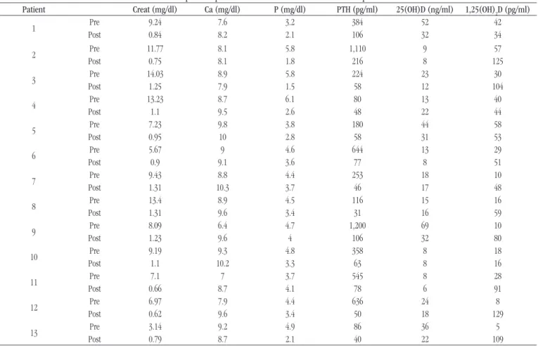
Individual results are shown in Table 1. Table 2 shows
29 ng/ml, and disability below 20 ng/ml(20); and 1,25(OH) 2D –
from 20 to 62.5 pg/ml(21).
DISCUSSION
At the time of transplant, all patients experienced biochemical changes consistent with renal failure: elevated serum creatinine, phosphate and PTH; and low levels of 25(OH)D and 1,25(OH)2D.
Three months after renal transplantation, serum creatinine concentrations indicate that these patients had recovery of renal function. In general, serum calcium levels increased in the irst two months after kidney transplantation, remaining within the normal range. Although all patients presented elevated PTH levels in the pre-transplantation period, only ive patients (38%) were hypocalcemic. This small number of patients with low calcium levels may be due to the dialysate composition with high calcium concentration, or the administration of phosphate binders containing calcium. Three months after transplantation, 12 of the 13 patients (92%) had higher
TABLE 1 − Serum concentrations of creatinine, calcium, phosphate, PTH, 25(OH)D, and 1,25(OH)
2D in immediate pre-transplantation and three months after renal transplantation
Patient Creat (mg/dl) Ca (mg/dl) P (mg/dl) PTH (pg/ml) 25(OH)D (ng/ml) 1,25(OH)2D (pg/ml)
1 Pre 9.24 7.6 3.2 384 52 42
Post 0.84 8.2 2.1 106 32 34
2 Pre 11.77 8.1 5.8 1,110 9 57
Post 0.75 8.1 1.8 216 8 125
3 Pre 14.03 8.9 5.8 224 23 30
Post 1.25 7.9 1.5 58 12 104
4 Pre 13.23 8.7 6.1 80 13 40
Post 1.1 9.5 2.6 48 22 44
5 Pre 7.23 9.8 3.8 180 44 58
Post 0.95 10 2.8 58 31 53
6 Pre 5.67 9 4.6 644 13 29
Post 0.9 9.1 3.6 77 8 51
7 Pre 9.43 8.8 4.4 253 18 10
Post 1.31 10.3 3.7 46 17 48
8 Pre 13.4 8.9 4.5 116 15 16
Post 1.31 9.6 3.4 31 16 59
9 Pre 8.09 6.4 4.7 1,200 69 10
Post 1.23 9.6 4 106 32 80
10 Pre 9.19 9.3 4.8 358 8 18
Post 1.1 10.2 3.3 63 8 16
11 Pre 7.1 7 3.7 545 8 28
Post 0.66 8.7 4.1 78 6 91
12 Pre 6.97 7.9 4.4 636 24 8
Post 0.62 9.6 3.4 50 18 129
13 Pre 3.14 9.2 4.9 86 36 5
Post 0.79 8.7 2.1 40 22 109
Creat: creatinine; Ca: calcium; P: inorganic phosphorus; PTH: intact parathyroid hormone.
TABLE 2 − Summary of statistical results of creatinine, calcium, phosphate, PTH, 25(OH)D, and 1,25(OH)2D measured in immediate pre-transplantation and three months after renal transplantation
Parameter Pre-TxR 3
rd month post-TxR
Range Median Mean (SD) Range Median Mean (SD) p value
Creatinine (mg/dl) 3.14-14.03 9.19 9.12 (3.256) 0.62-1.31 0.95 0.985 (0.247) < 0.0001 Calcium (mg/dl) 6.4-9.8 8.8 8.43 (0.98) 7.9-10.3 9.96 9.19 (0.81) 0.0644 Phosphate (mg/dl) 3.2-6.1 4.6 4.67 (0.85) 1.5-4.1 3.3 2.95 (0.86) < 0.0001
PTH (pg/ml) 80-1,200 358 447.4 (369.2) 31-216 58 75.2 (48.2) < 0.0001
25(OH)D (ng/ml) 8-69 18 25.5 (19.2) 6-32 17 17.8 (9.5) 0.3408
1,25(OH)2D (pg/ml) 5-58 28 27.2 (18) 39-298 59 72.5 (36.3) 0.0011
or sustained calcium levels; one patient, with previous calcium concentration within the normal range, had it decreased.
Only one patient with normal level of serum phosphate presented no reduction three months after transplantation, and four patients (31%) developed hypophosphatemia. Hypophosphatemia, accompanied by phosphaturia, is often described in kidney transplant patients, usually with resolution until the third month
after the surgery(8, 22). This change appears to be due to inhibition
of sodium gradient-dependent Na/P cotransport in the proximal tubule cells by elevated levels of PTH, low levels of 1,25(OH)2D, and high levels of ibroblast growth factor(23, 24).
In general, with the recovery of renal function, PTH levels are reduced by about 50% in the irst three to six months after transplantation, but about 25% of patients with high levels persist after one year. Vitamin D deiciency may be a factor contributing to increased PTH secretion, and its replacement is possibly important in prevention and control of secondary hyperparathyroidism. The persistence of hyperparathyroidism is the main cause of bone loss in stable transplant patients(15).
Van Cauter et al.(22), studying 49 renal transplant recipients,
described pre-transplantation PTH levels of 194.2 ± 273.5 pg/ml (mean ± SD), which stabilized at 71.5 ± 50.7 pg/ml six months after transplantation. In this study, pre-transplantation PTH levels were 447.4 ± 369.2 pg/ml, which stabilized at 75.2 ± 48.2 pg/ml three months after transplantation. All of our patients had elevated pre-transplant PTH levels, ive of which (38%) maintained this abnormality in the third month after transplantation. The pre-transplant levels of PTH in these ive patients were extremely high (two of them above 500, and two others above 1,000 pg/ml), suggesting maintenance of a state of hyperparathyroidism, even with recovery of renal function.
In addition to its action in maintaining homeostasis of calcium and phosphorus, vitamin D has been implicated as having relevance in maintaining muscle strength and in the prevention of some chronic diseases, including cardiovascular and neoplastic ones, type 1 diabetes, multiple sclerosis and rheumatoid arthritis,
among others(25, 26). For this reason, researchers started giving
more importance to vitamin D measurement, and the need for its supplementation in speciic populations, such as renal transplant patients(27). Serum levels of 25(OH)D are independent of renal
function and its endogenous production by exposure to sunlight, and by exogenous diet. The most important natural sources of vitamin D are fatty ish, such as salmon, and ish oil, including cod liver oil. The vitamin can also be found in supplemented foods such as milk and cereal(10). Low levels of 25(OH)D are described in patients
with pathologic fractures and even in control subjects, whereas subnormal concentrations of 1,25(OH)2D are usually only observed in some patients and not in controls. The activity of 25(OH)D is
about 1/500 of the activity of 1,25(OH)2D on the calcitriol receptor, but considering that the serum levels of 25(OH)D are 1,000 times greater than those of 1,25(OH)2D, the less active form can contribute to the overall effect, particularly in renal failure, when the levels of 1,25(OH)2D are decreased(28). Some authors(11, 17) have demonstrated
that up to 86% of the patients with end-stage renal disease may have the concentration of 25(OH)D below 30 ng/ml.
The incidence of vitamin D insuficiency or deiciency in transplanted patients has been reported to be about 50%, and its cause is multifactorial, including low sun exposure, use of sunscreens and increased catabolism due to immunosuppressive
drugs(8, 9). Mazzaferro et al.(29) found insuficiency or deiciency of
25(OH)D in 69.1% of renal transplant patients. In this study, nine of the 13 studied patients (69,2%) had levels of 25(OH)D considered deicient or insuficient, which remained unchanged three months after kidney transplantation, what may be due to the short time elapsed from transplantation and even to the rehabilitation status of these patients. As the use of immunosuppressants increase the risk of skin cancer, our patients are advised to use sunscreen and avoid direct sunlight. This condition, associated with the induction of increased catabolism of vitamin D by glucocorticoids(30), could
explain the high prevalence observed.
Stavroulopoulos et al.(24) reported that the prevalence of
deiciency or insuficiency of 25(OH)D is extremely high after kidney transplantation, affecting more than 90% of patients, especially in the irst year after the procedure. Some studies(14, 31, 32) reported
that patients with low levels of 25(OH)D maintain higher PTH levels compared to those patients who had their vitamin D levels normalized. An inverse correlation between PTH and 25(OH)D levels is described by Ghazali et al.(33) in dialysis patients. In a study
involving 129 kidney recipients, Reinhardt et al.(34) found an inverse
correlation between 25(OH)D and PTH after eight months of transplantation. Stavroulopoulos et al.(24) found no such correlation
in the recent post-transplant (less than one year) period, but it can be seen in the long-term transplant recipients. Just as some other
studies(35, 36), we found no signiicant association between PTH and
25(OH)D levels either in pre- or post-transplant measurements
(r = 0.219 and r = -0.133, respectively), what may be due to the short
time elapsed after transplantation and the recovery of these patients. Low levels of 1,25(OH)2D may occur with high prevalence in different populations due to a variety of reasons. Although the reduction of 1-alpha-hydroxylase activity is, in large part, responsible for them in patients with renal disease, other factors may be present, interfering with the action of the enzyme(37). The low levels of substrate
25(OH)D can contribute to the reduced synthesis of 1,25(OH)2D(38).
It is expected that with renal transplant, and, therefore, restoration of enzymatic activity, calcitriol levels increase. Van Cauter et al.(22),
± 9.1 pg/ml (mean ± SD), which normalized at 40 ± 19.6 pg/ml twelve months after transplantation. In this study, pre-transplantation calcitriol levels were 27.2 ± 18 pg/ml, and they stabilized at 72.5 ± 36.3 pg/ml three months after transplantation. Mazzaferro et al.(29),
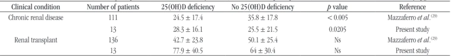
who studied two groups – one composed of 111 patients with chronic renal failure and another consisting of 136 patients who had received kidney transplantation –, found lower values of 1,25(OH)2D in patients with chronic renal failure and 25(OH)D deiciency compared to patients without deiciency of this vitamin. On the other hand, in the transplanted group there was no signiicant difference between patients with or without vitamin D deiciency. This inding suggests that deiciency of 25(OH)D seems to affect more the concentration of 1,25(OH)2D in patients with chronic kidney disease than in transplanted patients. In this study, albeit with a much smaller number of patients, we could demonstrate the same pattern, as shown
in Table 3. We detected reduced levels of 1,25(OH)
2D in six
pre-transplant patients (46%), and only one remained below the reference values three months after transplantation. This patient, in particular, had a very low pre-transplant level of 25(OH)D (8 ng/ml) that did not change after transplantation. One possible explanation for the lack of response in this case is the limited availability of the substrate 25(OH)D, and not the inactivity of the enzyme(24). Interestingly, two other patients,
with virtually the same post-transplant levels of 25(OH)D, had their concentration of 1,25(OH)2D normalized, what reinforces the inluence of other factors in this process. The average increase of post-transplant 1,25(OH)2D levels compared to pre-transplant levels was 266%. Although the immunoassay used for 1,25(OH)2D measurement is robust, it can measure 1-alpha-hydroxylated inactive metabolites apart from 1,25(OH)2D, justifying some very high values.
An issue that should be mentioned is the reference range used for both 25(OH)D and 1,25(OH)2D. The cut-off values for these parameters are not universally agreed in the literature. In addition to the possible inluences of general clinical conditions and environmental factors, intrinsic differences in study populations and differences in the used assays could be held responsible for the discrepancy.
CONCLUSION
The metabolic bone disease due to calcium and phosphorus metabolism disorders begins early in chronic kidney disease,
but the condition often remains underdiagnosed until it evolves with signiicant changes in calcium and phosphorus
levels(4, 39). Hyperphosphatemia stimulates PTH release and inhibits
the synthesis of 1,25(OH)2D, what leads to the development of secondary hyperparathyroidism and calcitriol deiciency. Our data, in agreement with the literature, conirms that even patients with good performance after renal transplantation may continue with some signiicant biochemical changes that result in deterioration of bone quality. Speciically in relation to vitamin D, our study reinforces the need for follow-up, which can be performed in the third month after transplantation. The main limitations of our study are the reduced number of patients and the short time elapsed after transplantation. Van Cauter et al.(22) analyzed 49 renal
transplant recipients; Mazzaferro et al.(29) studied 111 patients with
chronic renal disease and 136 transplanted patients. Reinhardt
et al.(34) studied 129 patients eight months after transplantation,
and Stavroulopoulos et al.(24) worked with two groups of patients,
one with 104 patients with less than one year of transplantation, and another with 140 patients over a year. Our study group is very limited and may not be representative of the majority of transplanted patients, especially in different stages after transplantation. Differences in sunlight exposure, pre-transplant conditions, therapeutic regimens, and variations in the consumption of fatty ish lead to differences in vitamin D status. The results of our study need to be conirmed in other transplanted cohorts.
APPROVAL
This study was approved by the Ethics Committee of the Universidade Federal de São Paulo on October 26, 2012, under number 132-031.
ACKNOWLEDGEMENTS
Thanks to Prof. Dr. Helio Tedesco Silva Júnior for their support in this work, to team of Clinical Biochemistry of the Hospital São Paulo Central Laboratory, especially, Celeste de Cássia Mendes and Tais Pavanelli Garcia for technical assistance and to Roche Diagnostics to enable graciously the dosages of 25-hydroxyvitamin D.
TABLE 3 − Concentration of 1,25(OH)
2D in patients with chronic kidney disease, and renal transplant patients, with and without 25(OH)D deiciency (pg/ml)
Clinical condition Number of patients 25(OH)D deiciency No 25(OH)D deiciency p value Reference
Chronic renal disease 111 24.5 ± 17.4 35.8 ± 17.8 < 0.005 Mazzaferro et al.(29)
13 28.3 ± 16.1 25.5 ± 21.5 0.0205 Present study
Renal transplant 136 42.7 ± 23.8 50.1 ± 25.4 Ns Mazzaferro et al.(29)
13 77.9 ± 40.5 64 ± 30.4 Ns Present study
RESUMO
Introdução: Função renal normal é condição para a manutenção do equilíbrio de cálcio e fósforo. A incidência de níveis
reduzidos de 1,25-di-hidroxivitamina D (1,25(OH)2D, ou calcitriol) em pacientes transplantados renais é de 50%. As causas são multifatoriais, incluindo baixa disponibilidade de 25-hidroxivitamina D (25(OH)D). Ainda que o transplante renal seja o tratamento de escolha, alguns pacientes mantêm alterações ósseas resultantes de vários fatores, como persistência de distúrbios minerais devido a disfunção do enxerto e ação dos imunossupressores. Objetivo: Avaliar a dinâmica de alguns parâmetros bioquímicos pós-transplante renal. Material e métodos: Treze pacientes, seguidos desde o pré-transplante até três meses pós- -transplante, com dosagens de creatinina, cálcio, fósforo, paratormônio (PTH), 25(OH)D e calcitriol. Resultados: A normalização do cálcio foi observada em 10 dos 13 pacientes (77%), e apenas um não teve os níveis de fósforo reduzidos; todos tinham PTH elevado no pré-transplante, quatro dos quais (31%) mantiveram essa alteração; nove (69%) tinham níveis baixos de 25(OH)D que se mantiveram praticamente inalterados pós-transplante. Baixos níveis de calcitriol foram observados em 46% dos pacientes no pré-transplante e permaneceram em apenas um deles. Esse paciente teve nível muito baixo de 25(OH)D pré-transplante (8 ng/ml), que não se alterou após a cirurgia. Uma possível explicação para a não normalização é a reduzida disponibilidade de substrato para a 1-alfa-hidroxilase. Conclusão: Nossos dados, em concordância com a literatura, confirmam que mesmo pacientes com boa evolução após o transplante renal podem continuar com alterações bioquímicas importantes associadas à deterioração da qualidade óssea.
Unitermos: transplante renal; 25-hidroxivitamina D; calcitriol; metabolismo ósseo.
REFERENCES
1. Llach F, Yudd M. Pathogenic, clinical, and therapeutic aspects of secondary hyperparathyroidism in chronic renal failure. Am J Kidney Dis. 1998; 32 (Suppl 2): S3-12.
2. Llach F, Velasquez F. Secondary hyperparathyroidism in chronic renal failure: pathogenic and clinical aspects. Am J Kidney Dis. 2001; 38(Suppl 5): S20-33.
3. Bastos MG, Bregman R, Kirsztajn GM. Doença renal crônica: frequente e grave, mas também prevenível e tratável. Rev Assoc Med Bras. 2010; 56: 248-53.
4. Hsu CY, Chertow GM. Elevations of serum phosphorus and potassium in mild to moderate chronic renal insuficiency. Nephrol Dial Transplant. 2002; 17: 1419-25.
5. Levi M. Post-transplant hypophosphatemia. Kidney Int. 2001; 59: 2377-87.
6. Block GA, Klassen PS, Lazarus JM, et al. Mineral metabolism, mortality, and morbidity in maintenance hemodialysis. J Am Soc Nephrol. 2004; 15: 2208-18.
7. Teng M, Wolf M, Ofsthun MN, et al. Activated injectable vitamin D and hemodialysis survival: a historical cohort study. J Am Soc Nephrol. 2005; 16: 1115-25.
8. Cunningham J. Post-transplantation bone disease. Transplantation. 2005; 79: 629-34.
9. Holick MF. Resurrection of vitamin D deiciency and rickets. J Clin Invest. 2006; 116: 2062-72.
10. Holick MF, Garabedian M. Vitamin D: photobiology, metabolism, mechanism of action, and clinical applications. In: Favus MJ, editor.
Primer on the metabolic bone diseases and disorders of mineral metabolism. 6th ed. Washington, DC: American Society for Bone and Mineral Research; 2006. p. 129-37.
11. Holick MF. Vitamin D: a D-Lightful health perspective. Nutrition Review. 2008; 10 Suppl 2: S182-94.
12. Vilarta CF. Prevalência de hipovitaminose D em pacientes transplantados renais. 2010. [dissertation]. São Paulo, Brasil: Universidade de São Paulo; 2010.
13. Braga Jr JWR, Neves, RMS, Pinheiro MM, et al. Prevalence of low trauma fracture in long-term kidney transplant patients with preserved renal function. Braz J Med Biol Res. 2006; 39: 137-47.
14. Giannini S, Sella S, Silva Netto F, et al. Persistent secondary hyperparathyroidism and vertebral fractures in kidney transplantation: role of calcium-sensing receptor polymorphisms and vitamin D deiciency. J Bone Min Res. 2010; 24: 841-8.
15. Gueiros APS, Neves CL, Sampaio EA, et al. Distúrbio mineral e ósseo após o transplante renal. J Bras Nefrol. 2011; 33: 189-247.
16. Nikkel LE, Hollenbeak CS, Fox EJ, et al. Risk of fractures after renal transplantation in the United States. Transplantation. 2009; 87: 1846-51. 17. González EA, Sachdeva A, Oliver DA, et al. Vitamin D insuficiency and deiciency in chronic kidney disease. A single center observational study. Am J Nephrol. 2004; 24: 503-10.
20. Holick MF, Binkley NC, Bischoff-Ferrari HA, et al. Evaluation, treatment, and prevention of vitamin D deiciency: an Endocrine Society clinical practice guideline. J Clin Endocrinol Metab. 2011; 96: 1911-30. 21. Lips P. Relative value of 25(OH) D and 1,25(OH)2D measurements.
J Bone Miner Res. 2007; 22: 1668-71.
22. Van de Cauter J, Sennesael J, Haentjens P. Long-term evolution of the mineral metabolism after renal transplantation: a prospective, single-center cohort study. Transplant Proc. 2011; 43: 3470-5.
23. Evenepoel P, Naesens M, Claes K, et al. Tertiary ‘hyperphosphatonism’ accentuates hypophosphatemia and suppresses calcitriol levels in renal transplant recipients. Am J Transplant. 2007; 7: 1193-2000.
24. Stavroulopoulos A, Cassidy MJ, Porter CJ, et al. Vitamin D status in renal transplant recipients. Am J Transplant. 2007; 7: 2546-52. 25. Holick MF. Vitamin D for health and in chronic kidney disease. Semin Dial. 2005; 18: 266-75.
26. Souberbielle JC, Body JJ, Lappe JM, et al. Vitamin D and musculoskeletal health, cardiovascular disease, autoimmunity and cancer: recommendations for clinical practice. Autoimmun Rev. 2010; 9: 709-15.
27. Vieth R, Bischoff-Ferrari H, Boucher BJ, et al. The urgent need to recommend an intake of vitamin D that is effective. Am J Clin Nutr. 2007; 85: 649-50.
28. Cunningham J, Makin H. How important is vitamin D deiciency in uraemia? Nephrol Dial Transplant. 1997; 12: 16-8.
29. Mazzaferro S, Pasquali M, Pugliese F, et al. Distinct impact of vitamin D insuficiency on calcitriol levels in chronic renal failure and renal transplant patients: a role for FGF23. J Nephrol. 2012; 25: 1108-18. 30. Levin A, Bakris GL, Molitch M, et al. Prevalence of abnormal serum vitamin D, PTH, calcium, and phosphorus in patients with chronic kidney
MAILING ADDRESS
Adagmar Andriolo
Escola Paulista de Medicina; Universidade Federal de São Paulo; Rua Barão do Triunfo, 142, apto 82 B; Brooklin Paulista; CEP: 04602-000; São Paulo-SP, Brazil; e-mail: [email protected].
disease: results of the study to evaluate early kidney disease. Kidney Int. 2007; 71: 31-8.
31. Boudville NC, Hodsman AB. Renal function and 25-hydroxyvitamin D concentrations predict parathyroid hormone levels in renal transplant patients. Nephrol Dial Transplant. 2006; 21: 2621-4.
32. Sadlier DM, Magee CC. Prevalence of 25(OH) vitamin D (calcidiol) deiciency at time of renal transplantation: a prospective study. Clin Transplant. 2007; 21: 683-8.
33. Ghazali A, Fardellone P, Pruna A, et al. Is low plasma 25-(OH) vitamin D a major risk factor for hyperparathyroidism and Looser’s zones independent of calcitriol? Kidney Int. 1999; 55: 2169-77.
34. Reinhardt W, Bartelworth H, Jockenhövel F, et al. Sequential changes of biochemical bone parameters after kidney transplantation. Nephrol Dial Transplant. 1998; 13: 436-42.
35. Lomonte C, Antonelli M, Vernaglione L, et al. Are low plasma levels of 25-(OH) vitamin D a major risk factor for hyperparathyroidism independent of calcitriol in renal transplant patients? J Nephrol. 2005; 18: 96-101.
36. Sezer S, Yavuz D, Canoz MB, et al. Vitamin D status, bone mineral
density, and inlammation in kidney transplantation patients. Transplant Proc. 2009; 41: 2823-5.
37. Andress DL. Vitamin D treatment in chronic kidney disease. Semin Dial. 2005; 18: 315-21.