Asynchronous expression of myeloid
antigens in leukemic cells in a
PML/RAR
α
α
α
α
α
transgenic mouse model
Divisão de Hematologia e Centro de Terapia Celular, Departamento de Clínica Médica,
Faculdade de Medicina de Ribeirão Preto,
Universidade de São Paulo, Ribeirão Preto, SP, Brasil B.A.A. Santana*, M.C. Pintão*,
R.S. Abreu e Lima, P.S. Scheucher, G.A.S. Santos, A.B. Garcia, R.P. Falcão and E.M. Rego
Abstract
Acute promyelocytic leukemia (APL) is characterized by the expan-sion of blasts that resemble morphologically promyelocytes and harbor a chromosomal translocation involving the retinoic acid recep-tor α (RARα) and the promyelocytic leukemia (PML) genes on
chromosomes 17 and 15, respectively. The expression of the PML/ RARα fusion gene is essential for APL genesis. In fact, transgenic
mice (TM) expressing PML/RARα develop a form of leukemia that
mimics the hematological findings of human APL. Leukemia is diagnosed after a long latency (approximately 12 months) during which no hematological abnormality is detected in peripheral blood (pre-leukemic phase). In humans, immunophenotypic analysis of APL blasts revealed distinct features; however, the precise immuno-phenotype of leukemic cells in the TM model has not been estab-lished. Our aim was to characterize the expression of myeloid antigens by leukemic cells from hCG-PML/RARα TM. In this study, TM
(N = 12) developed leukemia at the mean age of 13.1 months. Morphological analysis of bone marrow revealed an increase of the percentage of immature myeloid cells in leukemic TM compared to pre-leukemic TM and wild-type controls (48.63 ± 16.68, 10.83 ± 8.11, 7.4 ± 5.46%, respectively; P < 0.05). Flow cytometry analysis of bone marrow and spleen from leukemic TM identified the asynchronous co-expression of CD34, CD117, and CD11b. This abnormal pheno-type was rarely detected prior to the diagnosis of leukemia and was present at similar frequencies in hematologically normal TM and wild-type controls of different ages. The present results demonstrate that, similarly to human APL, leukemic cells from hCG-PML/RARα
TM present a specific immunophenotype.
Correspondence E.M. Rego
Departamento de Clínica Médica FMRP, USP
Av. Bandeirantes, 3900 14049-900 Ribeirão Preto, SP Brasil
Fax: +55-16-3633-6695 E-mail: [email protected]
Research supported by FAPESP (No. 01/08693-8). B.A.A. Santana was the recipient of a FAPESP fellowship (No. 01/07974-3).
*These authors contributed equally to this study.
Received July 29, 2005 Accepted December 2, 2005
Key words
•Acute promyelocytic leukemia
•Acute myelogenous leukemia
•Transgenic mice
•Flow cytometry
•Leukemogenesis
Acute promyelocytic leukemia (APL) is a distinct subtype of acute myelogenous leu-kemia invariably associated with recurrent chromosomal translocations involving the retinoic acid receptor α (RARα) locus on
chromosome 17 (1). In about 98% of APL cases, t(15;17) is detected and causes the fusion of RARα to the promyelocytic
PML/RARα fusion protein, which retains
the majority of the functional domains of the parental PML and RARα proteins (1).
An-other hallmark of APL is the block of differ-entiation at the promyelocytic stage pre-sented by the leukemic cells. Pharmacologi-cal doses of retinoic acid induce the degra-dation of the PML/RARα fusion protein and
disease remission (2). APL has a specific immunophenotypic profile, characterized by the co-expression of the pan-myeloid mark-ers CD13 (with a heterogeneous intensity of expression) and CD33 (homogeneous inten-sity of expression), and absence or low ex-pression of HLA-DR, CD11a and CD18 (3-5). APL is mostly CD34 negative and the low expression of this marker has been asso-ciated with the microgranular variant and with the expression of the bcr3 PML/RARα
isoform (6,7). In addition, a singular CD34 and CD15 pattern of expression has been reported, in which leukemic cells lose CD34 before they acquire CD15 expression, with the latter being never expressed at high lev-els (3). Finally, CD117 is frequently ex-pressed by APL cells, but with variable in-tensity (5). The study of the immunopheno-typic profile of APL has prompted some groups to develop flow cytometry methods for APL diagnosis and minimal residual dis-ease detection (3,4).
The generation of transgenic mouse (TM) models harboring the PML/RARα fusion
gene in their genome became a very useful tool for the study of APL pathogenesis. Sev-eral groups have used the human cathepsin G (hCG) minigene to drive the expression of PML/RARα to the promyelocytic stage of
myeloid differentiation, and these TM de-velop a lethal form of leukemia that closely resembles human APL (8,9). Overt acute leukemia occurred after a long pre-leukemic phase (12-15 months) and affected only 10 to 15% of the TM, suggesting that PML/ RARα is necessary but not sufficient for
full-blown leukemogenesis. In fact, Kogan et al. (10) demonstrated that double TM for
MRP8-PML/RARα and MRP8-BCL-2
de-veloped leukemia earlier than single MRP8-PML/RARα TM, suggesting that in this
mo-del BCL-2 cooperated with PML/RARα to
accelerate leukemogenesis and to block my-eloid differentiation.
Like human APL, leukemia in the hCG-PML/RARα TM responded to retinoic acid
treatment (8). Although leukemic cells of hCG-PML/RARα TM are morphologically
similar to human promyelocytes, their im-munophenotypic features have not been fully characterized. This characterization is an im-portant pre-requisite for studies aiming to analyze the gene and/or protein expression profiles, additional cytogenetic abnormali-ties, and proliferative and survival pathway deregulation in APL cells.
Thus, the objective of the present study was to determine if, similarly to human APL, leukemic cells from hCG-PML/RARα TM
also present a distinct immunophenotypic profile. If so, it was important to analyze TM prior to the development of leukemia and to establish if there is a progressive accumula-tion of immature myeloid cells during the pre-leukemic phase of the TM model or if the block of maturation is a late event in leukemogenesis.
hCG-PML/RARα TM were provided by
Prof. Pier Paolo Pandolfi (Memorial Sloan-Kettering Cancer Center, New York, NY, USA) and their generation has been described elsewhere (8). The mice were bred and main-tained under pathogen-free condition at the Animal Facility of the Fundação Hemocen-tro, Faculdade de Ciências Farmacêuticas de Ribeirão Preto, Universidade de São Paulo. Genotyping was performed by PCR analysis of tail genomic DNA (11) using the pair of primers C1 and D, and the reaction condi-tions described by van Dongen et al. (12). For monitoring peripheral blood (PB) counts, hCG-PML/RARα TM and their wild-type
using a Coulter T-890 counter (Coulter Cor-poration, Hialeah, FL, USA). PB differential counts were performed on Wright-Giemsa-stained smears. The diagnosis of leukemia was based on the following criteria: a) pres-ence of blasts/promyelocytes in the PB; b) leukocytosis (leukocyte counts >30 x 103/ µL), and c) anemia (hemoglobin <10 g/dL) or thrombocytopenia (platelet counts <500 x 103/µL). In the present study, hCG-PML/ RARα TM developed leukemia at the mean
age of 13.1 months. For brevity, hCG-PML/ RARα TM aged between 6 to 12 months
presenting PB counts within the normal range were named ‘pre-leukemic’.
Twelve leukemic hCG-PML/RARα TM,
6 pre-leukemic hCG-PML/RARα and 12
sex- and age-matched WT controls aged 6 to 12 months were sacrificed by CO2 asphyxi-ation at the time of diagnosis (leukemic), or at 4, 8, and 12 months of age (2 pre-leukemic and 4 WT controls at each time point). Bone marrow (BM) cells were obtained by flush-ing the bone cavity with RPMI 1640 (Gibco, Grand Island, NY, USA) and splenic cell suspensions by mechanical disruption of the spleen. The cells were washed once and the
pellet was resuspended in PBS at a concen-tration of 106/mL. Cytospin slides were pre-pared in a Cytospin 3 Cell Preparation Sys-tem (Shandon, Pittsburgh, PA, USA), air-dried, and Wright-Giemsa stained. A mini-mum of 200 cells were counted and myeloid cells were classified as immature, intermedi-ate or mature according to the Bethesda Proposals for Classification of non-lymphoid hematopoietic neoplasms in mice (13). The leukemic phase of the transgenic model was characterized by a marked increase of the number of white blood cells with a shift to the left and intense thrombocytopenia ac-companied by mild to moderate anemia (Table 1). The percentage of blasts in PB was only moderately elevated, in contrast to what was observed in BM and spleen (Table 1). The leukemic cells morphologically re-sembled human promyelocytes. Invariably, leukemic mice presented splenomegaly with a spleen weight (mean ± SD) 1.25 ± 0.9% of body weight. Along with the lack of abnor-malities in PB, a discrete, but nonsignificant, increase in spleen size was detected in pre-leukemic TM (0.27 ± 0.2 vs 0.1 ± 0.1% body
weight in pleukemic and WT mice,
re-Table 1. Peripheral blood, bone marrow, and spleenic cell counts of leukemic (N = 12), pre-leukemic (N = 6), and wild-type (N = 12) mice.
Genotype Hemoglobin White blood cells Platelets Immature Intermediate Differentiated Lymphocytes (g/dL) (x 103) (x 103) cells (%) cells (%) cells (%) (%)
Peripheral blood
LEU 9.90 ± 3.53* 107.78 ± 122.96* 250.32 ± 180.29* 17.90 ± 12.04* 15.65 ± 10.18* 36.00 ± 16.39 30.05 ± 20.27* PRE 15.02 ± 3.65 9.26 ± 4.51 1100.00 ± 514.87 0.00 ± 0.00 0.67 ± 1.03 24.33 ± 11.83 73.33 ± 12.69 WT 14.08 ± 3.23 7.41 ± 3.47 1055.13 ± 443.22 0.00 ± 0.00 0.70 ± 1.34 31.30 ± 17.18 66.80 ± 15.55
Bone marrow
LEU NA NA NA 48.63 ± 16.68* 32.32 ± 10.36* 12.00 ± 9.97* 6.84 ± 6.26* PRE NA NA NA 10.83 ± 8.11 21.33 ± 4.93 46.17 ± 21.48 17.50 ± 9.89 WT NA NA NA 7.40 ± 5.46 20.70 ± 4.79 41.10 ± 14.43 27.50 ± 9.96
Spleen
LEU NA NA NA 48.00 ± 20.88* 27.90 ± 18.25* 9.70 ± 6.38 20.67 ± 19.61* PRE NA NA NA 0.17 ± 0.41 0.50 ± 0.84 11.50 ± 7.77 87.83 ± 6.91 WT NA NA NA 0.44 ± 0.53 1.00 ± 0.71 2.56 ± 1.33+ 91.78 ± 11.21
Data are reported as mean ± SD. LEU = leukemic mice; PRE = pre-leukemic mice; WT = wild-type mice; NA = not applicable.
*P < 0.05 for LEU vs PRE and LEU vs WT comparison. +P < 0.05 for WT vs LEU and WT vs PRE comparison (one-way ANOVA followed by
spectively). The only significant difference between pre-leukemic and WT mice detected by cytomorphological analysis of PB, BM and spleen was a higher percentage of termi-nally mature myeloid cells in the spleen of the former.
For immunophenotypic analysis BM and splenic single cell suspensions were pre-pared as described above and 100 µL of the suspension were incubated with 5 µL of each monoclonal antibody (mAb) for 20 min at 4ºC protected from light. The following com-binations of mAbs directly conjugated with fluorochromes [fluorescein isothiocyanate (FITC), phycoerythrin (PE), peridin chloro-phyll protein (Per-CP)] were employed: CD117-PE/CD34-FITC/CD45-Per-CP, CD117-PE/CD11b-FITC/CD45-Per-CP, Gr-1-PE/CD11b-FITC/CD45-Per-CP and CD3-FITC/CD19-PE/CD45-Per-CP. Fluoro-chrome-conjugated isotypic antibodies of irrelevant specificity were used as negative controls. The anti-CD117 and anti-CD19 mAbs were obtained from Southern Bio-technology Associates (Birmingham, AL, USA); anti-CD3-ε from Santa Cruz
Bio-technology (Santa Cruz, CA, USA) and the remaining mAbs were purchased from BD Biosciences Pharmingen (San Diego, CA, USA). Red blood cells were lysed immedi-ately after labeling by incubation with 2 mL of FACS lysing solution (Becton Dickinson,
San Jose, CA, USA) as recommended by the manufacturer. A minimum of 10,000 events/ tube were acquired with a FACScan flow cytometer. Viable CD45-positive cells were gated and the percentage of each cell subset was determined using the CellQuest soft-ware (Becton Dickinson).
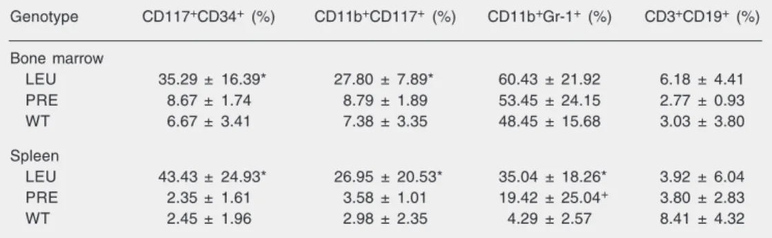
The spleen and BM of leukemic mice were infiltrated by CD117+CD34+ and CD117+CD11b+ cells, the former being the most frequent subset detected (Table 2). In contrast, the same phenotype was expressed by less than 3% of cells in spleen samples from WT and in pre-leukemic TM (Table 2). No differences were observed between pre-leukemic mice aged 4, 8 or 12 months. Im-portantly, a significant increase in the more mature phenotype CD11b+ Gr-1+ (19.42 ± 25.04 vs 4.29 ± 2.57%, P < 0.05) but not of
the immature subset CD34+ CD117+ CD11b+, was detected in the spleen of pre-leukemic TM compared to WT.
In the present study, the immunopheno-typic analyses demonstrated that BM and spleen of leukemic TM were infiltrated by CD117+CD34+ and CD117+CD11b+ cells, a phenotype that was seldom detected in the WT spleen. The CD117 antigen is rare in normal BM in both humans and mice, and is characteristic of the early stages of myeloid differentiation, decreasing after the colony-forming unit-granulocyte-monocyte stage
Table 2. Immunophenotypic profile of leukemic (N = 12), pre-leukemic (N = 6), and wild-type (N = 12) mice.
Genotype CD117+CD34+ (%) CD11b+CD117+ (%) CD11b+Gr-1+ (%) CD3+CD19+ (%)
Bone marrow
LEU 35.29 ± 16.39* 27.80 ± 7.89* 60.43 ± 21.92 6.18 ± 4.41 PRE 8.67 ± 1.74 8.79 ± 1.89 53.45 ± 24.15 2.77 ± 0.93 WT 6.67 ± 3.41 7.38 ± 3.35 48.45 ± 15.68 3.03 ± 3.80
Spleen
LEU 43.43 ± 24.93* 26.95 ± 20.53* 35.04 ± 18.26* 3.92 ± 6.04 PRE 2.35 ± 1.61 3.58 ± 1.01 19.42 ± 25.04+ 3.80 ± 2.83
WT 2.45 ± 1.96 2.98 ± 2.35 4.29 ± 2.57 8.41 ± 4.32
Data are reported as mean ± SD. LEU = leukemic mice; PRE = pre-leukemic mice; WT = wild-type mice. *P < 0.05 for LEU vs PRE and LEU vs WT comparison. +P < 0.05 for PRE vs LEU and PRE vs WT
(4,14). Similarly, CD34 is an important marker of human hematopoietic stem cells and is lost before the decline in CD117 expression (15). On the other hand, in adult mice, only 20% of the hematopoietic stem cells are CD34+ (16). Ishida et al. (17) have demonstrated the co-expression of different CD11b levels (an antigen expressed late dur-ing granulocytic differentiation) in a small fraction of CD34+ hematopoietic stem cells of normal adult mice. Therefore, in view of the rarity of the immunophenotypic profile the co-expression of CD34, CD11b and CD117 should be considered asynchronous. In fact, in human patients the expression of CD11b in association with immaturity mark-ers like CD34 and CD117 has been reported as an example of asynchronous antigen ex-pression characteristically observed in acute myelogenous leukemia (18).
Westervelt et al. (19), analyzing a knock-in model knock-in which the PML/RARα cDNA
was expressed under the control of the mu-rine cathepsin G locus, reported that 90% of these mice developed APL between 6 and 16 months of age. Interestingly, the leukemic cells presented an abnormal immunopheno-type co-expressing CD34 and Gr-1. Simi-larly, Pollock et al. (20) described the same immunophenotype in leukemic cells from double transgenic mice co-expressing the
bcr3 isoform PML/RARα and the reciprocal
product of the t(15;17), RARα/PML.
There-fore, the asynchronous myeloid antigen ex-pression is not dependent on the promoter region controlling PML/RARα expression,
on the PML/RARα isoform, or on the
pres-ence of the reciprocal.
In pre-leukemic TM, we did not detect an increase of the percentage of cells with the CD117+CD34+ and CD117+CD11b+ pheno-type compared to WT controls. In contrast, there was an infiltration by the more mature myeloid subset identified morphologically and immunophenotypically (CD11b+ Gr-1+) in the spleen of pre-leukemic and leukemic TM when compared to WT controls. Like-wise, the weight of the spleen increased in the same fashion. The follow-up of hCG-PML/RARα TM using the more sensitive
and specific immunophenotypic method, associated with morphological analysis, al-lowed us to demonstrate that the block of differentiation is a late event during the pre-leukemic phase, thus suggesting that it may depend on additional mutagenic events.
Acknowledgments
We are very grateful to Prof. Pier Paolo Pandolfi (Memorial Sloan Kettering Cancer Center, New York, NY, USA) who kindly provided the PML/RARα transgenic mice.
References
1. Brunning RD, Matutes E, Flandrin G et al. (2000). Acute myeloid leukaemia. In: Jaffe ES, Harris NL, Stein H et al. (Editors), Pathology and Genetics of Tumours of Haematopoietic and Lymphoid Tissues. IARC Press, Lyon, France, 75-108.
2. Rego EM, He LZ, Warrell Jr RP et al. (2000). Retinoic acid (RA) and As2O3 treatment in transgenic models of acute promyelocytic
leuke-mia (APL) unravel the distinct nature of the leukemogenic process induced by the PML/RARalpha and PLZF-RARalpha oncoproteins.
Proceedings of the National Academy of Sciences, USA, 97: 10173-10178.
3. Orfao A, Chillon MC, Bortoluci AM et al. (1999). The flow cytometric pattern of CD34, CD15 and CD13 expression in acute myeloblastic leukemia is highly characteristic of the presence of PML/RARalpha
gene rearrangements. Haematologica, 84: 405-412.
4. Rizzatti EG, Garcia AB, Portieres FL et al. (2002). Expression of CD117 and CD11b in bone marrow can differentiate acute promye-locytic leukemia from recovering benign myeloid proliferation. Ameri-can Journal of Clinical Pathology, 118: 31-37.
5. Paietta E, Goloubeva O, Bennett JM et al. (2002). A surrogate marker profile for acute promyelocytic leukaemia (APL) and the association of immunophenotypic markers with morphologic and molecular subtypes of APL. Blood, 100 (Suppl 1): 229b.
7. Rizzatti EG, Portieres FL, Martins SL et al. (2004). Microgranular and t(11;17)/PLZF-RARα variants of acute promyelocytic leukemia
also present the flow cytometric pattern of CD13, CD34 and CD15 expression characteristic of PML/RARα gene rearrangement.
Ameri-can Journal of Hematology, 76: 44-51.
8. He LZ, Tribioli C, Rivi R et al. (1997). Acute leukemia with promyelo-cytic features in PML/RARalpha transgenic mice. Proceedings of the National Academy of Sciences, USA, 94: 5302-5307.
9. Grisolano JL, Sclar GM & Ley TJ (1994). Early myeloid cell-specific expression of the human cathepsin G gene in transgenic mice.
Proceedings of the National Academy of Sciences,USA, 91: 8989-8993.
10. Kogan SC, Brown DE, Shultz DB et al. (2001). BCL-2 cooperates with promyelocytic leukemia retinoid acid receptor α chimeric
pro-tein (PMLRARα) to block neutrophil differentiation and initiate acute
leukemia. Journal of Experimental Medicine, 193: 531-543. 11. Sambrook J & Russell DW (2001). Molecular Cloning: A Laboratory
Manual 1. 3rd edn. Cold Spring Harbor Laboratory Press, New York, 6.26.
12. van Dongen JJ, MacIntyre EA, Gabert JA et al. (1999). Standardized RT-PCR analysis of fusion gene transcripts from chromosome aber-rations in acute leukemia for detection of minimal residual disease.
Leukemia, 13: 1901-1928.
13. Kogan SC, Ward JM, Anver MR et al. (2002). Bethesda proposals for classification of nonlymphoid hematopoietic neoplasms in mice.
Blood, 100: 238-245.
14. Macedo A, Orfao A, Martinez A et al. (1995). Immunophenotype of c-kit in normal human bone marrow: implications for the detection of minimal residual disease in AML. British Journal of Haematology, 89: 338-341.
15. Escribano L, Ocqueteau M, Almeida J et al. (1998). Expression of the c-kit (CD117) molecule in normal and malignant hematopoiesis.
Leukemia and Lymphoma, 30: 459-466.
16. Ito T, Tajima F & Ogawa M (2000). Developmental changes of D34 expression by murine hematopoietic stem cells. Experimental He-matology, 28: 1269-1273.
17. Ishida A, Zeng H & Ogawa M (2002). Expression of lineage markers by CD34+ hematopoietic stem cells of adult mice. Experimental
Hematology, 30: 361-365.
18. Macedo A, Orfao A, Vidriales MB et al. (1995). Characterization of aberrant phenotypes in acute myeloblastic leukemia. Annals of He-matology, 70: 189-194.
19. Westervelt P, Lane AA, Pollock JL et al. (2003). High-penetrance mouse model of acute promyelocytic leukemia with very low levels of PML/RARα expression. Blood, 102: 1857-1865.
20. Pollock JL, Westervelt P, Kurichety AK et al. (1999). A bcr-3 isoform of RARα-PML potentiates the development of PML/RARα-driven