ISSN 0100-879X
BIOMEDICAL SCIENCES
AND
CLINICAL INVESTIGATION
www.bjournal.com.br
www.bjournal.com.br
Volume 42 (11) 993-1118 November 2009
Faculdade de Medicina de Ribeirão Preto Campus
Ribeirão Preto
Institutional Sponsors
The Brazilian Journal of Medical and Biological Research is partially financed by
Braz J Med Biol Res, November 2009, Volume 42(11)1044-1049
Stem cell factor protects against neuronal apoptosis by
activating AKT/ERK in diabetic mice
Stem cell factor protects against neuronal
apoptosis by activating AKT/ERK in diabetic mice
J.-W. Li
1,3,4*, L.-L. Li
2*, L.-L. Chang
1, Z.-Y. Wang
3,4,5and Y. Xu
1,2,3,4,51Department of Neurology, Affiliated Drum Tower Hospital, Nanjing Medical University, Nanjing, Jiangsu, China 2Department of Neurology, Affiliated Drum Tower Hospital of Nanjing, School of Clinical Medicine,
Southeast University, China
3Department of Neurology, Affiliated Drum Tower Hospital, Nanjing University Medical School, Nanjing, Jiangsu, China 4Jiangsu Key Laboratory for Molecular Medicine, Nanjing, Jiangsu, China 5The State Key Laboratory of Pharmaceutical Biotechnology, Nanjing University,
Nanjing, Jiangsu, China
Abstract
Neuronal apoptosis occurs in the diabetic brain due to insulin deficiency or insulin resistance, both of which reduce the ex -pression of stem cell factor (SCF). We investigated the possible involvement of the activation of the MAPK/ERK and/or AKT
pathways in neuroprotection by SCF in diabetes.Male C57/B6 mice (20-25 g) were randomly divided into four groups of 10 animals each. The morphology of the diabetic brain in mice treated or not with insulin or SCF was evaluated by H&E staining
and TUNEL. SCF, ERK1/2 and AKT were measured by Western blotting. In diabeticmice treated with insulin or SCF, there was fewer structural change and apoptosis in the cortex compared to untreated mice. The apoptosis rate of the normal group, the diabetic group receiving vehicle, the diabetic group treated with insulin, and the diabetic group treated with SCF was 0.54 ± 0.077%, 2.83 ± 0.156%, 1.86 ± 0.094%, and 1.78 ± 0.095% (mean ± SEM), respectively. SCF expression was lower in the diabetic cortexthan in the normal cortex; however, insulinincreased the expression of SCF in the diabetic cortex.Furthermore,
expression of phosphorylated ERK1/2 and AKT was decreased in the diabetic cortex compared to the normal cortex. However, insulin or SCF could activate the phosphorylation of ERK1/2 and AKT in the diabetic cortex. The results suggest that SCF may
protect the brain from apoptosis in diabetes and that the mechanism of this protection may, at least in part, involve activation
of the ERK1/2 and AKT pathways. These results provide insight into the mechanisms by which SCF and insulin exert their
neuroprotective effects in the diabetic brain.
Key words: Stem cell factor; Diabetes; MAPK/ERK; AKT
Introduction
Correspondence: Y. Xu, Department of Neurology, Affiliated Drum Tower Hospital, Nanjing University Medical School, 321 Zhong
-Shan Road, Nanjing, Jiangsu, 210008, China. Fax: +86-25-8331-7016. E-mail: [email protected]
Research supported by the National Nature Science Foundation (#30470612, #30670739) of China, the Doctoral Program Founda-tion of the Ministry of EducaFounda-tion of China (#20060284044), and the InternaFounda-tional CooperaFounda-tion Program and Outstanding Researcher Program (#BZ2006045, #06-B-002, #RC2007006) of Jiangsu Province of China.
*These authors contributed equally to this study.
Received December 27, 2008. Accepted August 24, 2009. Available online October 5, 2009. Although structural abnormalities in neural tissue such
as synaptic and neuronal degeneration and neuronal apoptosis have been demonstrated in type 1 and type 2
diabetic rodents (1-3), very little is known about the mecha -nisms leading to these defects. Many researchers have suggested that neuronal apoptosis in diabetes is related to hyperglycemia (4), and insulin, proinsulin C peptide and
insulin-like growth factor (IGF) have anti-apoptotic effects (5,6). Furthermore, insulin or insulin-like growth factor-1 (IGF-1) induced the expression of stem cell factor (SCF)
and a lower expression of SCF was observed in the mouse
gut during diabetic gastroparesis (7,8). SCF, also termed Kit
ligand, steel factor, mast cell growth factor, or c-kit ligand,
is a receptor of tyrosine kinase. Both SCF and Kit support
neuronal survival and function and influence cell differentia -tion, migra-tion, and proliferation possibly by mutual
interac-tion (9,10). Knockdown of SCF results in abnormalities in
learning and memory in mice (11,12). SCF is a cytokine
Stem cell factor inhibited neuronal apoptosis in diabetes 1045
www.bjournal.com.br Braz J Med Biol Res 42(11) 2009
transduction may involve many signaling pathways. The cytoprotection induced by SCF is associated with activation of the AKT and/or MAPK signaling pathways (14,15). In the present study, we tested the hypothesis that SCF protects
neurons from apoptosis in diabetes via a mechanism that
partially involves the AKT and ERK pathways.
Material and Methods
Animal model
The animal study was conducted in accordance with National Regulations of Experimental Animal Administra
-tion. All experimental animal protocols were approved by the Committee of Experimental Animal Administration of Nanjing Medical University. Diabetes was induced in male C57/B6 mice (20-25 g) with a single intraperitoneal injec -tion of streptozotocin (100 mg/kg in 0.1 M citrate buffer,
pH 4.4; Sigma, USA); 72 h later the plasma glucose level of the mice (diabetes type I model) was above 300 mg/
dL (OneTouch® Ultra Blood Glucose Monitoring System, Johnson, USA) (16). Plasma glucose was evaluated weekly for 4 weeks (see plasma glucose level in Figure 1). Control mice received an equal volume of citrate buffer. Mice were
randomly divided into four groups of 10 animals each, i.e., normal control (CON), diabetic receiving vehicle (DM),
diabetic treated with insulin (DM + INS: 40 U·kg-1·day-1; Wanbang Bio-pharm, China), and diabetic treated with SCF (DM + SCF: 0.1 mg·kg-1·48 h-1; BD, USA). Four weeks
laterseven brains from each group were rapidly removed
and the cerebral cortices and hippocampi were isolated. Three brains from each group were transcardially perfused with cold 0.1 M phosphate buffer, pH 7.4, followed by 4.0% paraformaldehyde. The samples were stored at -80°C for later experiments.
HE staining and TUNEL assay
Brain sections were stained with hematoxylin and eosin
for histological assay. Cell damage was also assessed by TUNEL (Boster, Wuhan, China), which was performed as previously described (17). Briefly, 20-µm brain sections were incubated with TdT labeling buffer containing dUTP mix,
Mn2+ and TdT enzyme for 60 min at 37°C to incorporate biotinylated nucleotides at the sites of DNA breaks. Sections
were also incubated with diluted extraavidin-peroxidase link for 30 min at 37°C, followed by the addition of DAB (diaminobenzidine tetrahydrochloride) at room temperature
to develop a brown color. They were counterstained with modified Harris’ hematoxylin,changing to blue with TBS and observed microscopically. The number of cells exhibit
-ing a positive TUNEL signal was quantified with the Soft
Imaging System (Image Pro Plus 5.02, Media Cybernetics,
Inc., USA).
Western blotting
Total protein and protein expression by the cortex was assayed as described previously (17). Membranes were probed with primary antibodies against SCF (1:500, BD), c-kit (1:500, Santa Cruz Biotechnology, USA), phosphorated
ERK1/2, AKT (cell signaling, 1:1000), total ERK1/2, AKT
or GAPDH (cell signaling, 1:1000). Proteins were detected using horseradish peroxidase-conjugated goat or
anti-rabbit secondary antibodies and visualized by chemilumi-nescence using an ECL kit (Amersham Pharmacia Biotech,
USA) and exposure to film. The intensity of the blots was quantified by densitometry (AlphaEaseFC, Alpha Innotech
Corporation, USA). GAPDH, total ERK or total AKT were
used as loading controls.
Statistical analysis
Data are reported as means ± SEM. One-way ANOVA followed by multiple comparisons was used for statistical
comparisons. In all statistical procedures, a probability
value of P < 0.05 was used as a cutoff for statistical sig
-nificance.
Results and Discussion
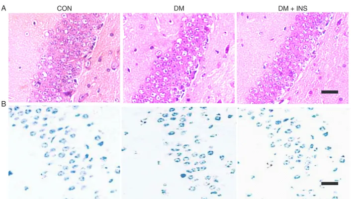
Cortical apoptosis appeared in the diabetic mice
Model type 1 diabetic mice (blood glucose level 300-450
mg/dL) were prepared using streptozotocin. To confirm pre
-vious studies, pathology and cell apoptosis were determined in the diabetic brains. Histological results confirmed strong
staining of the nuclei, an intensely eosinophilic cytoplasm
and widening of Virchow-Robin spaces. Along with these
changes, neuronal apoptosis occurred in the same areas
(Figure 2B,C). CON, DM + SCF or INS animals showed few pathological changes of this kind (Figure 2A). The pathological injuries in the cortex of diabetic mice, including structural changes and apoptosis, were fewerin animals
pretreated with insulin or SCF. However, in contrast to previous studies, the hippocampus did not show structural changes or apoptosis (Figure 3A,B). In other words, there
Figure 1. Plasma glucose curve. Type 1 diabetes was induced
in mice by a single intraperitoneal injection of streptozotocin (100 mg/kg), with plasma glucose levels exceeding 250 mg/dL 72 h later. Plasma glucose was then measured weekly. CON = nor
-mal mice injected with citrate buffer; DM = diabetic mice injected with vehicle; DM + INS = diabetic mice treated with insulin (40 U·
is a diabetes-related programmed neuronal cell death in
the hippocampus, which results in neuronal loss in type 1
diabetes (18,19). Different results might be obtained using
different models or at different times after experimental hyperglycemia. To test this model, we used a lower dose of streptozotocin and performed the experiment by the 4th week. The observed difference in structure changesmay have been related to milder diabetic damage.
SCF protects against apoptosis in the diabetic brain There is evidence that SCF expression levels can influence neuronal apoptosis in the diabetic brain. It is well known that diabetes results from insulin deficiency or variable degrees of insulin resistance. However, insulin or IGF-1 is a critical factor for inducing SCF expression and
maintaining SCF signaling (7,8). Reduced SCF resulted
in smooth muscle myopathy and loss of interstitial cells
of Cajal in murine diabetic gastroparesis (8). SCF and its receptor (c-kit) are expressed throughout the adult nervous
system. SCF includes both membrane-bound SCF (M-SCF) and soluble SCF (S-SCF), the latter consisting of cleaved
soluble forms of SCF (9,10). The present study showed
that the levels of both M-SCF and S-SCF in the diabetic
cortex were lower than that in the normal cortex (Figure
4) and that insulin can increase both M-SCF and S-SCF in the diabetic brain. Further study revealed that cortical apoptosis appeared in the diabetic mice and that SCF could attenuate it (Figure 2).
In an increasing number of studies, it has been shown that neuronal expression of SCF is induced in various forms of brain injury and that SCF expression after brain injury is correlated with neurogenesis and the mechanism of brain
Stem cell factor inhibited neuronal apoptosis in diabetes 1047
www.bjournal.com.br Braz J Med Biol Res 42(11) 2009
repair (14,20-22). SCF protects against camptothecin and glutamate-induced neuronal cell death (13,14). SCF and Kit are responsible for the survival of a variety of cells including neuronal cells and hematopoietic lineages (23). Targeted disruption of the kit gene leads to marked defects in learn-ing and memory (12), also a key manifestation of diabetic
encephalopathy. In the present study, no abnormal expres
-sion of c-kit was found in the diabetic brain. Kit alteration may possibly follow alterations in SCF. Our model using milder diabetic damage suggests that pathological injury in the diabetic brain may first occur in the cortex and only later extend to the hippocampus. Our model further sug
-gests that SCF/c-kit expression may be used to evaluate
the severity of diabetic encephalopathy.
Neuroprotection in diabetes by SCF via activation of AKT/ERK
To further investigate which signaling pathway is
involved in neuroprotection by SCF in diabetes, diabetic
mice were pretreated with insulin, SCF or vehicle and the
subsequent activation of MAPK/ERK1/2 and AKT in the
cortex was determined by Western blotting, as described in Figure 5. The results indicated that there was less activation of ERK1/2 and AKT in the cortex of the diabetic brain than in the normal cortex. However, the addition of SCF or insulin
to this model effectively activated MAPK and AKT. Both the
MAPK/ERK and PI-3K/Akt signaling pathways are essential
Figure 3. Morphology of the hippocampus. A, Hematoxylin and eosin staining. No significant structure change among the three groups
was found in the hippocampus. Bar = 40 µm.B, TUNEL assay. No apoptosis was observed in the hippocampus among the three groups. Bar = 25 µm.. For abbreviations, see legend to Figure 1.
Figure 4.A, Expression of M-SCF, S-SCF and c-kit by Western
blotting. B, Relative density of A. Expression of SCF
(membrane-bound SCF, M-SCF and soluble SCF, S-SCF) in the DM group
was lower than in the CON group; DM + INS increased the ex
-pression of SCF. There were no significant differences among the three groups in the expression of c-kit.
*
P < 0.05(one-wayin the prevention of neuronal apoptosis and are also im-plicated in the protection of the brain from ischemic stroke
and Alzheimer’s disease (14,17). SCF also suppressed
camptothecin and glutamate-induced neuronal apoptosis via MAPK/ERK and PI-3K/Akt activation. SCF-mediated signal transduction may involve a different cell signaling
pathway (14). Interestingly, SCF-mediated cytoprotection
in non-central nervous system cell types is also linked to
activation of the AKT and/or the MAPK signaling pathways
(24,25), suggesting that a similar mechanism of action of SCF/c-kit may be involved in neuroprotection and in the protection of other cell types.
Cortical neuronal apoptosis appeared in the diabetic
brain due to reduced SCF expression and this apoptosis could be inhibited by SCF. The mechanism by which this
occurred may be correlated to the activation of the ERK
and AKT pathways. Accordingly, SCF may hold promise
for development as a neuroprotectant for treating
diabetes-related brain injury.
Acknowledgments
We thank Mrs. Marilyn White for editing the manu-script.
References
1. Bestetti G, Rossi GL. Hypothalamic lesions in rats with
long-term streptozotocin-induced diabetes mellitus. A semiquantitative light- and electron-microscopic study. Acta Neuropathol 1980; 52: 119-127.
2. Mukai N, Hori S, Pomeroy M. Cerebral lesions in rats with
streptozotocin-induced diabetes. Acta Neuropathol 1980;
51: 79-84.
3. Mijnhout GS, Scheltens P, Diamant M, Biessels GJ, Wessels
AM, Simsek S, et al. Diabetic encephalopathy: A concept in
need of a definition. Diabetologia 2006; 49: 1447-1448.
Figure 5.A, C, Expression of phosphorylated AKT (P-AKT) and ERK (P-ERK) by Western blotting. B, D, Relative density of A and C.
There is decreased activation of AKT and ERK in DM; INS or SCF increased the expression of phosphorylated AKT and ERK.
*
P <Stem cell factor inhibited neuronal apoptosis in diabetes 1049
www.bjournal.com.br Braz J Med Biol Res 42(11) 2009
4. Srinivasan S, Stevens M, Wiley JW. Diabetic peripheral neuropathy: evidence for apoptosis and associated mito-chondrial dysfunction. Diabetes 2000; 49: 1932-1938.
5. Li ZG, Zhang W, Sima AA. C-peptide enhances insulin-mediated cell growth and protection against high glucose-induced apoptosis in SH-SY5Y cells. Diabetes Metab Res Rev 2003; 19: 375-385.
6. Kuemmerle JF. Endogenous IGF-I protects human intestinal smooth muscle cells from apoptosis by regulation of GSK-3
beta activity. Am J Physiol Gastrointest Liver Physiol 2005;
288: G101-G110.
7. Hsueh WA, Law RE. Insulin signaling in the arterial wall. Am J Cardiol 1999; 84: 21J-24J.
8. Horvath VJ, Vittal H, Lorincz A, Chen H, Almeida-Porada G,
Redelman D, et al. Reduced stem cell factor links smooth
myopathy and loss of interstitial cells of Cajal in murine dia -betic gastroparesis. Gastroenterology 2006; 130: 759-770. 9. Ashman LK. The biology of stem cell factor and its receptor
C-kit. Int J Biochem Cell Biol 1999; 31: 1037-1051.
10. Hollenbeck ST, Sakakibara K, Faries PL, Workhu B, Liu B, Kent KC. Stem cell factor and c-kit are expressed by and may affect vascular SMCs through an autocrine pathway. J Surg Res 2004; 120: 288-294.
11. Motro B, Wojtowicz JM, Bernstein A, van der Kooy D. Steel mutant mice are deficient in hippocampal learning but not
long-term potentiation. Proc Natl Acad Sci U S A 1996; 93: 1808-1813.
12. Katafuchi T, Li AJ, Hirota S, Kitamura Y, Hori T. Impairment
of spatial learning and hippocampal synaptic potentiation in c-kit mutant rats. Learn Mem 2000; 7: 383-392.
13. Manova K, Bachvarova RF, Huang EJ, Sanchez S, Pro
-novost SM, Velazquez E, et al. c-kit receptor and ligand expression in postnatal development of the mouse cerebel -lum suggests a function for c-kit in inhibitory interneurons. J Neurosci 1992; 12: 4663-4676.
14. Dhandapani KM, Wade FM, Wakade C, Mahesh VB, Brann
DW. Neuroprotection by stem cell factor in rat cortical neurons involves AKT and NFkappaB. J Neurochem 2005; 95: 9-19. 15. Matsui J, Wakabayashi T, Asada M, Yoshimatsu K, Okada M.
Stem cell factor/c-kit signaling promotes the survival, migra-tion, and capillary tube formation of human umbilical vein endothelial cells. J Biol Chem 2004; 279: 18600-18607.
16. Biessels GJ, Gispen WH. The impact of diabetes on cogni
-tion: what can be learned from rodent models? Neurobiol Aging 2005; 26 (Suppl 1): 36-41.
17. Xu Y, Zhang W, Klaus J, Young J, Koerner I, Sheldahl LC, et al. Role of cocaine- and amphetamine-regulated transcript in estradiol-mediated neuroprotection. Proc Natl Acad Sci U S A 2006; 103: 14489-14494.
18. Li ZG, Zhang W, Grunberger G, Sima AA. Hippocampal
neuronal apoptosis in type 1 diabetes. Brain Res 2002; 946: 221-231.
19. Sima AA, Li ZG. The effect of C-peptide on cognitive dys -function and hippocampal apoptosis in type 1 diabetic rats.
Diabetes 2005; 54: 1497-1505.
20. Kelly DJ, Zhang Y, Gow RM, Itescu S, Gilbert RE. Cells expressing the stem cell factor receptor, c-kit, contribute to
neoangiogenesis in diabetes. Diab Vasc Dis Res 2005; 2: 76-80.
21. Jin K, Mao XO, Sun Y, Xie L, Greenberg DA. Stem cell factor
stimulates neurogenesis in vitro and in vivo. J Clin Invest 2002; 110: 311-319.
22. Sun L, Lee J, Fine HA. Neuronally expressed stem cell factor induces neural stem cell migration to areas of brain injury. J Clin Invest 2004; 113: 1364-1374.
23. Gore BB, Wong KG, Tessier-Lavigne M. Stem cell factor functions as an outgrowth-promoting factor to enable axon exit from the midline intermediate target. Neuron 2008; 57:
501-510.
24. Edling CE, Pedersen M, Carlsson L, Ronnstrand L, Palmer
RH, Hallberg B. Haematopoietic progenitor cells utilise con -ventional PKC to suppress PKB/Akt activity in response to c-Kit stimulation. Br J Haematol 2007; 136: 260-268. 25. Kapur R, Chandra S, Cooper R, McCarthy J, Williams DA.