Etanercept protects rat cardiomyocytes against
hypertrophy by regulating in
fl
ammatory cytokines
secretion and cell apoptosis
Q. Li
1, Q. Yu
2, R. Na
2and B. Liu
2 1Zhejiang Province Hospital of Integrated Traditional Chinese and Western Medicine, Hangzhou, China 2
Affiliated Zhongshan Hospital of Dalian University, Dalian, China
Abstract
We aimed to investigate the effect of etanercept, a tumor necrosis factor-a(TNF-a) inhibitor, on rat cardiomyocyte hypertrophy and its underlying mechanism. Primary neonatal rat cardiomyocytes were isolated from Sprague-Dawley rats. The model of rat cardiomyocyte hypertrophy was induced by endothelin, and then treated with different concentrations of etanercept (1, 10, and 50mM). After treatment, cell counts, viability and cell apoptosis were evaluated. The mRNA levels of myocardial hypertrophy marker genes, including atrial natriuretic factor (ANF), matrix metalloproteinase (MMP)-9 and MMP-13, were detected by qRT-PCR, and the expressions of apoptosis-related proteins (Bcl-2 and Bax) were measured by western blotting. The protein levels of transforming growth factor-b1 (TGF-b1), interleukin (IL)-1b, IL-6, leukemia inhibitory factor (LIF) and cardiotrophin-1 (CT-1) were determined using enzyme linked immunosorbent assay (ELISA) kits. In the present study, TNF-alevel in cardiomyocytes with hypertrophy was significantly enhanced (Po0.05). Compared to the model group, cell number and viability were sig-nificantly increased and ratio of apoptotic cells was reduced by etanercept (Po0.05, Po0.01, or Po0.001). In addition, etanercept remarkably reduced the mRNA levels of ANF, MMP-9 and MMP-13, inhibited the expression of Bax, and increased the expression of Bcl-2 compared to the model group (Po0.05). ELISA results further showed that etanercept lowered the levels of IL-1b, IL-6, LIF and CT-1 but not TGF-b1 compared to the model group (Po0.05). Etanercept may protect rat cardiomyocytes from hypertrophy by inhibiting inflammatory cytokines secretion and cell apoptosis.
Key words: Tumor necrosis factor-ainhibitor; Endothelin; Myocardial hypertrophy; Cell apoptosis; Inflammatory response
Introduction
Cardiomyocyte hypertrophy is a common pathological change of progressive cardiac function failure, which can induce increased ventricular stress, declined cardiac func-tion, cardiomyocyte death and tissuesfibrosis (1). Cardio-myocyte hypertrophy is considered an adaptive response to preserve cardiac pump function under stress. However, prolonged hypertrophy could cause some adverse effects such as arrhythmias, functional decompensation, cardiac fibrosis or heart failure, and has been proven to be a major independent risk factor for cardiomyocyte hypertrophy (2). Therefore, it is urgent to search for effective therapeutic drugs and methods for cardiomyocyte hypertrophy.
Accumulating evidence has demonstrated the molecu-lar mechanisms of cardiomyocyte hypertrophy and heart failure are associated with various extracellular factors and signaling pathways (3). Previous studies have shown that adaptive cardiomyocyte hypertrophy is related to interleukin-6 (IL-6) cytokines, growth hormone/insulin-like growth factor-1, and biomechanical stretch signaling, as
well as the activation of the signal transducer and activator of transcription 3, the phosphatidylinositol 3-kinase/protein kinase B/glycogen synthase kinase 3bcascade, and extra-cellular signal-regulated kinases (4–7). Activations of these
signaling pathways have been proven to increase car-diomyocyte size and induce concentric hypertrophy in adaptive cardiomyocyte hypertrophy (4–7). In
maladap-tive cardiomyocyte hypertrophy, tumor necrosis factor-a
(TNF-a), angiotensin II and catecholamines (5,8), as well as c-Jun N-terminal kinase and p38 are all involved in cardiomyocyte apoptosis and cardiacfibrosis (9).
Etanercept, a human recombinant soluble tumor necro-sis factor-a(TNF-a) receptor protein, is considered to be an effective TNF-ainhibitor (10). It can competitively combine with TNF-aand then block the combination of TNF-aand its receptor, resulting in inhibition of TNF-a activity (10). Several studies have demonstrated that etanercept is used to treat TNF-a-related diseases, such as rheumatoid arthritis (11), Crohn’s disease (12), ankylosing spondylitis
Correspondence: Q. Li:<liqianxiao100@126.com>
(13), psoriasis (14), diabetes mellitus (15), Alzheimer dis-ease (16) and cancers (17). Notably, incrdis-eased level of TNF-ais found in patients with heart failure, and TNF-a
has been proven to serve as a prognostic factor of heart failure (18). A previous study has further revealed that overexpressed TNF-a in mice exerts a detrimental role and may promote the occurrence and development of cardiomyocyte hypertrophy and dilated cardiomyopathy (19). Thus, inhibition of TNF-aexpression may be bene-ficial for cardiomyocyte hypertrophy. However, few studies have investigated the role and mechanism of etanercept in cardiomyocyte hypertrophy.
In the present study, we aimed to evaluate treatment with different concentrations of etanercept on cell growth and cell apoptosis in a rat model of cardiomyocyte hyper-trophy l. Furthermore, we also analyzed the levels of myocardial hypertrophy marker genes as well as the expressions of inflammatory cytokines.
Material and Methods
Isolation and culture of cardiomyocytes
Healthy newborn (approximately 1–3-day old)
Sprague-Dawley rats were used to isolate rat cardiomyocytes fol-lowing previously reported procedures (20). In brief, rats were sacrificed by cervical dislocation and the heart was removed. Then, the cardiac apex was obtained and rinsed twice with pre-cooled phosphate buffered saline (PBS). After beingfinely chopped, fragments of the cardiac apex tissues were digested with 0.25% trypsin (Gibco, USA) 5–6 times at 37°C. Next, cardiomyocytes were collected
byfiltration and centrifugation at 8000gfor 5 min at 37°C, and maintained in Dulbecco’s modified Eagle’s medium (DMEM, Gibco) containing 10% fetal bovine serum (FBS, Gibco) and 50-bromodeoxyuridine (0.1 mM, Sigma, USA). On day 3, in order to remove the growth medium, the cardiomyocytes were incubated with serum-free DMEM for the next experiments.
Study design
Rat cardiomyocytes were stimulated with 100 nM endothelin (Sigma) for 4 days in serum-free DMEM to induce cardiomyocyte hypertrophy. To evaluate the effect of etanercept on cardiomyocyte hypertrophy, cardiomyo-cytes were then treated with different concentrations of etanercept (0, 1, 10, and 50mM, Sigma) for 48 h.
Cell counts
After treatment with etanercept, cell counts were performed using trypan blue staining. Cells were col-lected and resuspended in DMEM medium (1 mL), and then cultured with 0.4% trypan blue stain (100 mL, Gibco). Viable cells were counted in 5 randomly selected fields under inverted microscope (Olympus, Japan), by an investigator blind to the treatments. The ratio of viable cells was calculated as the following formula: (total cell
number–number of trypan blue) positive cells/total cell
number.
MTT assay
After treatment with etanercept, cell viability was evalu-ated by 3-(4, 5-dimethylthiazol-2-yl)-2, 5-diphenyltetrazo-lium bromide (MTT) assay. In brief, 103 cells/well were
seeded into a 96-well plate and cultured for 48 h. After 0, 12, 24, 48 h of incubation, 100mL of MTT solution (0.5 mg/mL, Sigma) was added into each well and the mixture was incubated at 37°C for 4 h. Then, the culture medium was removed and 150mL of dimethyl sulfoxide (DMSO, Sigma) was added to the cells to dissolve the formazan crystals. Afterwards, the absorbance at 570 nm was analyzed by a microplate reader (Bio-Rad, USA).
TUNEL assay
After treatment with etanercept, cells were developed with TUNEL stain kit (Thermo, USA) according to manu-facturer’s recommendations. In brief, cells were fixed in 10% formaldehyde, and then permeabilized with 0.2% Triton X-100. After treatment with equilibration buffer for 10 min, cells were incubated with terminal deoxynu-cleotidyl transferase reaction mix for 1 h at 37°C. Cells were then immersed in wash buffer for 15 min, stained with DAPI and finally covered in mounting medium. Positive cells (apoptotic cells) were counted in 5 randomly selectedfields under an invertedfluorescent microscope (Olympus).
qRT-PCR
After treatment with etanercept, the total RNA was extracted from cells using 800mL Trizol reagent (Invitro-gen, USA) and high-quality RNA was reversely tran-scribed into complementary DNA with the 5 All-in-One RT MasterMix (Applied Biological Materials Inc., Canada). Primers for atrial natriuretic factor (ANF), matrix metallo-proteinase (MMP)-9, MMP-13 and b-actin are shown in Table 1. PCR amplification was performed with SYBRs Premix Ex Taqt (TaKaRa, Japan). The PCR program was: 95°C for 3 min, 39 cycles of 95°C for 10 s, and 55°C for 30 s, using the ABI StepOne plus Real-time PCR system (Applied Biosystems, USA). The comparative threshold (Ct) cycle method (2–DDCt) was used to calculate
relative quantification. b-actin was used as the house-keeping gene.
Western blotting
monoclonal antibody (1:1000, Santa Cruz, USA), goat anti-rat Bcl-2 polyclonal antibody (1:500, Santa Cruz) and goat anti-rat Bax polyclonal antibody (1:500, Santa Cruz). After being washed for three times with PBS, the mem-branes were incubated with donkey anti-goat IgG (H+ L)-HRP (1:5000, Santa Cruz) for 2 h at 25°C. Ultimately, the proteins were detected with enhanced chemilumi-nescence (ECL, Applygen Technologies Inc., China) and analyzed by densitometry using the Image-J imaging anal-ysis software (NIH, USA).
Enzyme linked immunosorbent assay (ELISA)
After treatment with endothelin, the cell culture super-natant was collected to detect the concentrations of
TNF-a, transforming growth factor-b1 (TGF-b1), IL-1b, IL-6, leukemia inhibitory factor (LIF) and cardiotrophin-1 (CT-1), which were measured by an ELISA kit (RD, USA), fol-lowing the manufacturer’s protocol. A microplate reader (Bio-Rad) was used to read the absorbance at 450 nm.
Statistical analysis
Each experiment was repeatedfive times with at least three replicates. SPSS 19.0 statistical analysis software (SPSS Inc., USA) was used to analyze data. Continuous var-iables are reported as means±SD and analyzed by one-way ANOVA. Po0.05 was considered to be significant.
Results
Effect of etanercept on cell growth in
endothelin-induced cardiomyocyte hypertrophy
Compared with non-treated cardiomyocytes (control group), TNF-alevel was significantly enhanced in endothelin-treated cardiomyocytes (Po0.05, Figure 1A). Compared
with the cell model of endothelin-induced cardiomyocyte hypertrophy, 1, 10, and 50mM etanercept significantly ele-vated cell numbers (Po0.05, Figure 1B) and increased
cell viability at 12 h (Po0.05 or Po0.001), 24 h (Po0.01
or Po0.001), or 48 h (Po0.001) after stimulation (Figure 2C).
In addition, TUNEL results showed that after treatment with different concentrations of etanercept, the ratio of apoptotic
cells was markedly lowered than in endothelininduced cardiomyocyte hypertrophy model (Po0.05, Figure 1D).
The results also suggested that the optimal concentration of etanercept was 50mM, and thus, the experiments that followed were performed with 50mM etanercept.
Effect of etanercept on myocardial hypertrophy marker genes in endothelin-induced cardiomyocyte hypertrophy
To investigate the role of etanercept in myocardial hypertrophy, the mRNA levels of myocardial hypertrophy marker genes, including ANF, MMP-9 and MMP-13, were detected by qRT-PCR. The results showed that etanercept remarkably reduced the mRNA levels of these markers in comparison with endothelin-induced cardiomyocyte hyper-trophy cell model (Po0.05, Figure 2A–C).
Effect of etanercept on apoptosis-related proteins in endothelin-induced cardiomyocyte hypertrophy
The protein expression levels of apoptosis-associated proteins were assessed by western blotting. Compared to cardiomyocytes with endothelin-induced hypertrophy, etanercept significantly increased the protein level of anti-apoptotic Bcl-2 and decreased the expression of pro-apoptotic Bax (Po0.05, Figure 3).
Effect of etanercept on cell inflammatory response in endothelin-induced cardiomyocyte hypertrophy
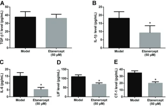
To investigate the mechanism of etanercept in rat car-diomyocyte hypertrophy, we detected the levels of infl am-matory factors using ELISA. As shown in Figure 4A, there was no significant effect of etanercept on TGF-b1 levels in cardiomyocytes. Conversely, etanercept significantly reduced the levels of IL-1b, IL-6, LIF and CT-1 compared to untreated cardiomyocytes with endothelin-induced hyper-trophy (Po0.05; Figure 4B–E).
Discussion
Although the pathophysiological mechanisms of car-diomyocyte hypertrophy remain unclear, the inflammatory
Table 1.Primer sequences for specific genes.
Gene Primer sequence
MMP-9 Forward: 50-AAGGATGGTCTACTGGCA-30 Reverse: 50-AGAGATTCTCACTGGGGC-30 MMP-13 Forward: 50-CCTGGGATTTCCAAAAGAGGT-30
Reverse: 50-TAACACCACAATAAGGAATTT-30 ANF Forward: 50-CCATATTGGAGCAAATCCTG-30 Reverse: 50-CGGCATCTTCTCCTCCAGG-30
b-actin Forward: 50-CCAAGGCCAACCGCGAGAAGATGAC-30 Reverse: 50-AGGGTACATGGTGGTGCCGCCAGAC-30
response has proved to be involved in the occurrence and development of cardiomyocyte hypertrophy (21). In the present study, we found that TNF-a level was obviously increased in cardiomyocytes with hypertrophy. Moreover, etanercept significantly promoted cell prolifera-tion and reduced the ratio of apoptotic cells in the cell model of cardiomyocyte hypertrophy. In addition, etaner-cept remarkably reduced the mRNA levels of myocardial hypertrophy marker genes, inhibited the expression of Bax but increased the expression of Bcl-2. Etanercept also lowered the levels of IL-1b, IL-6, LIF and CT-1, but not TGF-b1 compared with the cell model of cardiomyocyte hypertrophy.
A previous study has demonstrated that endothelin 1 is involved in the development of cardiomyocyte hyper-trophy (22), and the cell model of endothelin 1-induced cardiomyocyte hypertrophy had been reported in some experimental studies (23,24). Our study also successfully established the rat cell model of cardiomyocyte hyper-trophy by the stimulation of endothelin 1.
ANF is a hormone produced by the heart and mainly serves as a diuretic and hypotensor through inhibiting the secretion of renin and aldosterone (25). ANF is also over-expressed in cardiomyocyte hypertrophy and heart fail-ure. Anti-ANF has been considered an effective method to inhibiting cardiomyocyte hypertrophy by inducing the
Figure 2.mRNA levels of atrial natriuretic factor (ANF,A), matrix metalloproteinase (MMP)-9 (B) and MMP-13 (C) in cardiomyocytes with endothelin-induced hypertrophy treated with etanercept assessed by qRT-PCR. Data are reported as means±SD. *Po0.05 compared with model (ANOVA).
Figure 3.Protein levels of Bcl-2 and Bax in cardiomyocytes with endothelin-induced hypertrophy and then treated with 0 or 50mM etanercept assessed by western blotting. Data are reported as means±SD. *Po0.05 compared with model (ANOVA).
expression of mitogen-activated protein kinase phospha-tase-1 (26). In addition, cardiomyocyte hypertrophy is closely associated with ventricular remodeling and cardiac inflammation, which could contribute to heart failure (27). The production and release of MMPs play an important role during the ventricular remodeling, and MMP-9 and MMP-13 were overexpressed during the progression of remodeling (28). Our study showed that etanercept remark-ably reduced the mRNA levels of ANF, MMP-9 and MMP-13, which suggest that etanercept may have an inhibiting effect on cardiomyocyte hypertrophy.
In vitro research has suggested that TNF-a could induce down-regulation of sarcomeric proteins, apoptosis and depression of contractility in cardiomyocytes (29,30), indicating that it might play a key role in cardiomyocyte hypertrophy and heart failure. Therefore, the clearance of TNF-amight be a potential therapeutic strategy to improve cardiomyocyte hypertrophy and heart failure. Consistently, we used endothelin to induce cardiomyocyte hypertrophy and not surprisingly found the TNF-alevel was markedly increased. Etanercept, a well-known TNF-a inhibitor, is reported to play a pro-apoptotic role in many cells. Malaviya et al. (31) demonstrated that etanercept could induce dermal dendritic cell apoptosis in psoriatic plaques of responding patients. Fries et al. (32) showed the pro-apoptotic role of etanercept in experimental colitis. Avramidis et al. (33) also suggested that etanercept induced apop-tosis and reduced apopapop-tosis-inhibiting factors in psoriatic endothelial cells. Similarly, the present study demon-strated that etanercept reduced the ratio of apoptotic cells in cardiomyocytes with hypertrophy. It is well-known that the Bcl-2 family, which includes anti-apoptotic Bcl-2 and pro-apoptotic Bax, plays a regulatory role in cell apoptosis (34). Under normal conditions, the expressions of Bcl-2 and Bax are balanced. Under abnormal conditions, how-ever, overexpressed Bcl-2 could dissociate Bax dimers and promote the formation of complexes of Bax and Bcl-2, then inhibit cell apoptosis, and up-regulated Bax increases
the number of Bax dimers and promote cell apoptosis (34). Thus, the ratio of Bcl-2/Bax is considered a predictive index in regulating cell survival and death (34,35). Our study suggested that the down-regulated Bax and up-regulated Bcl-2 were consistent with the anti-apoptotic role of etanercept in the cell model of cardiomyocyte hypertrophy. Furthermore, a previous study has proved that up-regulated Bcl-2 could attenuate low ambient temperature-induced myocardial hypertrophyin vivo(36). Another study also proved that Cyclovirobuxinum D alle-viated cardiac hypertrophy in rats through repression of cardiac cell apoptosis (37). Taken together, the protective effect of etanercept on cardiomyocyte hypertrophy might be associated with cell apoptosis.
Furthermore, the present study found that etanercept inhibited the expressions of IL-1band proteins in the IL-6 family, including IL-6, LIF and CT-1, in the cell model of cardiomyocyte hypertrophy. Considering that IL-1band IL-6 family proteins possess potent pro-inflammatory actions, etanercept might exert an anti-inflammatory effect on cardiomyocyte hypertrophy. Consistently, Don et al. (38) revealed that etanercept could inhibit the systemic infl am-matory response in chronic hemodialysis patients. Galarraga et al. (39) also reported that etanercept could improve arterial stiffness by inhibiting inflammatory response in rheumatoid arthritis. To further determine anti-inflammatory signaling pathway of etanercept in cardiomyocyte hyper-trophy, we detected the expression of TGF-b1. Unfortu-nately, etanercept did not change the level of TGF-b1 in cardiomyocyte hypertrophy, which suggests that TGF-b1 might not be involved in the anti-inflammatory role of etanercept in cardiomyocyte hypertrophy. Thus, further investigation is essential to understand the signaling path-way underlying TNF-a-induced inflammatory response in cardiomyocyte hypertrophy.
In summary, etanercept may improve rat cardiomyo-cyte hypertrophy by inhibiting inflammatory cytokines secretion and cell apoptosis.
References
1. Shimizu I, Minamino T. Physiological and pathological car-diac hypertrophy.J Mol Cell Cardiol 2016; 97: 245–262, doi: 10.1016/j.yjmcc.2016.06.001.
2. Eder P, Molkentin JD. TRPC channels as effectors of cardiac hypertrophy.Circ Res 2011; 108: 265–272, doi: 10.1161/ CIRCRESAHA.110.225888.
3. Hilfiker-Kleiner D, Landmesser U, Drexler H. Molecular mechanisms in heart failure: focus on cardiac hypertrophy, inflammation, angiogenesis, and apoptosis.J Am Coll Cardiol 2006; 48: A56–A66, doi: 10.1016/j.jacc.2006.07.007. 4. Bueno OF, De Windt LJ, Tymitz KM, Witt SA, Kimball TR,
Klevitsky R, et al. The MEK1–ERK1/2 signaling pathway promotes compensated cardiac hypertrophy in transgenic mice. EMBO J 2000; 19: 6341–6350, doi: 10.1093/emboj/ 19.23.6341.
5. Dorn GW, Force T. Protein kinase cascades in the regulation of cardiac hypertrophy.J Clin Invest 2005; 115: 527–537, doi: 10.1172/JCI24178.
6. Hilfiker-Kleiner D, Hilfiker A, Drexler H. Many good reasons to have STAT3 in the heart. Pharmacol Ther 2005; 107: 131–137, doi: 10.1016/j.pharmthera.2005.02.003.
7. Selvetella G, Hirsch E, Notte A, Tarone G, Lembo G. Adaptive and maladaptive hypertrophic pathways: points of convergence and divergence. Cardiovasc Res 2004; 63: 373–380, doi: 10.1016/j.cardiores.2004.04.031.
8. Dorn GW, Brown JH. Gq signaling in cardiac adaptation and maladaptation.Trends Cardiovasc Med1999; 9: 26–34, doi: 10.1016/S1050-1738(99)00004-3.
cultured myocytes and animal models.J Mol Cell Cardiol 2003; 35: 1385–1394, doi: 10.1016/j.yjmcc.2003.10.001. 10. Murdaca G, Spanò F, Contatore M, Guastalla A, Magnani O,
Puppo F. Pharmacogenetics of etanercept: role of TNF-a
gene polymorphisms in improving its efficacy.Expert Opin Drug Metab Toxicol 2014; 10: 1703–1710, doi: 10.1517/ 17425255.2014.970165.
11. Neovius M, Arkema E, Olsson H, Eriksson J, Kristensen LE, Simard J, et al. Drug survival on TNF inhibitors in patients with rheumatoid arthritis comparison of adalimumab, eta-nercept and infliximab.Ann Rheum Dis2015; 74: 354–360, doi: 10.1136/annrheumdis-2013-204128.
12. Tichy M, Hercogova J. Manifestation of Crohn’s disease in a young woman during the course of treatment for severe form of chronic plaque psoriasis with etanercept.Dermatol Ther 2014; 27: 211–214, doi: 10.1111/dth.12119.
13. Baraliakos X, Haibel H, Fritz C, Listing J, Heldmann F, Braun J, et al. Long-term outcome of patients with active ankylos-ing spondylitis with etanercept-sustained efficacy and safety after seven years.Arthritis Res Ther2013; 15: 1, doi: 10.1186/ ar4244.
14. Knight C, Mauskopf J, Ekelund M, Singh A, Yang S, Boggs R: Cost-effectiveness of treatment with etanercept for pso-riasis in Sweden.Eur J Health Econ 2012; 13: 145–156, doi: 10.1007/s10198-010-0293-8.
15. Olivieri AN, Iafusco D, Mellos A, Zanfardino A, Mauro A, Granato C, et al. Refractory rheumatoid factor positive polyarthritis in a female adolescent already suffering from type 1 diabetes mellitus and Hashimoto’s thyroiditis suc-cessfully treated with etanercept.Ital J Pediatr2013; 39: 1, doi: 10.1186/1824-7288-39-64.
16. Butchart J, Brook L, Hopkins V, Teeling J, Püntener U, Culliford D, et al. Etanercept in Alzheimer disease A ran-domized, placebo-controlled, double-blind, phase 2 trial. Neu-rology2015; 84: 2161–2168, doi: 10.1212/WNL.000000000 0001617.
17. Waters JP, Pober JS, Bradley JR. Tumour necrosis factor and cancer. J Pathol 2013; 230: 241–248, doi: 10.1002/ path.4188.
18. Mann DL. Recent insights into the role of tumor necrosis factor in the failing heart. In: Anonymous,The role of infl am-matory mediators in the failing heart. Springer; 2001. p 3–12, doi: 10.1007/978-1-4615-1449-7.
19. Sivasubramanian N, Coker ML, Kurrelmeyer KM, MacLellan WR, DeMayo FJ, Spinale FG, et al. Left ventricular remod-eling in transgenic mice with cardiac restricted overexpres-sion of tumor necrosis factor.Circulation2001; 104: 826–831, doi: 10.1161/hc3401.093154.
20. Shyu K-G, Chen C-C, Wang B-W, Kuan P: Angiotensin II receptor antagonist blocks the expression of connexin43 induced by cyclical mechanical stretch in cultured neonatal rat cardiac myocytes.J Mol Cell Cardiol2001; 33: 691–698, doi: 10.1006/jmcc.2000.1333.
21. Smeets PJ, Teunissen BE, Planavila A, de Vogel-van den Bosch H, Willemsen PH, van der Vusse GJ, et al. Infl am-matory pathways are activated during cardiomyocyte hyper-trophy and attenuated by peroxisome proliferator-activated receptors PPARa and PPARd. J Biol Chem 2008; 283: 29109–29118, doi: 10.1074/jbc.M802143200.
22. Ito H, Hirata Y, Adachi S, Tanaka M, Tsujino M, Koike A, et al. Endothelin-1 is an autocrine/paracrine factor in the
mechanism of angiotensin II-induced hypertrophy in cul-tured rat cardiomyocytes.J Clin Invest1993; 92: 398, doi: 10.1172/JCI116579.
23. Zhu W, Zou Y, Shiojima I, Kudoh S, Aikawa R, Hayashi D, et al. Ca2+/calmodulin-dependent kinase II and calcineurin play critical roles in endothelin-1-induced cardiomyocyte hypertrophy. J Biol Chem 2000; 275: 15239–15245, doi: 10.1074/jbc.275.20.15239.
24. Dulce RA, Hurtado C, Ennis IL, Garciarena CD, Alvarez MC, Caldiz C, et al. Endothelin-1 induced hypertrophic effect in neonatal rat cardiomyocytes: involvement of Na+/H+and Na+/Ca2+exchangers.J Mol Cell Cardiol2006; 41: 807
– 815, doi: 10.1016/j.yjmcc.2006.05.016.
25. De Bold AJ. Atrial natriuretic factor: a hormone produced by the heart.Science 1985; 230: 767–770, doi: 10.1126/ science.2932797.
26. Hayashi D, Kudoh S, Shiojima I, Zou Y, Harada K, Shimoyama M, et al. Atrial natriuretic peptide inhibits car-diomyocyte hypertrophy through mitogen-activated protein kinase phosphatase-1. Biochem Biophysl Res Commun 2004; 322: 310–319, doi: 10.1016/j.bbrc.2004.07.119. 27. Tomaselli GF, Marbán E. Electrophysiological remodeling in
hypertrophy and heart failure. Cardiovasc Res 1999; 42: 270–283, doi: 10.1016/S0008-6363(99)00017-6.
28. Hayashidani S, Tsutsui H, Shiomi T, Suematsu N, Kinugawa S, Ide T, et al. Fluvastatin, a 3-hydroxy-3-methylglutaryl coenzyme a reductase inhibitor, attenuates left ventricular remodeling and failure after experimental myocardial infarc-tion.Circulation2002; 105: 868–873, doi: 10.1161/hc0702. 104164.
29. Müller-Werdan U, Schumann H, Fuchs R, Reithmann C, Loppnow H, Koch S, et al.: Tumor necrosis factor-a(TNFa) is cardiodepressant in pathophysiologically relevant con-centrations without inducing inducible nitric oxide-(NO)-synthase (iNOS) or triggering serious cytotoxicity. J Mol Cell Cardiol 1997; 29: 2915–2923, doi: 10.1006/jmcc. 1997.0526.
30. Patten M, Krämer E, Bünemann J, Wenck C, Thoenes M, Wieland T, et al. Endotoxin and cytokines alter contractile protein expression in cardiac myocytes in vivo. Pflügers Archiv2001; 442: 920–927, doi: 10.1007/s004240100612. 31. Malaviya R, Sun Y, Tan JK, Wang A, Magliocco M, Yao M,
et al. Etanercept induces apoptosis of dermal dendritic cells in psoriatic plaques of responding patients. J Am Acad Dermat 2006; 55: 590–597, doi: 10.1016/j.jaad.2006. 05.004.
32. Fries W, Muja C, Crisafulli C, Costantino G, Longo G, Cuzzocrea S, et al. Infliximab and etanercept are equally effective in reducing enterocyte apoptosis in experimental colitis.Int J Med Sci2008; 5: 169–180, doi: 10.7150/ijms. 5.169.
33. Avramidis G, Krüger-Krasagakis S, Krasagakis K, Fragia-daki I, Kokolakis G, Tosca A. The role of endothelial cell apoptosis in the effect of etanercept in psoriasis. Br J Dermatol 2010; 163: 928–934, doi: 10.1111/j.1365-2133. 2010.09935.x.
34. Vaux D, Cory S, Adams J. Bcl-2 and cell survival.Nature 1988; 335: 440–442, doi: 10.1038/335440a0.
upregulation PI3K/Akt signaling pathway.J Neurol Sci2014; 344: 100–104, doi: 10.1016/j.jns.2014.06.033.
36. Liang J, Yin K, Cao X, Han Z, Huang Q, Zhang L, et al. Attenu-ation of low ambient temperature-induced myocardial hyper-trophy by atorvastatin via promoting Bcl-2 expression.Cell Physiol Biochem2017; 41: 286–295, doi: 10.1159/000456111. 37. Wu J-b, Zhou Y, Liang C-l, Zhang X-j, Lai J-m, Ye S-f, et al. Cyclovirobuxinum D alleviates cardiac hypertrophy in hyper-thyroid rats by preventing apoptosis of cardiac cells and inhibiting the p38 mitogen-activated protein kinase signaling
pathway.Chin J Integr Med2016; 1–9, doi: 10.1007/s11655-015-2299-7.
38. Don BR, Kim K, Li J, Dwyer T, Alexander F, Kaysen G. The effect of etanercept on suppression of the systemic inflammatory response in chronic hemodialysis patients. Clin Nephrol2010; 73: 431–438, doi: 10.5414/CNP73431. 39. Galarraga B, Khan F, Kumar P, Pullar T, Belch JJ.