ISSN 0100-879X
BIOMEDICAL SCIENCES
AND
CLINICAL INVESTIGATION
www.bjournal.com.br
www.bjournal.com.br
Volume 45 (3) 179-290 March 2012
Braz J Med Biol Res, March 2012, Volume 45(3) 256-263
doi: 10.1590/S0100-879X2012007500018
Cardiopulmonary exercise testing variables as predictors of
long-term outcome in thoracic sarcoidosis
A.J. Lopes, S.L.S. Menezes, C.M. Dias, J.F. Oliveira, M.R.M. Mainenti and F.S. Guimarães
Institutional Sponsors
The Brazilian Journal of Medical and Biological Research is partially financed by
Faculdade de Medicina de Ribeirão Preto Campus
Ribeirão Preto
Explore High - Performance MS Orbitrap Technology In Proteomics & Metabolomics
Cardiopulmonary exercise testing
variables as predictors of long-term
outcome in thoracic sarcoidosis
A.J. Lopes
1,2, S.L.S. Menezes
2,3, C.M. Dias
2, J.F. Oliveira
2, M.R.M. Mainenti
2and F.S. Guimarães
2,31Setor de Provas de Função Respiratória, Universidade do Estado do Rio de Janeiro, Rio de Janeiro, RJ, Brasil 2Programa de Pós-Graduação em Ciências da Reabilitação, Centro Universitário Augusto Motta,
Rio de Janeiro, RJ, Brasil
3Curso de Fisioterapia, Universidade Federal do Rio de Janeiro, Rio de Janeiro, RJ, Brasil
Abstract
Cardiopulmonary exercise testing (CPET) plays an important role in the assessment of functional capacity in patients with interstitial lung disease. The aim of this study was to identify CPET measures that might be helpful in predicting the vital capacity and diffu-sion capacity outcomes of patients with thoracic sarcoidosis. A longitudinal study was conducted on 42 nonsmoking patients with thoracic sarcoidosis (median age = 46.5 years, 22 females). At the first evaluation, spirometry, the measurement of single-breath carbon monoxide diffusing capacity (DLCOsb) and CPET were performed. Five years later, the patients underwent a second evalu-ation consisting of spirometry and DLCOsb measurement. After 5 years, forced vital capacity (FVC)% and DLCOsb% had decreased
significantly [95.5 (82-105) vs 87.5 (58-103) and 93.5 (79-103) vs 84.5 (44-102), respectively; P < 0.0001 for both]. In CPET, the peak oxygen uptake, maximum respiratory rate, breathing reserve, alveolar-arterial oxygen pressure gradient at peak exercise (P(A-a)O2), and Δ SpO2 values showed a strong correlation with the relative differences for FVC% and DLCOsb% (P < 0.0001 for all). P(A-a)O2 ≥22 mmHg and breathing reserve ≤40% were identified as significant independent variables for the decline in pulmo
-nary function. Patients with thoracic sarcoidosis showed a significant reduction in FVC% and DLCOsb% after 5 years of follow-up. These data show that the outcome measures of CPET are predictors of the decline of pulmonary function.
Key words: Sarcoidosis; Exercise; Respiratory function tests; Respiratory mechanics
Introduction
Correspondence: A.J. Lopes, Rua Araguaia, 1266, Bloco 1/405, Frequesia/Jacarepaguá, 22745-271 Rio de Janeiro, RJ, Brasil. E-mail: [email protected]
Received July 2, 2011. Accepted February 6, 2012. Available online February 17, 2012. Published March 19, 2012.
Sarcoidosis is a multisystem granulomatous disease of unknown origin that occurs in mediastinal and pulmonary sites in 90% of cases. The clinical course varies widely. Parenchymal abnormalities often resolve spontaneously, but progress toward pulmonary fibrosis in 20-25% of cases (1).
Clinicians frequently find it difficult to manage patients with thoracic sarcoidosis due to the significant variability
in disease manifestation and multiple, non-specific symp
-toms. Pulmonary function tests (PFTs) are important for measuring initial lung impairment and providing a baseline to assess improvement or deterioration of lung disease. In clinical practice, the most common parameters that indicate functional impairment are forced vital capacity (FVC) and single-breath diffusion of carbon monoxide across the lung
(DLCOsb); these measures are often combined with
radio-logical assessment. However, the management of some patients requires a thoracic computed tomography scan and further physiological measurements, including exer-cise testing (2). Cardiopulmonary exerexer-cise testing (CPET) provides an accurate assessment of functional capacity in patients with interstitial lung disease, allowing the clinician to grade the severity of the disease. Many sarcoidosis pa-tients experience significant symptoms only with exertion and remain asymptomatic at rest (3,4).
state-Exercise tests as predictors of outcome in thoracic sarcoidosis 257
ment recommends that a chest radiograph and resting PFTs be performed regularly. Although these exams are considered to be sufficient to follow the evolution of the disease, they provide no information on the patients’ func-tional capacity.
Therefore, in the present study, we sought to identify CPET measures that may be helpful in predicting the vital capacity and diffusion capacity outcomes of patients with thoracic sarcoidosis.
Patients and Methods
Patients
This was a longitudinal study involving 57 nonsmoking patients with thoracic sarcoidosis determined by a chest radiograph. The criteria for the diagnosis of sarcoidosis included compatible clinical, radiographic, and laboratory findings; histological evidence of noncaseating granuloma; the absence of mycobacterial infection, and no exposure to aerocontaminants or medication known to cause granu-lomatous disorders (2). Subjects with a medical history or laboratory findings of concomitant respiratory, cardiac or neuromuscular disease were excluded from the study. Participants were previously informed about the objective of the study and gave written informed consent according to current ethical standards. The study protocol was approved by the Research Ethics Committee of Hospital Universitário Pedro Ernesto, Universidade do Estado do Rio de Janeiro (Process No. 2273-CEP/HUPE).
Study protocol and measurements
The study consisted of two evaluations. At the first evalu
-ation, in addition to chest radiographs, spirometry and the
measurement of DLCOsb and CPET were performed on the
patients. The severity of dyspnea was estimated using a standard 5-point scale (Medical Research Council): grade 0 = absent; grade 1 = hurrying on hills or after two flights of stairs; grade 2 = hurrying on flat ground or after one
flight of stairs; grade 3 = breathlessness on minimal exer
-tion, and grade 4 = breathlessness at rest (5). Five years later, the patients underwent the second evaluation of the study, consisting of another round of PFTs (spirometry and
measurement of DLCOsb).
Each pair of chest radiographs was examined by two radiologists, who then reached a consensus about the final classification. The following classification was used: stage 0 = no radiographic abnormalities; stage 1 = bilateral hilar adenopathy without parenchymal abnormalities; stage 2 =
bilateral hilar adenopathy with interstitial parenchymal infil
-trates; stage 3 = interstitial parenchymal infiltrates without hilar adenopathy, and stage 4 = cicatricial changes (6).
PFTs were completed using the Collins Plus Pulmonary Function Testing Systems (Warren E. Collins, USA) fol-lowing the ATS standards for both the procedure and the interpretation (7). Maximum voluntary ventilation (MVV)
was performed by coaching the patients to hyperventilate as vigorously as possible for 10 s; the goal was a minimum frequency of 80 breaths/min, while the mobilized volume was recorded in liters per minute (8). The results are re-ported as a percent of the predicted values for the Brazilian population (9,10).
Each subject performed symptom-limited maximal exer-cise testing using an electronically braked cycle ergometer connected to the Collins Plus Pulmonary Function Testing System. Following the collection of 2 to 5 min of resting data, subjects pedaled for 3 min at 60 rpm without resistance, after which the work rate (WR) was incremented by 10 to 25 W each minute. The WR increment for each ramped exercise test was individualized based on each patient’s pretest activity level; the objective was to achieve 8 to 12 min of progressive exercise before stopping (11). Heart rate (HR) was monitored continuously using a 12-lead electrocardiogram monitor (Marquette Electronics, USA), and blood pressure measurements (Hewlett Packard, USA) were obtained every 2 min throughout the exercise and
recovery periods. Oxygen saturation (SpO2) was monitored
noninvasively with a fiber optic earpiece oxymeter (Nonin Medical, USA). Oxygen desaturation during exercise was defined as a reduction of oxygen saturation greater than 4%
from baseline values (12). Oxygen uptake (VO2), carbon
dioxide output (VCO2), minute ventilation (VE), and related
variables were calculated breath-by-breath. Arterial blood gas and lactate levels were measured (Roche Diagnostics, Brazil) at rest and at the end of the exercise period (peak performance) using samples taken from an indwelling radial arterial line. Breathing reserve was calculated as the dif-ference between the measured resting MVV and the peak VE and is reported as a percentage of MVV [1 - (VE / MVV) x 100]. Heart rate reserve was calculated as the difference between the peak and resting heart rates [(220 - age) - peak
HR]. The VO2 at the anaerobic threshold (VO2θL) was
estimated using both the modified V-slope and ventila
-tor methods (13). The alveolar-arterial oxygen pressure
gradient (P(A-a)O2) was calculated using the measured
respiratory exchange ratio (VCO2/VO2 or RER) values. The
values were compared to those predicted by Neder et al. (14) for the adult Brazilian population.
Data analysis
Data were analyzed using the SAS 6.11 software (SAS Inc., USA). Data are reported as the median and interquartile range values or frequencies (percentage). The Kolmogorov-Smirnov test was used to check the homogeneity of the
sample. Peak VO2 was compared to various parameters
using nonparametric testing, including the Mann-Whitney test, and Kruskal-Wallis analysis of variance followed by Dunn’s multiple comparisons post-test. The relative variation
of FVC and DLCOsb over the 5-year period was evaluated
using the Wilcoxon signed-rank test. Correlations between
period and the outcome measures of the CPET were studied using Spearman’s rank correlation, with the exception of the correlation between breathing reserve and relative variations of FVC (logarithmic equation).
The clinical potential of the decline in pulmonary function to detect the potential factors related to outcome measures of the CPET was evaluated by means of receiver operating charac-teristic (ROC) analyses. Logistic regression was used for joint analysis to determine the factors that were independently re-lated to the decreased pulmonary function. Only those variables found to be significant were retained in the model. Differences were considered to be significant when P < 0.05.
Results
Fifty-seven outpatients were evaluated. Fifteen were excluded due to a history of smoking (10), concomitant respi-ratory disease (2) and cardiac (2) or neuromuscular disease (1). Of the 42 patients studied, 22 (52.4%) were females. Median age was 46.5 (39-53) years. When white subjects were considered separately from non-white subjects
(Mu-latto or Black), only 11 (26.2%) described themselves as white. The time since the diagnosis of sarcoidosis in the 42 outpatients ranged from 1 to 360 months. Of the 42 subjects, 32 (76.2%) had criteria for chronic disease (presence of the disease for 2 or more years) (2). Thirty-three patients (78.6%) were receiving systemic therapy (corticosteroids alone or in combination with an immunosuppressive drug), which reflects the policy of the tertiary care clinic. However, no subject presented evidence of steroid toxicity.
Data from the pulmonary function studies and the CPET results at the first evaluation are summarized in Table 1. Thirty-seven of the 42 patients (88.1%) failed to reach at
least 80% of their predicted peak VO2. Arterial oxygen
desaturation occurred with exercise in 20 patients (47.6%), while a breathing reserve of less than 25% was observed in 17 patients (40.5%). Fifteen patients (35.7%) had a P(A-a)
O2 (mmHg) value greater than 35 mmHg.
The characteristics of the patients, along with the peak
VO2 value for the different patient groups, are listed in Table
2. Females reached a lower peak VO2 than males, but the
difference was not statistically significant. While patients
diagnosed with higher chest roentgenogram stage
gener-ally displayed a lower peak VO2, the association between
the radiographic stage and peak VO2 was not statistically
significant. Patients receiving therapy tended to achieve
a lower peak VO2, but the difference was not statistically
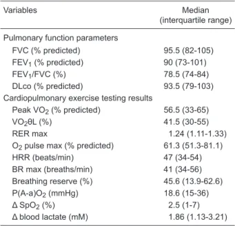
significant. Table 1. Pulmonary function parameters and outcome measures
of the cardiopulmonary exercise testing.
Variables Median
(interquartile range)
Pulmonary function parameters
FVC (% predicted) 95.5 (82-105) FEV1 (% predicted) 90 (73-101)
FEV1/FVC (%) 78.5 (74-84)
DLco (% predicted) 93.5 (79-103) Cardiopulmonary exercise testing results
Peak VO2 (% predicted) 56.5 (33-65)
VO2θL (%) 41.5 (30-55)
RER max 1.24 (1.11-1.33) O2 pulse max (% predicted) 61.3 (51.3-81.1) HRR (beats/min) 47 (34-54) BR max (breaths/min) 41 (34-56) Breathing reserve (%) 45.6 (13.9-62.6) P(A-a)O2 (mmHg) 18.6 (15-36)
Δ SpO2 (%) 2.5 (1-7)
Δ blood lactate (mM) 1.86 (1.13-3.21)
Data are reported as median (interquartile range) for 42 patients. FVC = forced vital capacity; FEV1 = forced expiratory volume in
one second; DLco = carbon monoxide diffusing capacity;peak VO2 = peak oxygen uptake; VO2θL = % peak VO2 at the
estimat-ed lactate threshold; RER max = maximum respiratory exchange ratio (VCO2/VO2); O2 pulse max = maximum oxygen pulse (VO2/ heart rate); HRR = heart rate reserve; BR max = maximum re -spiratory rate; P(A-a)O2 = alveolar-arterial oxygen pressure gra-dient at peak exercise; Δ SpO2 = difference between peak and resting oxygen saturation; Δ blood lactate = difference between peak and resting blood lactate.
Table 2. Characteristics of the patients studied as well as the differences in peak oxygen uptake.
Characteristics N (%) Peak VO2
(% predicted)
Gender
Male 20 (47.6) 59 (36-67.5) Female 22 (52.4) 44 (31-62) Degree of dyspnea
0 14 (33.3) 59 (38-65)
1-2 24 (57.2) 55 (31-63) 3-4 4 (9.5) 41 (29-58.5) Receiving systemic therapy
Yes 33 (78.6) 48.5 (29-64) No 9 (21.4) 56.5 (33-65) Chest roentgenogram stage
1 5 (11.9) 60 (60-61)
2 9 (21.4) 51 (35-62)
3 23 (54.8) 62 (32-69)
4 5 (11.9) 33 (26-37)
Exercise tests as predictors of outcome in thoracic sarcoidosis 259
Five years after the first evaluation, FVC% and DLCOsb%
were significantly decreased [95.5 (82-105) vs 87.5
(58-103) and 93.5 (79-(58-103) vs 84.5 (44-102), respectively; P <
0.0001]. The relative variation of FVC and DLCOsb between
the first and the second assessment was -5.1% (-23.1-0%) and -2.5% (-44.4-0.93%), respectively. The median interval between measurements was 61.5 months (range: 55-67 months). In this second evaluation, 26 patients (61.9%) were under systemic therapy (treatment decisions were taken according by the treating physician). The relative
variations of FVC and DLCOsb did not differ significantly
between subjects receiving therapy or not at the time of follow-up evaluation.
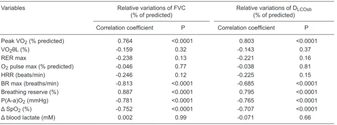
We compared the relative variations of the pulmonary function tests with the CPET measures. The results of uni-variate analysis are given in Table 3 and Figure 1. Among
the variables studied, peak VO2, maximum respiratory rate
(BR max), breathing reserve, P(A-a)O2 max, and Δ SpO2
values showed a better correlation.
A significant decline in pulmonary function (a decrease of ≥10% from the initial FVC values or a decrease of
≥15% from the values initially measured in the DLCOsb)
was observed in 18 outpatients; a decrease in both FVC
and DLCOsb was observed in 17 patients. According to
this criterion, the values for the area under the ROC curve
identified the following optimal cut-off points: peak VO2
≤50% of the predicted value; BR max ≥40 breaths/min;
breathing reserve ≤40%; P(A-a)O2 ≥22 mmHg, and Δ
SpO2 ≥4%. Table 4 shows the baseline CPET results for
42 patients according to the decline in pulmonary function after a 5-year follow-up.
We also investigated whether outcome measures of the CPET would demonstrate an independent role in predict-ing the decline of pulmonary function. All variables were assessed in a forward stepwise logistic regression model.
In the model, a P(A-a)O2 ≥22 mmHg and a breathing re
-serve ≤40% were identified as significant and independent variables (Table 5).
Discussion
This study evaluated the role of CPET as a predictor of the decline in pulmonary function after a 5-year follow-up in a sample of 42 nonsmoking patients with sarcoidosis. To our knowledge, no study has previously conducted a similar evaluation in this patient subset.
A 2-year follow-up period has been considered a stan-dard time to determine the outcome of sarcoidosis (15,16). However, in order to define the clinical phenotypes of the disease, WASOG developed a recent task force using a 5-year follow-up period (17). Compared to the 2-year follow-up outcome, nearly one-quarter of the patients had a different clinical outcome after 5 years. Thus, the 5-year follow-up period is an important methodological character-istic of our study.
The main findings of this research were as follows.
First, we observed that FVC and DLCOsb significantly de
-creased after a 5-year follow-up in patients with thoracic
Table 3. Univariate analysis comparing the relative variations of pulmonary function tests and cardiopulmonary exercise testing measures.
Variables Relative variations of FVC (% of predicted)
Relative variations of DLCOsb
(% of predicted)
Correlation coefficient P Correlation coefficient P
Peak VO2 (% predicted) 0.764 <0.0001 0.803 <0.0001
VO2θL (%) -0.159 0.32 -0.143 0.37
RER max -0.238 0.13 -0.221 0.16
O2 pulse max (% predicted) -0.046 0.77 -0.038 0.81
HRR (beats/min) -0.246 0.12 -0.225 0.15
BR max (breaths/min) -0.813 <0.0001 -0.685 <0.0001 Breathing reserve (%) 0.887 <0.0001 0.795 <0.0001 P(A-a)O2 (mmHg) -0.781 <0.0001 -0.765 <0.0001
Δ SpO2 (%) -0.752 <0.0001 -0.707 <0.0001
Δ blood lactate (mM) 0.002 0.99 -0.071 0.66
FVC = forced vital capacity; DLCOsb = single-breath diffusion of carbon monoxide across the lung; peak VO2 = peak oxygen
up-take; VO2θL = % peak VO2 at the estimated lactate threshold; RER max = maximum respiratory exchange ratio (VCO2/VO2); O2
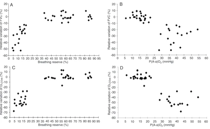
Figure 1. Relationship of relative variation of forced vital capacity (FVC) over a 5-year period with the breathing reserve (r2 = 0.887; P
< 0.0001) (Panel A) and the alveolar-arterial oxygen pressure gradient [P(A-a)O2] at peak exercise (ρ = -0.781; P < 0.0001) (Panel B)
obtained by cardiopulmonary exercise testing. Relationship of relative variation of single-breath diffusion of carbon monoxide across the lung (DLCOsb) over a 5-year period with the breathing reserve (ρ = 0.795; P < 0.0001) (Panel C) and the P(A-a)O2 at peak exercise (ρ = -0.765; P < 0.0001) (Panel D) obtained by cardiopulmonary exercise testing. Correlation was determined by univariate analysis using Spearman’s rank correlation (Panels B, C and D), with the exception of the correlation between breathing reserve and relative variations of FVC (logarithmic equation; Panel A).
Table 4. Results of cardiopulmonary exercise testing according to the decline in pulmonary func-tion after a 5-year follow-up in thoracic sarcoidosis patients.
Significant variables* Decline in pulmonary function+
Present (N = 18) Absent (N = 24) RR 95%CI
N % N %
Peak VO2 ≤50% of predicted 17 94.4 2 8.3 20.6 3.01-140.8 BR max ≥40 breaths/min 16 88.9 6 25.0 7.27 1.91-27.8 Breathing reserve ≤40% 17 94.4 2 8.3 20.6 3.01-140.8 P(A-a)O2 ≥22 mmHg 17 94.4 1 4.2 22.7 3.32-154.9
Δ SpO2 ≥4% 17 94.4 3 12.5 18.7 2.73-128.0
RR = relative risk; 95%CI = 95% confidence interval; peak VO2 = peak oxygen uptake; BR max =
maximum respiratory rate; P(A-a)O2 = alveolar-arterial oxygen pressure gradient at peak exercise; Δ SpO2 = difference between peak and resting oxygen saturation. *Significant variables resulting
from Spearman’s rank correlation between the relative variations of pulmonary function tests and cardiopulmonary exercise testing measures (optimal cut-off points of the area under the ROC curve). +Decrease >10% in relation to the initial values of forced vital capacity or single-breath
Exercise tests as predictors of outcome in thoracic sarcoidosis 261
sarcoidosis (P < 0.0001 for both). Second, a spectrum of ventilatory and pulmonary gas exchange abnormalities obtained in the CPET was associated with the reduction
in FVC and DLCOsb five years later. In addition, we deter
-mined the cut-off of the CPET measures responsible for a significant decline in pulmonary function and demonstrated
that P(A-a)O2 and breathing reserve were independent
predictors of functional impairment.
Winterbauer and Hutchinson (18) reported a reduction
in FVC and DLCOsb in more than two thirds of patients
(dis-cordant changes occurred in fewer than 5% of patients). However, in contrast to these authors, we found a higher
relative variation of DLCOsb when considering the two
study time points. In sarcoidosis, the reduction of DLCOsb
may reflect impairments of the gas exchange area, barrier thickness, or ventilation-perfusion-diffusion mismatching of
the lung (19). Interestingly, DLCOsb has also been found to
be fairly correlated with gas exchange abnormalities dur-ing exercise and in particular to be the best predictive and
sensitive index of a fall in PaO2 (8).
Exercise testing is a remarkably robust and versatile tool that provides valuable diagnostic and prognostic infor-mation regarding patients with pulmonary diseases (20). A
low peak VO2 is usually the starting point in the evaluation
of reduced exercise capacity. In our study, approximately
90% of patients failed to reach at least 80% of peak VO2 (%
predicted) at the initial evaluation. Interestingly, peak VO2
showed a significant correlation with the relative variations of both FVC and DLco (P < 0.0001). However, changes in
VO2 can be due to multiple factors, including gas exchange
across the lung, the oxygen content of blood, oxygen delivery to tissues, and the oxygen uptake in the tissues (13).
Patients with significant interstitial lung disease often display low breathing reserve and arterial desaturation during exercise. In the present study, these two variables correlated with the decline in pulmonary function at the 5-year follow-up. Because the majority of patients in this sample (66.7%) had radiographic stages 3 and 4 at the initial evaluation may reflect a limitation by ventilatory mechanisms (21). Moreover, ventilatory abnormalities dur-ing CPET have been reported in up to 47% of sarcoidosis
patients (11,22). The ventilatory stress (demand/capacity) for a given work rate is much greater in patients with inter-stitial lung disease than in normal subjects. Additionally, it is possible that the exercise limitation in these patients is due to the marked arterial oxygen desaturation, which usually develops during exercise. Arterial hypoxemia can impair exercise tolerance because of inadequate oxygen delivery to working muscles (23).
The persistence of abnormal pulmonary gas exchange is related to a poor prognosis and is an important criterion indicating long-term steroid therapy (2). Eklund et al. (24) showed that diffusion limitation contributed up to 50% of
the P(A-a)O2 widening during exercise in sarcoidosis
pa-tients; these investigators found only a moderate degree of ventilation-perfusion mismatch. In the present study,
we found a significant correlation between P(A-a)O2 and
the relative variations of both FVC and DLco (P < 0.0001). At rest, the apical regions of the lung contribute relatively little to ventilation and gas exchange; however, during exercise, apical pulmonary circulation is normally recruited and contributes significantly to the capacity of the lung to increase the rate of gas exchange. Because the lesions of sarcoidosis favor the upper lobes of the lungs, lung function measured during exercise may have a higher sensitivity for detecting the presence and progression of parenchymal disease (25).
Using logistic regression analysis of the independent factors affecting the decline of pulmonary function at the
5-year follow-up, the only predictive factors were P(A-a)O2
≥22 mmHg and breathing reserve ≤40%, in that order. In
fact, the P(A-a)O2 value during exercise has been found to
be the best pulmonary gas exchange impairment index in sarcoidosis patients, showing associations with both DLco and FVC (8,12). Kollert et al. (26) performed a retrospective study to determine the extent to which the gas exchange measurement during exercise reflects disease activity and the clinical course of sarcoidosis. They showed that the
P(A-a)O2 during exercise represents disease activity and
its extent and is associated with a prolonged need for im-munosuppressive treatment during follow-up care in patients with pulmonary sarcoidosis. In interstitial lung disease, a
Table 5. Forward stepwise regression analysis: relationship between the decline in pulmonary function and study vari-ables.
Outcome variable Independent variables Unstandardized coefficient P RR 95%CI
B SE
Decline in pulmonary function* P(A-a)O2 ≥22 mmHg 4.25 1.60 0.001 70.0 3.03-161.3 Breathing reserve ≤40% 3.03 1.63 0.014 20.8 0.85-507.6
smaller pulmonary vascular bed reserve is available for recruitment, limiting the increase in lung surface area for gas exchange during exercise. Hence, both mixed venous oxygen tension and arterial oxygen tension drop with the progressively increasing workload and increased drop of peripheral oxygen uptake (8). Regarding the breathing reserve, patients with more advanced stages of interstitial lung disease often display higher levels of minute ventilation than those observed in normal subjects during exercise. These higher levels are largely due to the increased dead space ventilation; however, other contributing mechanisms responsible for this heightened ventilatory response may include inputs from mechanoreceptors and chemoreceptors and mediators of inflammation (3).
The present investigation has some limitations. First, there was, by definition, a selection bias because the study was performed at a tertiary hospital to which patients with severe complaints and advance stages of sarcoidosis are more likely to be referred. In fact, 32 patients (76.2%) had criteria for the definition of chronic disease. Second, in 17
of 42 patients (40.1%), the VO2θL was low, and the O2
pulse max was abnormal, suggesting cardiocirculatory
dysfunction (12).Although some patients may have had
cardiac sarcoidosis or pulmonary hypertension, we did not directly determine cardiac circulatory status; thus, this can be considered a limitation of the study. A new CPET in the second evaluation could also have helped to define the cases difficult to interpret. Finally, the conclusion was based on a small number of patients, resulting in high values for relative risk and longer intervals for the 95%
confidence interval in the logistic regression model. Care
-fully controlled prospective studies conducted on a larger sample are required to confirm these preliminary results and to validate our prediction model. In future studies, a number of potential prognostic variables, including clinical and radiological findings, may also be tested along with the CPET variables.
Patients with thoracic sarcoidosis showed significant
reductions in FVC and DLCOsb at the 5-year follow-up.
Additionally, the outcome measures of the CPET (P(A-a)
O2 and breathing reserve) were predictors of a decline
in pulmonary function. Determining the CPET measures may be helpful in predicting the outcomes of patients with thoracic sarcoidosis.
References
1. Abehsera M, Valeyre D, Grenier P, Jaillet H, Battesti JP, Brauner MW. Sarcoidosis with pulmonary fibrosis: CT pat -terns and correlation with pulmonary function. AJR Am J Roentgenol 2000; 174: 1751-1757.
2. Statement on sarcoidosis. Joint Statement of the American Thoracic Society (ATS), the European Respiratory Society (ERS) and the World Association of Sarcoidosis and Other Granulomatous Disorders (WASOG) adopted by the ATS Board of Directors and by the ERS Executive Committee, February 1999. Am J Respir Crit Care Med 1999; 160: 736-755. 3. Marciniuk DD, Gallagher CG. Clinical exercise testing in
in-terstitial lung disease. Clin Chest Med 1994; 15: 287-303. 4. Weisman IM, Zeballos RJ. Clinical exercise testing. Clin
Chest Med 2001; 22: 679-701, viii.
5. Xaubet A, Rodriguez-Roisin R, Bombi JA, Marin A, Roca J, Augusti-Vidal A. Correlation of bronchoalveolar lavage and clinical and functional findings in asbestosis. Am Rev Respir Dis 1986; 133: 848-854.
6. Scadding JG. Prognosis of intrathoracic sarcoidosis in Eng-land. A review of 136 cases after five years’ observation. Br Med J 1961; 2: 1165-1172.
7. Standardization of Spirometry, 1994 Update. American Thoracic Society. Am J Respir Crit Care Med 1995; 152: 1107-1136.
8. Medinger AE, Khouri S, Rohatgi PK. Sarcoidosis: the value of exercise testing. Chest 2001; 120: 93-101.
9. Pereira CA, Sato T, Rodrigues SC. New reference values for forced spirometry in white adults in Brazil. J Bras Pneumol
2007; 33: 397-406.
10. Neder JA, Andreoni S, Peres C, Nery LE. Reference val-ues for lung function tests. III. Carbon monoxide diffusing capacity (transfer factor). Braz J Med Biol Res 1999; 32:
729-737.
11. Sietsema KE, Kraft M, Ginzton L, Sharma OP. Abnormal oxygen uptake responses to exercise in patients with mild pulmonary sarcoidosis. Chest 1992; 102: 838-845. 12. Gibbons WJ, Levy RD, Nava S, Malcolm I, Marin JM, Tardif
C, et al. Subclinical cardiac dysfunction in sarcoidosis. Chest
1991; 100: 44-50.
13. Barros WG, Neder JA, Pereira CA, Nery LE. Clinical, radiographic and functional predictors of pulmonary gas exchange impairment at moderate exercise in patients with sarcoidosis. Respiration 2004; 71: 367-373.
14. Neder JA, Nery LE, Castelo A, Andreoni S, Lerario MC, Sachs A, et al. Prediction of metabolic and cardiopulmonary responses to maximum cycle ergometry: a randomised study. Eur Respir J 1999; 14: 1304-1313.
15. Baughman RP, Shipley R, Eisentrout CE. Predictive value of gallium scan, angiotensin-converting enzyme level, and bronchoalveolar lavage in two-year follow-up of pulmonary sarcoidosis. Lung 1987; 165: 371-377.
16. Judson MA, Baughman RP, Thompson BW, Teirstein AS, Terrin ML, Rossman MD, et al. Two year prognosis of sar-coidosis: the ACCESS experience. Sarcoidosis Vasc Diffuse Lung Dis 2003; 20: 204-211.
17. Baughman RP, Nagai S, Balter M, Costabel U, Drent M, du Bois R, et al. Defining the clinical outcome status (COS) in sarcoidosis: results of WASOG Task Force. Sarcoidosis Vasc Diffuse Lung Dis 2011; 28: 56-64.
18. Winterbauer RH, Hutchinson JF. Use of pulmonary function tests in the management of sarcoidosis. Chest 1980; 78: 640-647.
Exercise tests as predictors of outcome in thoracic sarcoidosis 263
diffusion capacity of the lung for carbon monoxide in pulmo-nary sarcoidosis: relation to exercise gas exchange. Chest
2004; 125: 2061-2068.
20. Arena R, Sietsema KE. Cardiopulmonary exercise testing in the clinical evaluation of patients with heart and lung disease. Circulation 2011; 123: 668-680.
21. Baughman RP, Teirstein AS, Judson MA, Rossman MD, Yeager H Jr, Bresnitz EA, et al. Clinical characteristics of patients in a case control study of sarcoidosis. Am J Respir Crit Care Med 2001; 164: 1885-1889.
22. Miller A, Brown LK, Sloane MF, Bhuptani A, Teirstein AS. Cardiorespiratory responses to incremental exercise in sar-coidosis patients with normal spirometry. Chest 1995; 107: 323-329.
23. Marciniuk DD, Watts RE, Gallagher CG. Dead space load-ing and exercise limitation in patients with interstitial lung disease. Chest 1994; 105: 183-189.
24. Eklund A, Broman L, Broman M, Holmgren A. V/Q and al -veolar gas exchange in pulmonary sarcoidosis. Eur Respir J 1989; 2: 135-144.
25. Arcasoy SM, Christie JD, Pochettino A, Rosengard BR, Blu-menthal NP, Bavaria JE, et al. Characteristics and outcomes of patients with sarcoidosis listed for lung transplantation.
Chest 2001; 120: 873-880.