Aggre can structure in am phibian
cartilage
Departamentos de 1Biologia Celular and
2Bioquímica, Universidade Estadual de Campinas,
Campinas, SP, Brasil D.Z. Covizi1,2 and
H.F. Carvalho1
Abstract
The structure of the large proteoglycan present in the bullfrog epiphy-seal cartilage was studied by immunochemical and biochemical meth-ods. The isolated monomer showed a polydisperse behavior on Sepha-rose CL2B, with a peak at Kav = 0.14. Chondroitin sulfate chains were identified by HPLC analysis of the products formed by chondroitinase digestion and mercuric acetate treatment. These chains have approxi-mately 38 disaccharides, a Di45:Di68 ratio of 1.6 and GalNAc4S + GalNAc4,6S are the main non-reducing terminals. Keratan sulfate was identified by the use of two monoclonal antibodies in Western blots after chondroitinase ABC treatment. A keratan sulfate-rich region (~110 kDa) was isolated by sequential treatment with chondro-itinase ABC and proteases. We also employed antibodies in Western blotting experiments and showed that the full length deglycosylated core protein is about 300 kDa after SDS-PAGE. Domain-specific antibodies revealed the presence of immunoreactive sites correspond-ing to G1/G2 and G3 globular domains and the characterization of this large proteoglycan as aggrecan. The results indicate the high conserva-tion of the aggrecan domain structure in this lower vertebrate. Co rre spo nde nce
H.F. Carvalho
Departamento de Biologia Celular UNICAMP
Caixa Postal 6109 13083-970 Campinas, SP Brasil
Fax: + 55-19-788-7821 E-mail: hern@ unicamp.br
Research supported by FAPESP (Nos. 95/6683-2, 95/6684-9, and 97/4925-4) and CNPq (No. 520582/95-0).
Received December 22, 1999 Accepted September 14, 2000
Ke y wo rds ·Aggrecan
·Bullfrog
·Cartilage
·Chondroitin sulfate
·Epiphyseal cartilage
Intro ductio n
Cartilage is a resilient tissue able to resist tension and pressure forces (1). These prop-erties are thought to result from a highly coordinated array of various components, amongst which are type II collagen, large and small proteoglycans, and other non-col-lagenous glycoproteins (2,3). The large pro-teoglycan aggrecan consists of a core protein to which the glycosaminoglycans (GAGs) chondroitin sulfate (CS) and keratan sulfate (KS) and N- and O-linked oligosaccharides are attached (4). The GAGs have a high
negative fixed-charge density, conferring on aggrecan its characteristic osmotic activity (5) which, in addition to the capacity to aggregate with hyaluronan and participate in a concerted interplay with type II collagen fibrils, endows the tissue with the ability to withstand compressive forces and to distrib-ute the load (6).
domains, is a proteolytically sensitive region of the molecule, centrally involved in aggre-can catabolism (10-14). The G2 domain (15) consists of two subdomains also present in G1. The function of G2 is still unclear. The two GAG substitution domains correspond to a KS-rich region with about 50 chains (16), and a CS-rich region with about 100 chains (17). The G3 domain consists of EGF-like, lectin-like (18) and CRP-like motifs, which also occur together in some cell adhe-sion molecules (19).
We have studied the bullfrog epiphyseal cartilage and reported that it differs from its mammalian counterpart by lacking a colum-nar arrangement of the chondrocytes in the growth cartilage. Chondrocyte hypertrophy is not associated with matrix calcification or endochondral ossification. Moreover, there is no secondary center of ossification (20).
This led us to believe that the identifica-tion and characterizaidentifica-tion of the macromol-ecules in the bullfrog epiphyseal cartilage would be important for the understanding of the physiology of this tissue at the cellular and molecular levels.
Since aggrecan function is essential to the physiology and structure of cartilage, we used immunochemical tests to elucidate some of the characteristics of the core protein and biochemical assays to determine some as-pects of the attached GAGs. Considering
that amphibians are lower vertebrates, the results presented here demonstrate a high conservation of the domain structure of aggrecan.
Mate rial and Me tho ds
Pre paratio n o f the large pro te o glycan fro m the bullfro g fe mo ral distal e piphyse al cartilage
One-year-old bullfrogs, Rana catesbeia-na, were purchased from a farm in Atibaia
(SP, Brazil). The femoral distal epiphyseal cartilages were dissected out and the proteo-glycans were extracted with 4 M guanidine-HCl in the presence of protease inhibitors and then dialyzed against 0.4 M guanidine-HCl to attain associative conditions before associative cesium chloride gradient cen-trifugation (21). The high buoyant density fraction (1.70 mg/ml, A1 fraction) was cen-trifuged again under dissociative conditions to obtain A1D1 fractions. A1D1 fractions were sequentially dialyzed against 1 M NaCl and water prior to subsequent analyses.
Hydro dynamic size o f pro te o glycan m o no m e rs
Proteoglycans (300 µg) were dialyzed against 0.5 M sodium acetate, pH 8.0, and subjected to gel filtration on a Sepharose CL2B column (0.5 x 110 cm) eluted with the same solution at a flow rate of 0.5 ml/h. Fractions (1 ml) were assayed for sulfated GAG content by the dimethylmethylene blue (DMMB) procedure (22).
Iso latio n o f CS chains and Supe ro se 6 chro m ato graphy
Large proteoglycans were subjected to ß-elimination and reduction for 24 h at 45oC in
1 M sodium borohydride in 50 mM NaOH, neutralized with acetic acid on ice, and vacuum dried after the addition of an equal Figure 1 - Schematic draw ing of
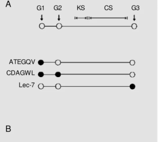
the aggrecan core protein (A) and the sites of reactivity of the different antibodies employed in the present study. G1 and G2 are 21 nm apart from each other. Betw een G2 and G3 are the keratan sulfate (KS)-rich region and the chondroitin sulfate (CS) substitution region. The anti-ATEGQV antibody reacts w ith a peptide present in G1. The anti-CDAGWL antibody reacts w ith a peptide sequence present in both G1 and G2. The Lec-7 anti-body recognizes a segm ent found in the G3 domain. B is a schematic draw ing of a chon-droitin sulfate chain attached to a serine residue (Ser) in the core protein. The filled circles-open circles-open circles-open squares sequence represents the tet-rasaccharide xylose-galactose-galactose-uronic acid linkage. The filled square/open square symbols represent the repeating disaccharides (DDi4S, DDi6S or
DDi0S) and the dotted circle corresponds to the non-reducing terminal (GalNAc4S; GalNAc4,6S; Di4S or Di6S).
ATEGQV
CDAGWL
Lec-7
Ser-G1 G2 KS CS G3 A
volume of methanol (23). The residues were washed three times with 500 µl of methanol to remove borate salts, dissolved in 500 µl of 4 M guanidine-HCl in 50 mM acetate buffer, pH 6.0, and subjected to chromatography on Superose 6 (1 x 24 cm) (Pharmacia, Uppsala, Sweden). The column was eluted with the same solution at a flow rate of 0.5 ml/min. Fractions (0.5 ml) were collected and as-sayed for sulfated GAG content by the DMMB assay (22).
Fluo re sce nt de rivatizatio n o f cho ndro itinase dige stio n pro ducts with 2-amino pyridine
The large proteoglycans from the A1D1 fraction (10 µg of sulfated GAGs) were di-gested with 5 mU of chondroitinase ABC (Seikagaku America, Falmouth, MA, USA) in 50 µl of 100 mM ammonium acetate, pH 7.4, for 2 h at 37oC. Released products were
collected into the filtrate of prewashed MicroCon 3 filters. Ammonium acetate was removed in vacuo. The residue was dis-solved in 500 µl of water and redried. Diges-tion products were derivatized with 2-aminopyridine (AP) (23). AP was freshly prepared before use by adding 500 mg to 100 µl of glacial acetic acid, which had been heated to and maintained at 65oC, with
re-peated vortexing until it was completely dis-solved. This reagent (20 µl) was added to 20-100 nmol of the chondroitinase digestion products. After incubation of the mixture for 24 h at 37oC, 5 µl of 6 M borane
dimethyl-amine in glacial acetic acid was added and allowed to stand overnight at 37o
C. Acid was removed by speed-vac evaporation and samples were stored at -20oC until the time
for analysis. Mono- and disaccharide stan-dards were also subjected to the chondroiti-nase digestion procedure before fluorescent derivatization. AP-derivatized unsaturated disaccharides were decomposed by treat-ment with 120 µl of 35 mM mercuric acetate, pH 5.0. Mercuric ions were removed in vacuo before chromatography.
HPLC se paratio n o f AP-de rivatize d cho ndro itinase dige stio n pro ducts
AP derivatives (23) were dissolved in 100 µl of water and passed over a 100 µl bed volume of Dowex H+ (BioRad Laboratories,
Richmond, CA, USA) immediately before loading onto an AS4A Ion Pac column equili-brated with 1 mM sodium trifluoroacetate, pH 7.0, in a Dionex AI-450 HPLC system. Samples were eluted at a flow rate of 1 ml/ min with a step gradient of trifluoroacetate (10 mM, 6 min; 10-50 mM, 4 min; 50-150 mM, 17 min; 150-250 mM, 13 min; 250-500 mM, 10 min). The eluant was monitored by fluorescent detection with an excitation wave-length of 310 nm and emission wavewave-length of 410 nm, using an in-line fluorimeter (Shimadzu Corporation, Tokyo, Japan).
Ide ntificatio n o f KS and characte rizatio n o f the KS-rich re gio n
The large proteoglycans (1.0 mg of sul-fated GAGs) were loaded onto a Sepharose CL6B column (Pharmacia) (0.6 x 120 cm) and eluted with 4 M guanidine-HCl in 50 mM sodium acetate, pH 6.0, at a flow rate of 5 ml/h. The peak eluted in the void volume of the column was digested with chondroiti-nase ABC (Seikagaku America) (24) (10 mU/mg of sulfated GAG), subjected to SDS-PAGE on a 3-16% gradient gel (25) and assayed by Western blotting with the mono-clonal antibodies MST1 (produced by im-munization with the large proteoglycan from the hammerhead shark cartilage; non-diluted culture medium) (26) and 4-A-4 (1:1000) (27) against KS, as described below. An-other gel was run under the same conditions and stained with 0.25% Alcian blue in 3% acetic acid. For the isolation of the KS-rich region, the large proteoglycan (7.5 mg of sulfated GAG) was digested with chondroi-tinase ABC (10 mU/mg) in 0.1 M Tris-ace-tate buffer, pH 7.3, for 18 h at 37o
subse-quently digested with trypsin and then with chymotrypsin (Sigma Chemical Co., St. Louis, MO, USA) at concentrations of 2 µg enzyme/mg of the initial sulfated GAG, for 10 h at 37oC in the same buffer. The material
was then chromatographed on a Sepharose CL6B column (0.6 x 120 cm) in 0.5 M sodium acetate, pH 7.0, and eluted at a flow rate of 5 ml/h as previously described by Heinegård and Axelsson (28). A peak con-taining material that showed metachromasy with DMMB and was devoid of uronic acid (as detected by the orcinol reaction) (29) was found. Fractions of this peak were pooled and electrophoresed on 3-16% gradient SDS-PAGE for 3 h at 25 mA, with or without prior digestion with keratanase (Seikagaku Ame-rica). The gel was stained with Alcian blue as described above.
D e glyco sylatio n, SD S-PAGE and We ste rn blo tting o f the co re pro te in
A1D1 fractions were deglycosylated by sequential treatment with chondroitinase ABC (proteinase free; Seikagaku America), keratanase II (Seikagaku America; 0.7 mU/ 100 µg of sulfated GAG) and endo-ß-gly-cosidase (Seikagaku America; 0.7 mU/100 µg of sulfated GAG). Thirty micrograms of the initial sulfated GAGs were subjected to SDS-PAGE on precast 4-12% gels (Novex, San Diego, CA, USA) for 90 min at 125 V
(24). The material was electrotransferred onto nitrocellulose using an XCELL transfer unit (Novex) (30). Membranes were blocked with 5% (w/v) skim milk powder in Tris-buffered saline (0.5 M NaCl and 20 mM Tris, pH 7.5), containing 0.1% Tween 20 (TST), for 1 h at room temperature. The polyclonal antibod-ies used were anti-ATEGQV (1:5000) (which reacts with the IgG loop of the G1 domain of the aggrecan; Kenagy A, Wight T and Sandy JD, unpublished results), anti-CDAGWL (1:3000) (which detects the protein tandem repeat loops of the G1 domain, obtained from Dr. Steve S. Carlson, University of Washington, Seattle, WA, USA) and Lec-7 (raised against a peptide contained in the lectin-like motif of the G3 domain, obtained from Dr. Kurt Doege, Shriners Hospital for Children, Portland, OR, USA) (1:5000) poly-clonal antibodies (Figure 1). The anti-chon-droitin 4-sulfate (C4S) stub (2B6 clone, 1:750) (31) and anti-chondroitin 6-sulfate (C6S) stub (3B3 clone, 1:5000) (32) mono-clonal antibodies and the peroxidase-conju-gated secondary antibody (1:5000) were all diluted in 1% (w/v) milk powder in TST. Immunoreactivity was developed using the ECL detection kit (Amersham Corporation, Arlington Heights, IL, USA) and exposure to Hyperfilm (Amersham). Rat chondrosar-coma aggrecan (13) was included for com-parison.
Re sults
The hydro dynamic size o f the pro te o glycan mo no me r and o f the CS chains
Figure 2 shows the chromatographs ob-tained for the proteoglycan monomers extracted from the bullfrog epiphyseal carti-lage by gel filtration on Sepharose CL2B. Kav was 0.14 and Kav range (50% of the peak) was 0 to 0.34. Glycosaminoglycan chains released by ß-elimination of the pro-teoglycans present in the A1D1 fraction were chromatographed on Superose 6 gel. The 100
% 50
0
-0.4 -0.2 0.0 0.2 0.4 0.6 0.8 1.0 Kav
chains eluted as a single peak with Kav = 0.40 (Figure 3).
Fluo re sce nt HPLC analysis o f cho ndro itinase ABC dige sts
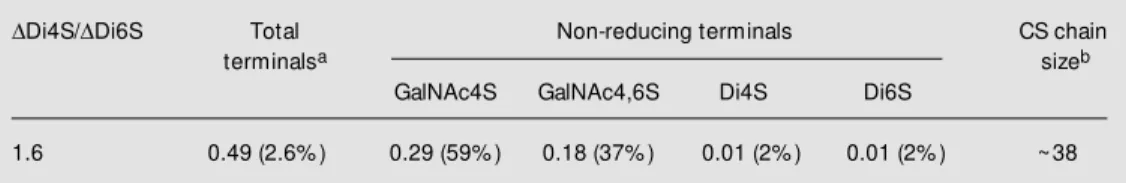
AP-derivatized chondroitinase digestion products were submitted to ion-exchange HPLC analysis with measurement of all in-ternal disaccharides and non-reducing ter-minals (33). Most of the fluorescent prod-ucts present in the digests were sulfated
Ddisaccharides (DDi, unsaturated disaccha-ride) (97.4%) (Figure 4A and Table 1). The
DDi4S:DDi6S ratio was 1.6. GalNAc4S, GalNAc4,6S and the saturated disaccharides GlcA-GalNAc4S (Di4S) and Glc-GalNAc6S (Di6S) were identified as the non-reducing termini of the CS chains (Figure 4) and corresponded to 2.6% of the total
chondroi-tinase digestion products. The average num-ber of repeating disaccharides per chain was estimated by the ratio of interior DDi to non-reducing termini (Table 1). The analyses also showed that sulfated GalNAc residues were the most abundant termini in the bull-frog CS chain, representing 96% of the total non-reducing termini (Figure 4B and Table 1)
Figure 3 - Hydrodynamic size of large proteoglycan chondroitin sulfate. A 250-µg aliquot of large proteoglycans w as treated w ith sodium borohydrate and NaOH to release the composing gly-cosaminoglycan (GAG) chains. The released GAGs w ere chro-matographed on Superose 6 and fractions w ere assayed for sulfated GAGs w ith DM M B.
R
e
la
ti
v
e
c
o
n
te
n
t
o
f
s
u
lf
a
te
d
G
A
G
100
80
60
40
20
0
-0.4 -0.2 0.0 0.2 0.4 0.6 0.8 1.0 Kav
m
V
120 100 80 60 40 20 0
m
V
30
20
10
0 1
3
4
2 5 6
2
0 5 10 15 20 25 30 Elution time (min)
0 5 10 15 20 25 30 Elution time (min)
A B
Figure 4 - AS4A ion chromatography-derivatized products from a chondroitinase-digested large proteoglycan. A and
B, The samples w ere digested w ith chondroitinase ABC and derivatized, and portions (200 ng) w ere chromatographed by AS4A HPLC. Column effluents w ere monitored for fluorescence (B). Other portions of derivatized products w ere treated w ith mercuric acetate prior to HPLC fractionation and monitoring. 2-Aminopyridine-derivatized (AP) products w ere identified by comparison of their elution times relative to standards (1, GalNAc4S-AP; 2, GalNAc4,6S-AP; 3, DDi4S-AP; 4, DDi6S-AP; 5, Di4S-AP; Di6S-AP).
Table 1 - Quantitative data on chondroitin sulfate (CS) chain internal and non-reducing terminal disaccharide composition.
aData are reported as the total area under the corresponding peaks and the percent of the total chondroitinase
digestion products. bData are reported as the number of disaccharides calculated from the ratio betw een the
amount of internal Ddisaccharides (DDi) and the amount of non-reducing terminals.
DDi4S/DDi6S Total Non-reducing terminals CS chain
terminalsa sizeb
GalNAc4S GalNAc4,6S Di4S Di6S
in the form of GalNAc4S and GalNAc4,6S, which corresponded to 59 and 37%, respec-tively. Both Di4S and Di6S termini were present in equal amounts (2% each of the total) (Figure 4B and Table 1), and hence 4% of the CS chains terminated as GlcA.
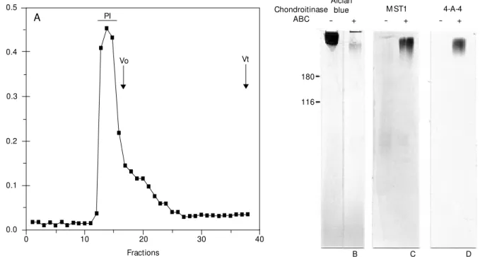
Ide ntificatio n o f KS and characte rizatio n o f the KS-rich re gio n
The initial guanidine-HCl extract was chromatographed on Sepharose CL6B (Fig-ure 5A). The peak eluted at the void volume was treated with chondroitinase ABC. The digestion products were electrophoresed by SDS-PAGE. Components with high relative molecular mass were developed after stain-ing with Alcian blue. The non-digested pro-teoglycan was in the stacking gel and the chondroitinase ABC-treated proteoglycan migrated as a high molecular mass compo-nent at the top of the separating gel (Figure
5B). Other samples were electroblotted onto nitrocellulose membranes and incubated with the monoclonal antibody MST1 (Figure 5C) or 4-A-4 (Figure 4D). Immunoreactivity for both MST1 and 4-A-4 was found in high molecular mass components only after chon-droitinase digestion. Non-digested proteo-glycans did not react with either monoclonal antibody. To isolate the KS-rich region, the monomers of A1D1 were digested first with chondroitinase ABC and then with a trypsin/ chymotrypsin combination. The digestion products were chromatographed on a Sepha-rose CL6B column (Figure 6A). The peak detected by metachromasy with DMMB was devoid of uronic acid, as detected by the orcinol reaction. This product migrated as a polydisperse band centered at 110 kDa in a 3-16% gradient SDS-PAGE, after Alcian blue staining (Figure 6B). This Alcian blue-posi-tive band was digested by keratanase II (Fig-ure 6B).
0.5
0.4
0.3
0.2
0.1
0.0
A
b
s
o
rb
a
n
c
e
a
t
5
2
5
n
M
0 10 20 30 40
Fractions PI
Vo Vt
A
Alcian blue Chondroitinase
ABC
M ST1 4-A-4 - + - +
- +
180
116
B C D
Ide ntificatio n o f G1/G2, G3, C4S stubs and C6S stubs in a large pro te o glycan
The reactivity of the large proteoglycan core protein to anti-ATEGQV, anti-CDAGWL, anti-Lec-7, anti-C4S stubs (2B6) and anti-C6S stubs (3B3) was examined by Western blot analysis. The anti-ATEGQV antibody reacted with a single band at about 300 kDa in the A1D1 fraction (Figure 7A, lane 4) and with fast moving species in A1 fractions (Figure 7A, lane 3). The anti-CDAGWL detected at least five species with molecular mass be-tween 130 and 300 kDa in the A1 fractions (Figure 7B, lane 3). The A1D1 fraction pre-sented the same molecular species in different amounts and a further 250-kDa band, besides the 300-kDa full core protein. A variety of molecular species reactive to the Lec-7 anti-body was found in the A1 fraction. The 300-kDa component showed strong immunoreac-tivity to this antibody (Figure 7C, lanes 3 and 4). Immunoreactivity to the 3B3 and 2B6 mono-clonal antibodies was shown for many bands in the A1 and A1D1 fractions obtained by ultracentrifugation. Three main species of 60, 75 and 100 kDa were found in the A1 fraction (lane 3 in Figure 7D and E). Other bands in the
A1 and A1D1 fractions migrated between 130 and 250 kDa (Figure 7D and E, lanes 3 and 4). The 300-kDa band also showed reactivity to these two antibodies.
D iscussio n
The bullfrog epiphyseal cartilage shows a unique morphology and has a distinctive role in long bone growth, as compared to the mammalian and avian models (19). We used immunochemical and biochemical assays for the identification of structural and composi-tional characteristics of the large proteogly-can found in the bullfrog epiphyseal carti-lage, as compared to the classical model for aggrecan.
Chondroitin sulfate chains were isolated and shown to be about 38 disaccharides long and to have DDi4S as the predominant form of sulfation (DDi4S:DDi6S = 1.6). Consider-ing the number of internal disaccharides, the tetrasaccharide linkage and 300 Da as the mean molecular mass for the non-reducing terminal (23,34), it appears that the CS chains in the young adult bullfrog epiphyseal carti-lage have a molecular mass of about 19,524 Da.
0.5
0.4
0.3
0.2
0.1
0.0
A
b
s
o
rb
a
n
c
e
a
t
5
2
5
n
M
0 10 20 30 40
Fractions
Vo Vt
A - +
200
100 KS-rich region
0.14
0.12
0.10
0.08
0.06
0.04
0.02
0.00
A
b
s
o
rb
a
n
c
e
a
t
6
7
0
n
M
50
Keratanase B
0.6
0.7 Figure 6 - Characterizationof the
Since DDi0S was not detected by the HPLC procedure employed in the present study, it is possible that these chains are longer. However, the presence of nonsulfated disaccharides needs to be confirmed by complementary methodology.
These structural characteristics of the CS chains correspond to those found in young
human aggrecan, with respect to the chain length, and to the aged human aggrecan, with respect to the structure of the non-reducing terminal, considering the presence of GalNAc4,6S, which is only found in older individuals (33). It is worth mentioning that GalNAc4S is the only form of GalNAc at the non-reducing terminal of CS in the newborn
Figure 7 - Western blot of A1 and A1D1 proteoglycans w ith t he ant ATEGQV (A), ant i-CDAGWL (B), Lec-7 (C), 3B3 (D) and 2B6 (E) antibodies. A1 (lanes 3) and A1D1 (lanes 4) extracts w ere deglycosylated, electro-phoresed on 4-12% gradient gels and blotted onto nitrocellu-lose for the reaction w ith the dif-ferent antibodies. Lanes 1, M o-lecular mass markers. Lanes 2, Rat chondrosarcoma aggrecan.
200 200
97
68
1 2 3 4
1 2 3 4
1 2 3 4
97
68
200
97
68
Anti-ATEGQV Anti-CDAGWL Lec-7
1 2 3 4 1 2 3 4
200
97
68 200
97
68
46
E
A B C
D
and young human aggrecan (33).
Keratan sulfate was also identified as a component of the bullfrog large proteogly-can using two monoclonal antibodies (4-A-4 and MST1 clones) on Western blots. Anti-body reactivity is hindered by the presence of the CS chains, as indicated by the fact that the reaction with either antibody is only achieved after chondroitinase treatment. The isolation of a KS-rich region suggests further similarity with the mammalian aggrecan. We have shown by SDS-PAGE that the KS-rich region is about 110 kDa. This is really close to the 122 kDa determined for the KS-rich region of the bovine aggrecan (28). Though it is well known that KS concentrates in a KS-rich region after the G2 domain and before the CS substitution region in all aggrecan molecules already described, ex-cept for the rat chondrosarcoma aggrecan, which lacks this KS-rich region (35,36), its exact location in the bullfrog molecule still needs to be determined.
The use of different antibodies has re-vealed important aspects of the large proteo-glycan structure. The 2B6 and 3B3 antibod-ies, besides showing the existence of C4S and C6S stubs in the core protein after chon-droitinase digestion, also revealed that the deglycosylated full core protein is about 300 kDa. On the other hand, the positive reac-tions to the ATEGQV and Lec-7 anti-bodies are strong evidence for the existence of G1 and G3 domains. Though CDAGWL is a sequence found in G2, it is also present in G1 and, as such, cannot be used as an indicator of the existence of this globular domain. However, transmission electron
microscopy of rotary shadowed isolated molecules has demonstrated morphologically the existence of the G2 domain besides G1 and G3 (Covizi DZ, Keene DR, Plaas AHK, Sandy JD and Carvalho HF, unpublished results).
The presence of molecular species with molecular mass below 300 kDa may repre-sent degradation products containing the globular domains (11-14). Whether they rep-resent normal catabolic products of the ag-grecan remains to be ascertained. Since pro-tease inhibitors were added to the extracting buffer, we do not consider the possibility of artifactual degradation of the proteoglycan monomer during extraction.
The characteristics of the large proteo-glycan of the bullfrog epiphyseal cartilage described in this paper permit us to suggest that it is aggrecan. Furthermore, they also reveal a high conservation of its domain structure and glycosaminoglycan substitu-tion and that the unique characteristics of the bullfrog epiphyseal cartilage are not attribut-able to differences in its aggrecan structure.
Ackno wle dgm e nts
Parts of this work were developed in the Laboratories of Cell Biology and Biochem-istry of the Shriners Hospital for Children, Tampa Unit, Tampa, FL, USA. The authors are thankful to Anna H.K. Plaas and John D. Sandy for their hospitality, help and free access to equipment and reagents during D.Z. Covizis stay in Tampa. The MST1 antibody was a kind donation of Dr. Y. Michelacci.
Re fe re nce s
1. M aroudas A (1979). Physico-chemical properties of articular cartilage. In: Free-man M AR (Editor), Adult Articular Carti-lage. 2nd edn. Pitman, Tunbridge Wells, England, 215-290.
2. Hascall VC (1988). Proteoglycans: the chondroitin sulfate/keratan sulfate
proteo-glycan of cartilage. ISI Atlas of Science: Biochemistry, 1: 189-198.
3. Heinegård D & Hascall VC (1979). The effects of dansylation and acetylation on the interaction betw een hyaluronic acid and the hyaluronic acid-binding region of cartilage proteoglycans. Journal of
Biologi-cal Chemistry, 254: 921-926.
4. Heinegård D & Sommarin Y (1987). Isola-tion and characterizaIsola-tion of proteoglycans.
M ethods in Enzymology,144: 319-372. 5. Carney SL & M uir H (1988). The structure
Arthri-tis and RheumaArthri-tism,26: 1111-1119. 6. Buckw alter J, Huntikor E, Rosenberg L,
Cortts R, Adams M & Eyre D (1988). Ar-ticular cartilage: injury and repair. In: Woo SY & Buckw alter J (Editors), Injury and Repair of M uscle Skeletal Soft Tissues. American Academy of Orthopaedic Sur-geons, Park Ridge, IL, 405-425.
7. Buckw alter JA, Poole AR, Reiner A & Rosenberg LC (1982). Im m unoferritin binding to proteoglycan monomers. An electron microscopic study. Journal of Biological Chemistry, 257: 10529-10532. 8. Hardingham TE & M uir H (1972).
Biosyn-thesis of proteoglycans in cartilage slices: fractionation by gel chromatography and equilibrium density gradient centrifuga-tion. Biochemical Journal,126: 791-803. 9. Hascall VC & Heinegård D (1974).
Aggre-gation of cartilage proteoglycans. II. Oli-gosaccharide competitors of the proteo-glycan-hyaluronic acid interaction. Journal of Biological Chemistry, 249: 4242-4249. 10. Sandy JD, Boynton RE & Flannery CR
(1991). Analysis of the catabolism of aggrecan in cartilage explants by quantita-tion of peptides from the three globular domains. Journal of Biological Chemistry, 266: 8198-8205.
11. Sandy JD, Neame PJ, Boynton RE & Flannery CR (1991). Catabolism of aggre-can in cartilage explants. Identification of a major cleavage site w ithin the inter-globular domain. Journal of Biological Chemistry, 266: 8683-8685.
12. Flannery CR, Lark M W & Sandy JD (1992). Identification of a stromelysin cleavage site w ithin the interglobular domain of human aggrecan. Evidence for proteoly-sis at this site in vivo in human articular cartilage. Journal of Biological Chemistry, 267: 1008-1014.
13. Lark M W, Gordy JT, Weidner JR, Avala J, Kimura JH, Williams HR, M umford RA, Flannery CR, Carlson SS, Iw ata M & Sandy JD (1995). Cell-mediated catabolism of aggrecan: Evidence that cleavage at the “ aggrecanase” site (Glu 373- Ala 374) is a primary event in proteolysis of the inter-globular domain. Journal of Biological Chemistry, 270: 2550-2556.
14. Arner EC, Pratta M A, Decicco CP, Xue CB, New ton RC, Trzaskos JM , M agolda RL & Tortorella M D (1999). Aggrecanase. A target for the design of inhibitors of cartilage degradation. Annals of the New York Academy of Sciences, 878: 92-107. 15. Fosang AJ & Hardingham TE (1989).
Iso-lation of the N-terminal globular protein domains from cartilage proteoglycans.
Biochemical Journal, 261: 801-809. 16. Antonsson P, Heinegård D & Oldberg A
(1989). The keratan sulfate-enriched re-gion of bovine cartilage proteoglycan con-sists of a consecutively repeated hexa-peptide motif. Journal of Biological Chem-istry, 264: 16170-16173.
17. Paulsson M , M orgelin M , Wiedemann H, Beardmore-Gray M , Dunham D, Harding-ham TE, Heinegård D, Timpl R & Engel J (1987). Extended and globular protein do-mains in cartilage proteoglycans. Bio-chemical Journal,245: 763-772. 18. Halberg DF, Proulx G, Doege K, Yamada Y
& Drickamer K (1988). A segment of the cartilage core protein has lectin-like activ-ity. Journal of Biological Chemistry, 263: 9486-9490.
19. Flannery CR, Stanescu V, M orgelin M , Boynton R, Gordy J & Sandy JD (1992). Variability in the G3 domain content of bovine aggrecan from cartilage extracts and chondrocyte cultures. Archives of Bio-chemistry and Biophysics, 297: 52-60. 20. Felisbino SL & Carvalho HF (1999). The
epiphyseal cartilage and grow th of long bones in Rana catesbeiana. Tissue and Cell, 31: 301-307.
21. Heinegård D (1972). Extraction, fraction-ation and characterizfraction-ation of proteogly-cans from bovine tracheal cartilage. Bio-chimica et Biophysica Acta,285: 181-192. 22. Farndale RW, Buttle DJ & Barret AJ (1986). An improved quantitation and dis-crimination of sulfated glycosaminogly-cans by use of dimethylmethylene blue.
Biochimica et Biophysica Acta,883: 173-177.
23. Plaas AHK, Hascall VC & M idura RJ (1996). Ion exchange HPLC microanalysis of chondroitin sulfate: quantitative deriva-tization of chondroitin lyase digestion products w ith 2-aminopyridine. Glycobiol-ogy, 6: 823-829.
24. Shibata S, M idura RJ & Hascall VC (1992). Structural analysis of the linkage region oligosaccharides and unsaturated disac-charides from chondroitin sulfate using CarboPac PA1. Journal of Biological Chemistry, 267: 6548-6555.
25. Laemmli UK (1979). Cleavage of struc-tural proteins during the assembly of the head of bacterial phage T4. Nature,227: 680-685.
26. Alves M LM , Straus AH, Takahashi HK & M ichelacci YM (1995). M onoclonal anti-body directed to shark cartilage proteo-glycans that recognizes keratan sulfate chains. XXIV Annual M eeting of the Bra-zilian Society for Biochemistry and M
olec-ular Biology, Caxambu, M G, Brazil,M ay 6-9, 148 (Abstract).
27. Thonar EJ, Lenz M E, Klint w ork GK, Caterson B, Pachman EM , Glickman P, Kartz R, Huff J & Kuettner KE (1985). Quantification of keratan sulfate in blood as a marker of cartilage catabolism. Arthri-tis and RheumaArthri-tism,28: 1367-1376. 28. Heinegård D & Axelsson I (1977).
Distri-bution of keratan sulfate in cartilage pro-teoglycans. Journal of Biological Chemis-try,252: 1971-1979.
29. Brow n AH (1946). Determination of pen-toses in the presence of large quantities of glucose. Archives of Biochemistry and Biophysics,11: 269-278.
30. Larsson T, Sommarin Y, Paulsson M , Antonsson P, Hedbom E, Wendel M & Heinegård D (1991). Cartilage matrix pro-teins. A basic 36-kDa protein w ith a re-stricted distribution to cartilage and bone.
Journal of Biological Chem istry, 266: 20428-20433.
31. Caterson B, Christner JE, Bakert JR & Couchman JR (1985). Production and characterization of monoclonal antibodies directed against connective tissue proteo-glycans. Federation Proceedings, 44: 386-393.
32. Caterson B, M ohmoodian F, Sorrell JM , Hardingham TE, Bayliss M T, Carney SL, Ratcliff A & M uir HM (1990). M odulation of native chondroitin sulfate structure in tissue development and disease. Journal of Cell Science, 97: 411-417.
33. Plaas AHK, Wong-Palms S, Roughley PJ, M idura RJ & Hascall VC (1997). Chemical and immunological assay of the nonre-ducing terminal residues of chondroitin sulfate from human aggrecan. Journal of Biological Chemistry,272: 20603-20610. 34. M idura RJ, Salust ri A, Calabro A,
Yanagishita M & Hascall VC (1994). High-resolution separation of disaccharides and oligosaccharide alditols from chondroitin sulfate, dermatan sulfate and hyaluronan using CarboPac PA1 chromatography.
Glycobiology,4: 333-342.
35. Choi HU, M eyer K & Sw arm R (1971). M ucopolysaccharide and protein-polysac-charide of a transplantable rat chondrosar-coma. Proceedings of the National Acade-my of Sciences, USA, 68: 877-879. 36. Oegema TR, Hascall VC & Dziew iatkow ski