Low expression of antigen-presenting
and costimulatory molecules by lung
cells from tuberculosis patients
1Laboratório Multidisciplinar de Pesquisa, Instituto de Doenças do Tórax,
Hospital Universitário Clementino Fraga Filho, Universidade Federal do Rio de Janeiro, Rio de Janeiro, RJ, Brasil
2Division of International Medicine and Infectious Diseases,
Weill Medical College of Cornell University, New York, NY, USA V.C.S. Flores-Batista1,
N. Boechat1, P.M. Lago1,
L.C. Lazzarini1,
L.R. Pessanha1,
A.S. Almeida1, T.T. Mafort1,
A.L. Kritski1, J.L. Ho2
and J.R. Lapa-e-Silva1,2
Abstract
Costimulatory and antigen-presenting molecules are essential to the initiation of T cell immunity to mycobacteria. The present study analyzed by immunocytochemistry, using monoclonal antibodies and alkaline phosphatase-anti-alkaline phosphatase method, the frequency of costimulatory (CD86, CD40, CD40L, CD28, and CD152) and antigen-presenting (MHC class II and CD1) molecules expression on human lung cells recovered by sputum induction from tuberculosis (TB) patients (N = 22) and non-TB controls (N = 17). TB cases showed a statistically significant lower percentage of HLA-DR+ cells than control subjects (21.9 ± 4.2 vs 50.0 ± 7.2%, P < 0.001), even though similar proportions of TB cases (18/22) and control subjects (16/17, P = 0.36) had HLA-DR-positive-stained cells. In addition, fewer TB cases (10/22) compared to control subjects (16/17) possessed CD86-expressing cells (P = 0.04; OR: 0.05; 95%CI = 0.00-0.51), and TB cases expressed a lower percentage of CD86+ cells (P = 0.04). Moreover, TB patients with clinically limited disease (≤1 lobe) on chest X-ray exhibited a lower percentage of CD86-bearing cells compared to patients with more extensive lung disease (>1 lobe) (P = 0.02). The lower expression by lung cells from TB patients of HLA-DR and CD86, molecules involved in antigen presentation and activa-tion of T cells, may minimize T cell recogniactiva-tion of Mycobacterium tuberculosis, fostering an immune dysfunctional state and active TB. Correspondence
J.R. Lapa-e-Silva
Laboratório Multidisciplinar de Pesquisa
Hospital Universitário Clementino Fraga Filho
Instituto de Doenças do Tórax, UFRJ Av. Brigadeiro Trompovski, s/n 20541-590 Rio de Janeiro, RJ Brasil
Fax: +55-21-2290-3520 E-mail: [email protected] Research supported by Fogarty/NIH 3 D43 TW000018-16S3 and 5 U2R TW006883-02 grants, Instituto Millenium/CNPq (No. 480269/2003-3) and PRONEX/FAPERJ/CNPq (No. E-26/171.203/2003).
Received October 20, 2006 Accepted May 9, 2007
Key words
•Tuberculosis
•Costimulatory molecules •CD86
•MHC class II •HLA-DR
Introduction
One of the most intriguing aspects of tuberculosis (TB), a disease that kills over two million people every year (1), is the finding of a functionally down-modulated lung state despite a demonstrable Th1 type lung immune response (2,3). TB is also
CD86 (B7.2), and antigen peptides presented on MHC class II molecules (for a recent review, see Ref. 7). Specifically, develop-ment of Mycobacterium tuberculosis- spe-cific T cells and interferon-γ (IFN-γ) in-volves binding of MHC class II molecule/ mycobacterial antigen peptide to T cell re-ceptors, along with CD80/86 coupling to CD28 and CD40 coupling to CD40L (8). The ability to evade or suppress the host’s immune response is a property of several pathogens, indicating that an immune eva-sion mechanism provides an advantage for the spreading and the persistence of infec-tion (9). Clinical and experimental data sup-port the concept that active TB is dependent in part on a “shutting-off” of the host’s pro-inflammatory immune response, with de-creased production of IFN-γ and interleukin 12 (IL-12) concomitant with increased pro-duction of immunosuppressive cytokines, IL-10 and TGF-ß (2,3,10,11), that may alter the interaction of antigen-presenting cells with T cells. IL-10 was shown to diminish costimulatory molecules in vitro upon M. tuberculosis infection of monocytes-macro-phages from healthy individuals (8). Neu-tralizing endogenous IL-10 resulted in up-regulation of CD86 expression on target cells from TB patients (8). In vitro infection of human macrophages with M. bovis BCGand M. tuberculosis attenuated MHC class II surface expression (12). Moreover, treat-ment of the infected macrophages with neu-tralizing anti-IL-10 antibodies restored the expression of class II molecules on cell sur-face (12).
Prior study from our group demonstrated increased levels of IL-10 and TGF-ß in the bronchoalveolar fluid, as well as increased co-expression of TGF-ß receptors I and II by lung cells from TB patients (2). We also showed that a culture filtrate protein of M. tuberculosis (designated as CFP32) was pres-ent in the sputum of TB patipres-ents whose level positively correlated with the amounts of IL-10 but not with IFN-γ (3). Moreover CFP32
is a potent inducer of IL-10 production by human monocytes in vitro (Huard RC, Weill Medical College of Cornell University, New York, USA, personal communication). The promotion of IL-10 secretion by M. tubercu-losis may ultimately function as an evasion strategy, favoring the development of a rela-tively down-modulated lung milieu. In sum-mary, it is possible that by direct M. tubercu-losis infection or by indirect paracrine im-munosuppressive cytokines, M. tuberculo-sis will foster lower expression of surface costimulatory and antigen-presenting mol-ecules.
Therefore, we investigated whether ac-tive TB is also associated with diminished expression of costimulatory (CD86, CD40, CD150 (CD40L), CD28, and CD152 (CTL-4)), and antigen-presenting (MHC class II DR and CD1a) molecules by lung cells and, if this is the case, uncontrolled proliferation of M. tuberculosis and active disease are favored. We compared lung cells recovered from patients presenting active pulmonary TB, from patients with pulmonary diseases other than TB and from healthy control indi-viduals.
Patients and Methods
Study design and ethics
ba-cilli in spontaneous sputum. Excluded were patients with HIV seropositivity, diabetes mellitus, cardiac failure, liver failure, renal failure, chronic obstructive pulmonary dis-ease, current use of corticosteroids, immu-nodepressive agents, or anti-tuberculous therapy for more than 1 week. Twenty-three patients with culture-proven pulmonary TB were enrolled; 1 TB patient was excluded from the final data analysis due to positive HIV-1 serology. Thus, the final study group consisted of 22 patients, 13 males, aged 17 to 58 years (mean: 33.91 ± 2.2 years). A control group of 17 subjects (9 males, 19-70 years old (mean 35.82 ± 4.5)) composed of healthy volunteers (hospital staff, students and others, N = 10) and patients with non-TB pulmonary diseases (respiratory tract in-fections, N = 6, and lung cancer, N = 1) was also studied. All participants agreed to take part in the study and signed informed con-sent forms. The University Hospital Institu-tional Revision Board approved the study.
Clinical, radiological, and bacteriological data
After signing informed consent, the sub-jects answered a standardized questionnaire on symptoms and signs related to their con-ditions, were submitted to a chest X-ray, blood tests, including HIV serology, and sputum induction.
Sputum induction and processing
Sputum was induced as described by Pin et al (12).Briefly, patients were pretreated with 200 µg inhaled salbutamol to inhibit airway constriction. Hypertonic saline (3%) aerosol was administered to patients and healthy controls during two periods of 10 min each using an ultrasonic nebulizer (De Vilbiss, London, UK). After each inhalation period, subjects were asked to blow their nose, rinse their mouth, swallow water (to minimize contamination with postnasal drip-ping and saliva) and finally, to expectorate
into a sterile container. At the end of the procedure, peak flow measurement was per-formed and any fall in peak flow was re-corded. In case of bronchospasm the nebuli-zation was interrupted and bronchodilator drugs were administered. The resulting spu-tum was processed within 2 h. Half of the sputum was sent to the Mycobacteriology Laboratory for routine diagnosis: Ziehl-Nielsen staining, microscopic examination for acid-fast bacilli and culture in Lowenstein Jensen medium. All positive cultures were tested by biochemical methods and drug sensitivity was determined by the propor-tionate method (13). For the second sputum aliquot, the dense and viscid portions free of salivary contamination were placed in a 15-mL conical polystyrene tube (Falcon 2097, Becton Dickinson, Franklin Lakes, NJ, USA), mixed and treated with a 0.1% dithiothreitol in distilled water (Sigma, St. Louis, MO, USA) to dissociate disulfide bonds in mu-cus. This mixture was vortexed for 15 s and further mixed by gentle aspiration in and out with a Pasteur pipette. The contents in coni-cal tubes were rocked for 15 min in a bench rocker to which 4 volumes of phosphate-buffered saline were added, and further rocked for 5 min. The cell suspension was centrifuged (1500 rpm, 20 min at 4ºC; Becton Dickinson) and the cell pellet was further washed twice in phosphate-buffered saline each time after cell resupension. Total leu-kocytes were counted with a Neubauer hemocytometer. Cell viability was deter-mined by Trypan blue exclusion. The total recovered cells numbered from 5.3 x 10⁶ to
1.4 x 10⁷ and viability was >80%. The lung
cells were suspended at 1.0 x 10⁶ per mL.
Cytospins containing ≥5 x 10⁵ were
Immunocytochemistry
Cytospins were fixed in cold acetone for 10 min, air dried at room temperature, wrapped in plastic film, and kept at -20ºC prior to use. Alkaline phosphatase-anti-al-kaline phosphatase staining was performed using the following primary monoclonal anti-bodies: CD1a (dendritic cells), HLA-DR (MHC class II) both from PharMingen/ Becton Dickinson (Franklin Lakes, NJ, USA), CD86 (B7.2, costimulatory molecule), CD40L (CD154, costimulatory molecule), both from Dako AS (Glostrup, Norway), CD40 (costimulatory molecule), CD152 (CTLA-4, costimulatory molecule), both from Santa Cruz Biotechnology (Santa Cruz, CA, USA), CD28 (costimulatory molecule), from R&D Systems (Minneapolis, MN, USA). After 1-h incubation, the slides were washed in Tris-buffered saline, followed by anti-rat immunoglobulin rabbit Ig (D455, Dako A/S, Glostrup, Denmark) and by alka-line phosphatase-streptavidin AP (D0396, Dako). The reaction was developed with the substrates Fast Red TR and naphthol AS MX
phosphate (Sigma) and light hematoxylin counterstaining. Negative controls were gen-erated by substituting the primary antibodies for Tris-buffered saline. Slides were stained, coded and read in a “blind” fashion. Posi-tively stained cells (pink to red color) were counted in at least 10 fields at a magnifica-tion of 400X, with enumeramagnifica-tion of ≥200 cells. The results are reported as number of positive cells per total number of macro-phages or total number of cells counted on the slide.
Statistical analysis
Statistical analysis was performed with the GraphPad Prism 4 software (GraphPad Software, Inc., San Diego, CA, USA). The Student t-test was used for comparisons be-tween TB cases and controls for parametric data and the Mann-Whitney test for non-parametric analysis. The Fisher exact test was used when the expected value was less than 5. Level of significance was 5%.
Results
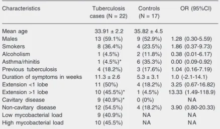
Twenty-two pulmonary TB cases and 17 non-TB controls were studied (healthy vol-unteers, N = 10, and patients with non-TB pulmonary diseases (respiratory tract infec-tions that were cured by short-course antibi-otics, N = 6, and primary lung cancer, N = 1). The demographic and clinical character-istics of the case and control groups were similar except for a past history of asthma and allergy (Table 1). Table 1 also describes the radiologic and bacteriologic findings. TB cases were further subdivided into “lim-ited disease” or “extensive disease” defined as lung involvement of less than one lobe or more than one lobe, respectively, on chest X-ray at first medical visit; as having cavi-tary or non-cavicavi-tary disease by the presence of cavitation, and as having low or high mycobacterial load (defined as ≤20 colonies or >20 colonies in the culture media). Table 1. Demographic, clinical, and radiologic features of tuberculosis cases and
controls.
Characteristics Tuberculosis Controls OR (95%CI)
cases (N = 22) (N = 17)
Mean age 33.91 ± 2.2 35.82 ± 4.5
Males 13 (59.1%) 9 (52.9%) 1.28 (0.30-5.59)
Smokers 8 (36.4%) 4 (23.5%) 1.86 (0.37-9.73)
Alcoholism 1 (4.5%) 2 (11.8%) 0.38 (0.01-6.17)
Asthma/rhinitis 1 (4.5%)* 6 (35.3%) 0.00 (0.09-0.92)
Previous tuberculosis 4 (18.2%) 3 (17.6%) 1.04 (0.16-7.19)
Duration of symptoms in weeks 11.3 ± 2.6 5.3 ± 3.1 1.0 (-2.1-14.1)
Extension <1 lobe 11 (50%) 4 (18.2%) 3.25 (0.67-16.82)
Extension >1 lobe 10 (45.5%)* 1 (4.5%) 13.33 (1.49-118.9)
Cavitary disease 9 (40.9%)* 0 (0%) N A
Non-cavitary disease 12 (54.5%) 4 (18.2%) 3.90 (0.80-20.33)
Low mycobacterial load 9 (40.9%) NA N A
High mycobacterial load 10 (45.5%) NA N A
Data are reported as mean ± SEM or number with percent in parentheses. Extension = number of pulmonary lobes affected by the disease; cavitary tuberculosis = lung cavitation on the chest X-ray; OR = odds ratio; 95%CI = 95% confidence interval; NA = not applicable.
Lung cells obtained by the induced sputum technique for evaluation of immune cell markers
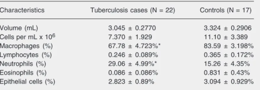
The volume of induced sputum, cell yield and differential cell counts are described in Table 2. Significantly lower percentages of macrophages and higher percentages of neu-trophils were present in sputum from TB cases than in controls. The cell yield and low percentage of squamous cells suggest the utility of induced sputum for studying lung immune response in pulmonary TB.
Expression of antigen-presenting molecules revealed that MHC class II HLA-DR are diminished in tuberculosis patients
Figure 1 shows the percent distribution of positive cells in the two study groups related to total cell or to total macrophage numbers. A statistically significant lower percentage of cells possessing HLA-DR was found for both total lung cells and lung macrophages among TB cases. However, the proportions of cases and controls having HLA-DR-positive cells were similar, 18 of 22 among TB cases and 16 of 17 among control subjects. Further analysis of HLA-DR expression in TB cases by extent of disease (limited versus extensive (≥1 lobe)), by lung cavitation, and by mycobacterial load on culture plates (<20 or ≥20) showed no statistically significant differences. In a similar analysis, CD1a expression was simi-lar between TB cases and controls (Table 3).
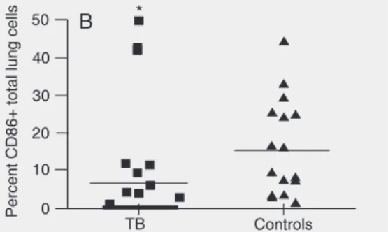
Expression of the costimulatory molecule CD86 (B7.2) is decreased in tuberculosis patients
Fewer TB patients expressed detectable CD86-positive cells, 10 of 22 versus 16 of 17 control subjects (P < 0.042). Figure 2 shows the expression of CD86 by total lung cells or by macrophages. TB cases showed a statistically significant lower CD86
expres-Table 2. Characteristics of the induced sputum.
Characteristics Tuberculosis cases (N = 22) Controls (N = 17)
Volume (mL) 3.045 ± 0.2770 3.324 ± 0.2906
Cells per mL x 106 7.370 ± 1.929 11.10 ± 3.389
Macrophages (%) 67.78 ± 4.723%* 83.59 ± 3.198%
Lymphocytes (%) 0.246 ± 0.089% 0.365 ± 0.172%
Neutrophils (%) 29.06 ± 4.99%* 15.26 ± 4.35%
Eosinophils (%) 0.086 ± 0.086% 0.831 ± 0.43%
Epithelial cells (%) 2.823 ± 0.89% 3.094 ± 0.929%
Data are reported as means ± SEM.
*P < 0.05 compared to controls (Student t-test).
Figure 1. HLA class II DR expres-sion by total lung cells or macro-phages from tuberculosis (TB) cases and non-TB controls. The horizontal lines are the means. A, Percentage of macrophages, and B, percentage of total lung cells expressing HLA-DR. Cells from induced sputum were prepared from TB cases (N = 22) and non-TB controls (N = 17) consisting of healthy volunteers (N = 10), non-TB pulmonary infection (N = 6), and a patient with lung cancer (N = 1). *P < 0.05 compared to con-trols (Student t-test). One result for the controls was equal to zero and is not shown.
Table 3. CD1a and costimulatory molecules.
Markers Positive staining Positive staining % Positive cells % Positive cells
in tuberculosis in controls in tuberculosis in controls
(N = 22) (N = 17) (N = 22) (N = 17)
CD1a 3 2 0.80 ± 0.49% 0.23 ± 0.19%
CD40 10 12 4.03 ± 1.17% 4.78 ± 1.3%
CD40L 3 4 0.22 ± 0.14% 0.82 ± 0.41%
CD28 5 3 2.55 ± 1.19% 1.03 ± 0.53%
CD152 2 2 0.88 ± 0.76% 0.29 ± 0.24%
sion as determined by total lung cells or by lung macrophages compared to controls. TB cases were further subdivided by disease severity. Figure 2C shows that patients with limited TB (<1 lobe) exhibited a significant-ly lower CD86 expression by lung macro-phages (P = 0.021). However, no statisti-cally significant difference was observed for CD86 expression when TB cases were ana-lyzed by presence of cavitary disease or by bacillary load. CD80 could not be evaluated because of an insufficient number of slides containing cytopreparation of cells from TB patients and controls. In contrast to CD86, other costimulatory molecules evaluated, CD40, CD150 (CD40L), CD28 and CD152 (CTL-4), failed to show differences between TB cases and controls (Table 3).
Discussion
Experimental models of M. tuberculosis infection have advanced our understanding of the pathogenesis of TB, shedding light on host resistance/susceptibility mechanisms, mycobacterial virulence factors and evasion
strategies. Nevertheless, neither in vivo nor in vitro animal models of TB mimic human disease. Therefore, understanding TB still requires studies on human or non-human primates. It is noteworthy that the majority of the studies conducted on humans have used cells from peripheral blood or pleural effusion. These studies may not mirror the immune response in the lung, the site of direct host and M. tuberculosis interaction in over 80% of TB cases.
Investigation of the lung compartment is hampered by the relative inaccessibility of the lung parenchyma. Bronchoalveolar lav-age, considered to be the gold standard for these analyses, is expensive and invasive, and is associated with occasional serious adverse events. Despite these serious limita-tions, our group has extensively used bron-choalveolar lavage to investigate lung im-mune responses in human TB (2,14,15). Al-ternatively, induced sputum has proven abil-ity to enhance the culture diagnosis of pul-monary TB even in the case of pleural dis-ease (16,17). Previous studies using induced sputum have suggested its usefulness in the
Figure 2. CD86 expression by total lung cells or macrophages from tuberculosis (TB) cases and non-TB controls. The horizontal lines are the means. The thick line over TB represents the individual results equal to zero. A, Percentage of macrophages expressing CD86. B, Percentage of total lung cells expressing CD86 cells from induced sputum were prepared from TB cases (N = 22) and non-TB con-trols (N = 17). One result for the concon-trols was equal to zero and is not shown. C, TB patients were segre-gated by extent of disease on chest X-ray: limited (<1 lobe involvement) and extensive (>1 lobe in-volvement). *P < 0.05 compared to controls in A and B and to limited disease in C (Student t-test).
A B
study of lung inflammation and TB (3,18-20). The application of this technique is a valuable strategy for the investigation of TB pathogenesis because of its safety, noninva-sive nature, lower cost, and its ability to obtain sufficient viable lung cells from the diseased portion of the lung.
Human TB is amongst several diseases in which an altered balance in the Th1-type and Th2-type immune response is thought to foster active disease. Studies of the periph-eral compartment of cells have found sup-pressed T cell proliferation, reduced IL-2 production, and impaired delayed type hy-persensitivity reaction to the tuberculin skin test (4-6). Among the proposed contributory mechanisms to this down-modulated lung immune state is that M. tuberculosis anti-gens act either directly or via a paracrine effect by the promotion of excessive “anti-inflammatory/down-modulatory” cytokines such as IL-10 that diminish antigen presen-tation and/or co-stimulation (8,21,22). IL-10 has been shown in vitro to diminish co-stimulatory and MHC class II molecule ex-pression on peripheral blood immune cells (8,12). Alternatively, excessive apoptosis of CD4+ T cells due to low expression of Bcl-2 and excessive expression of TGF-ß, Fas ligand, and TNF-α may further contribute to the immune dysregulation (23). Our previ-ous study in the lung bridges the two con-cepts. First, we identified increased amounts of IL-10 and bioactive TGF-ß, as well as increased co-expression of TGF-ß receptors I and II required for a cell response to TGF-ß (2,3). Second, we noted in the lung of patients with active TB detectable levels of CFP32, an M. tuberculosis-specific antigen that positively correlated with amounts of IL-10 but not IFN-γ (3). However, we did not examine in these patients whether ex-pression of antigen presentation and costimu-latory molecules were disturbed in active TB. Our present results revealed that active TB is associated with significantly lower expression of antigen presentation and
co-stimulatory molecules, MHC class II HLA-DR and CD86. Both molecules (MHC class II and CD86) play pivotal roles in T cell activation and differentiation through the presentation of antigen peptides to T cell receptors and the concomitant co-stimula-tory signaling (7,24,25). Therefore, our data on lung immune cells during active TB pro-vide the in vivo missing link for many in vitro mechanistic studies, in which attenua-tion of MHC class II expression by mono-nuclear phagocytes infected with virulent M. tuberculosis has been described in past years to involve several mechanisms (26,27). M. tuberculosis molecules, such as 19-kDa lipoprotein, antigen 85B and LpsG, interfere with the production of MHC class II mol-ecules in the phagosomes of macrophages and dendritic cells (21,28,29). The 19-kDa lipoprotein was also shown to selectively inhibit IFN-γ-induced class transactivator expression to diminish MHC class II expres-sion (30). Both IFN-γ and IL-10 also play a role in MHC class II. Inhibition of IFN-γ by up-regulation of histone deacetylation in the promoter region of the IFN-γ gene leads to attenuation of MHC class II expression in M. tuberculosis-infected macrophage-like cells (31). M. tuberculosis-induced IL-10 secre-tion leads to inhibisecre-tion of cathepsin S, result-ing in lower export of peptide-loaded class II molecules to the cell surface and diminished antigen presentation (12). Furthermore, Chang et al. (32), using mathematical mod-eling, concluded that M. tuberculosis uti-lizes multiple mechanisms to interfere with antigen presentation, allowing a continuous inhibition of MHC class II antigen presenta-tion to effector cells.
activated state (25). In murine TB, two stud-ies correlated lower levels of CD86 expres-sion with persistence of M. tuberculosis or disease progression (22,36). Garcia-Romo et al. (37) found delayed expression of co-stimulatory molecules by dendritic cells in mice mediastinal lymph nodes after intratra-cheal infection by M. tuberculosis, leading to delayed adaptive immune responses and progression to disease in the lung. The pres-ent results found diminished CD86 expres-sion in human lung cells and specifically in macrophages during active pulmonary TB. This is one of the earliest human in vivo results to confirm the prior reports on mu-rine TB (22,37). In one other study of co-stimulatory molecules in human TB, Soler et al. (38) failed to show differences in the expression of CD80/CD86 costimulatory molecules in tubercle granulomas caused by M. tuberculosis. This finding is in apparent contrast to our current findings. However, on close inspection, even though we ob-served an overall diminished CD86 expres-sion by all cells, the most significant de-crease was seen in patients with limited dis-ease (≤1 lobe). We interpret these findings to suggest that diminished CD86 expression occurs preferentially in earlier stages of clini-cal TB and might contribute to disease es-tablishment and progression in the lungs.
Down-modulation of CD86 becomes less apparent with advanced disease or in con-tained infection, as in the case of the tubercle granuloma.
In conclusion, our ex vivo findings indi-cate that HLA-DR and CD86, key elements for antigen-specific T cell immune responses against M. tuberculosis, are diminished on the lung cells of pulmonary TB patients. Decreased expression of HLA-DR and CD86 on the surface of lung immune cells of TB patients might result from direct cell infec-tion by M. tuberculosis or from a response to antigens released by M. tuberculosis, or may be indirectly mediated by immunosuppres-sive cytokines, IL-10 and/or TGF-ß pro-moted by M. tuberculosis. The diminished levels of HLA-DR and CD86 molecules on lung immune cells of TB patients may limit T cell immune responses that, in turn, favor M. tuberculosis persistence and progression to active disease.
Acknowledgments
The authors thank Dr. Richard D. Huard, Weill Medical College of Cornell Univer-sity, NewYork, USA, for personal commu-nication on the induction of IL-10 by human monocytes stimulated in vitro by CFP32.
References
1. http://www.who.int/tb/en. Accessed May 10, 2006.
2. Bonecini-Almeida MG, Ho JL, Boechat N, Huard RC, Chitale S, Doo H, et al. Down-modulation of lung immune responses by interleukin-10 and transforming growth factor beta (TGF-beta) and analysis of TGF-beta receptors I and II in active tuberculosis. Infect Immun 2004; 72: 2628-2634.
3. Huard RC, Chitale S, Leung M, Lazzarini LC, Zhu H, Shashkina E, et al. The Mycobacterium tuberculosis complex-restricted gene cfp32 encodes an expressed protein that is detectable in tuberculosis patients and is positively correlated with pulmonary interleukin-10. Infect Immun 2003; 71: 6871-6883.
4. Ellner JJ. Suppressor adherent cells in human tuberculosis. J Immunol 1978; 121: 2573-2579.
5. Toossi Z, Kleinhenz ME, Ellner JJ. Defective interleukin 2
produc-tion and responsiveness in human pulmonary tuberculosis. J Exp Med 1986; 163: 1162-1172.
6. Schauf V, Rom WN, Smith KA, Sampaio EP, Meyn PA, Tramontana JM, et al. Cytokine gene activation and modified responsiveness to interleukin-2 in the blood of tuberculosis patients. J Infect Dis 1993; 168: 1056-1059.
7. Sharpe AH, Freeman GJ. The B7-CD28 superfamily. Nat Rev Immunol 2002; 2: 116-126.
8. de la Barrera S, Aleman M, Musella R, Schierloh P, Pasquinelli V, Garcia V, et al. IL-10 down-regulates costimulatory molecules on Mycobacterium tuberculosis-pulsed macrophages and impairs the lytic activity of CD4 and CD8 CTL in tuberculosis patients. Clin Exp Immunol 2004; 138: 128-138.
STAT protein interference and suppression of cytokine signal trans-duction by measles virus V protein. J Virol 2003; 77: 7635-7644. 10. Murray PJ, Young RA. Increased antimycobacterial immunity in
interleukin-10-deficient mice. Infect Immun 1999; 67: 3087-3095. 11. Hirsch CS, Hussain R, Toossi Z, Dawood G, Shahid F, Ellner JJ.
Cross-modulation by transforming growth factor beta in human tu-berculosis: suppression of antigen-driven blastogenesis and inter-feron gamma production. Proc Natl Acad Sci U S A 1996; 93: 3193-3198.
12. Pin I, Gibson PG, Kolendowicz R, Girgis-Gabardo A, Denburg JA, Hargreave FE, et al. Use of induced sputum cell counts to investi-gate airway inflammation in asthma. Thorax 1992; 47: 25-29. 13. American Thoracic Society/Centers for Disease Control and
Pre-vention/Infectious Diseases Society of America: controlling tuber-culosis in the United States. Am J Respir Crit Care Med 2005; 172: 1169-1227.
14. Nicholson S, Bonecini-Almeida MG, Lapa e Silva JR, Nathan C, Xie QW, Mumford R, et al. Inducible nitric oxide synthase in pulmonary alveolar macrophages from patients with tuberculosis. J Exp Med 1996; 183: 2293-2302.
15. Lapa e Silva JR, Linhares C, Boechat N, Rego L, Almeida MG, Kriski AL, et al. Phenotypes of lung mononuclear phagocytes in HIV sero-negative tuberculosis patients: evidence for new recruitment and cell activation. Mem Inst Oswaldo Cruz 1996; 91: 389-394. 16. Conde MB, Soares SL, Mello FC, Rezende VM, Almeida LL,
Reingold AL, et al. Comparison of sputum induction with fiberoptic bronchoscopy in the diagnosis of tuberculosis: experience at an acquired immune deficiency syndrome reference center in Rio de Janeiro, Brazil. Am J Respir Crit Care Med 2000; 162: 2238-2240. 17. Conde MB, Loivos AC, Rezende VM, Soares SL, Mello FC, Reingold
AL, et al. Yield of sputum induction in the diagnosis of pleural tuberculosis. Am J Respir Crit Care Med 2003; 167: 723-725. 18. Brightling CE. Clinical applications of induced sputum. Chest 2006;
129: 1344-1348.
19. Pavord ID, Pizzichini MM, Pizzichini E, Hargreave FE. The use of induced sputum to investigate airway inflammation. Thorax 1997; 52: 498-501.
20. Almeida A, Boechat N, Lago P, Flores-Batista V, Santos AR, Nociari M, et al. Analysis of immunomodulatory molecules in lung cells of tuberculosis patients before and after chemotherapy by RT-PCR. Proc Am Thorac Soc 2006; 3: 391.
21. Fulton SA, Reba SM, Pai RK, Pennini M, Torres M, Harding CV, et al. Inhibition of major histocompatibility complex II expression and antigen processing in murine alveolar macrophages by Mycobacte-rium bovis BCG and the 19-kilodalton mycobacterial lipoprotein. Infect Immun 2004; 72: 2101-2110.
22. Bonato VL, Medeiros AI, Lima VM, Dias AR, Faccioliti LH, Silva CL. Downmodulation of CD18 and CD86 on macrophages and VLA-4 on lymphocytes in experimental tuberculosis. Scand J Immunol 2001; 54: 564-573.
23. Hirsch CS, Johnson JL, Okwera A, Kanost RA, Wu M, Peters P, et al. Mechanisms of apoptosis of T-cells in human tuberculosis. J Clin Immunol 2005; 25: 353-364.
24. Bhatia S, Edidin M, Almo SC, Nathenson SG. B7-1 and B7-2: similar costimulatory ligands with different biochemical, oligomeric and
sig-naling properties. Immunol Lett 2006; 104: 70-75.
25. Rogers NJ, Game DS, Camara NO, Jackson IM, Lombardi G, Lechler RI. Distinct effects of CD86-mediated costimulation on rest-ing versus activated human CD4+ T cells. Eur J Immunol 2005; 35: 2909-2919.
26. Hmama Z, Gabathuler R, Jefferies WA, de Jong G, Reiner NE. Attenuation of HLA-DR expression by mononuclear phagocytes infected with Mycobacterium tuberculosis is related to intracellular sequestration of immature class II heterodimers. J Immunol 1998; 161: 4882-4893.
27. Noss EH, Harding CV, Boom WH. Mycobacterium tuberculosis in-hibits MHC class II antigen processing in murine bone marrow macrophages. Cell Immunol 2000; 201: 63-74.
28. Ramachandra L, Noss E, Boom WH, Harding CV. Processing of Mycobacterium tuberculosis antigen 85B involves intraphagosomal formation of peptide-major histocompatibility complex II complexes and is inhibited by live bacilli that decrease phagosome maturation. J Exp Med 2001; 194: 1421-1432.
29. Gehring AJ, Dobos KM, Belisle JT, Harding CV, Boom WH. Myco-bacterium tuberculosis LprG (Rv1411c): a novel TLR-2 ligand that inhibits human macrophage class II MHC antigen processing. J Immunol 2004; 173: 2660-2668.
30. Pai RK, Convery M, Hamilton TA, Boom WH, Harding CV. Inhibition of IFN-gamma-induced class II transactivator expression by a 19-kDa lipoprotein from Mycobacterium tuberculosis: a potential mech-anism for immune evasion. J Immunol 2003; 171: 175-184. 31. Wang Y, Curry HM, Zwilling BS, Lafuse WP. Mycobacteria inhibition
of IFN-gamma induced HLA-DR gene expression by up-regulating histone deacetylation at the promoter region in human THP-1 mono-cytic cells. J Immunol 2005; 174: 5687-5694.
32. Chang ST, Linderman JJ, Kirschner DE. Multiple mechanisms allow Mycobacterium tuberculosis to continuously inhibit MHC class II-mediated antigen presentation by macrophages. Proc Natl Acad Sci U S A 2005; 102: 4530-4535.
33. Greenwald RJ, Freeman GJ, Sharpe AH. The B7 family revisited. Annu Rev Immunol 2005; 23: 515-548.
34. Teft WA, Kirchhof MG, Madrenas J. A molecular perspective of CTLA-4 function. Annu Rev Immunol 2006; 24: 65-97.
35. Vijayakrishnan L, Slavik JM, Illes Z, Greenwald RJ, Rainbow D, Greve B, et al. An autoimmune disease-associated CTLA-4 splice variant lacking the B7 binding domain signals negatively in T cells. Immunity 2004; 20: 563-575.
36. Ordway D, Harton M, Henao-Tamayo M, Montoya R, Orme IM, Gonzalez-Juarrero M. Enhanced macrophage activity in granuloma-tous lesions of immune mice challenged with Mycobacterium tuber-culosis. J Immunol 2006; 176: 4931-4939.
37. Garcia-Romo GS, Pedroza-Gonzalez A, Aguilar-Leon D, Orozco-Estevez H, Lambrecht BN, Estrada-Garcia I, et al. Airways infection with virulent Mycobacterium tuberculosis delays the influx of den-dritic cells and the expression of costimulatory molecules in medias-tinal lymph nodes. Immunology 2004; 112: 661-668.