ISSN 0100-879X
BIOMEDICAL SCIENCES
AND
CLINICAL INVESTIGATION
www.bjournal.com.br
www.bjournal.com.br
Volume 43 (11) 1010-1134 November 2010
Institutional Sponsors
The Brazilian Journal of Medical and Biological Research is partially financed by
Hotsite of proteomics metabolomics developped by:
Braz J Med Biol Res, November 2010, Volume 43(11) 1116-1122
doi: 10.1590/S0100-879X2010007500103
Prognosis of colorectal cancer with liver metastasis: value of a
prognostic index
Prognosis of colorectal cancer with
liver metastasis: value of a prognostic index
Y. Wang
1,2, Y.F. Liu
1, Y. Cheng
1, D.H. Yi
1, P. Li
1, W.Q. Song
1,
D.Z. Fu
1and X. Wang
11Department of General Surgery, The First Hospital of China Medical University,
Shen Yang, Liao Ning Province, China
2Department of Surgery, Shen Zhou Hospital, Shen Yang Medical College,
Shen Yang, Liao Ning Province, China
Abstract
The objective of the present study was to explore the factors related to the prognosis of colorectal cancer (CRC) and to
estab-lish a prognostic model for the selection of patients who might benefit from hepatic resection for metastatic CRC. A total of 293
patients undergoing liver resection for metastatic CRC (172 males and 80 females ranging in age from 26 to 80 years) were selected and clinical, pathological and outcome data were examined in this retrospective study. The prognostic index (PI) of
the patients was calculated on the basis of results of multivariate analysis. Patients were stratified into different groups, with
survival curves projected according to PI. The 1-, 3-, and 5-year overall survival rates were 58.3, 26.4, and 11.3%, respectively. Univariate analysis indicated that degree of primary tumor differentiation, resection margin, preoperative carcinoembryonic
an-tigen (CEA) level, number of liver metastases, and resection of liver metastases were associated with prognosis (P < 0.05). In
multivariate analysis, the last three factors were found to be independent prognostic factors. The resection of liver metastases
was a favorable factor. Patients were classified into three groups according to PI, which differed significantly in survival rate (P < 0.05). The individual survival rate was evaluated based on PI. Resection of hepatic colorectal metastases may produce
long-term survival and cure. The proposed PI was easy to use, was highly predictive of patient outcome, and permitted catego-rization of patients into treatment groups.
Key words: Colorectal cancer; Liver metastasis; Prognosis
Introduction
Correspondence: Y.F. Liu, Department of General Surgery, The First Hospital of China Medical University, Shen Yang, 110001, Liao Ning Province, China. E-mail: [email protected]
Received April 7, 2010. Accepted September 16, 2010. Available online October 8, 2010. Published November 12, 2010.
Colorectal carcinoma (CRC) is a common malignancy of the digestive system and up to 50% of affected patients will develop metastases during the course of their disease (1,2). Radical resection offers the only chance of long-term survival, with 30 to 40% of patients surviving 5 years. Recent advances in imaging modality, indication of hepatic
resec-tion, technical refinement of hepatectomy and perioperative
care, together with the expansion of criteria for surgery, have increased the number of patients suitable for hepatic resection and the safety of the procedure (3,4). However, the prognosis for patients with liver metastases from CRC is still poor. Because of this, only 30 to 58% of patients who undergo a curative liver resection with complete extirpation of liver metastases are alive at 5 years (5,6). The present study retrospectively analyzed 293 patients subjected to resection of liver metastases in the Department of Surgery,
the First Hospital of China Medical University and Shen Zhou Hospital of Shen Yang Medical College, China, from January 1993 to January 2006. Several variables were analyzed by univariate and multivariate methods to deter-mine independent prognostic factors in order to calculate the prognostic index (PI) and then to establish a prognostic
scoring system to identify patients most likely to benefit
from surgery.
Patients and Methods
Patients
A total of 293 patients with histologically proven liver
Colorectal cancer prognosis 1117
Medical College, from January 1993 to January 2006.
The diagnosis of metastatic CRC was confirmed by his -topathological assessment. Patients were considered to have resectable disease if there was local control of the primary cancer, no extrahepatic disease existed, and com-plete removal of all hepatic lesions was expected, leaving enough hepatic parenchyma to prevent liver failure. No ablative strategies were used along with resection in any of these patients. Intraoperative ultrasound of the liver was performed in all patients to assign the location of the lesions to anatomical structures and to rule out additional lesions in the remnant liver.
The treatment policy for synchronous liver metastases was simultaneous resection regardless of their number and extent and the location of the primary cancer. Data for these patients were extracted from the hospital database and interviews, including gender, age, vascular invasion, hepatic lymph node metastases, extrahepatic metastases, type of hepatic resection, location of the primary tumor,
pre-operative carcinoembryonic antigen (CEA) levels, degree
of primary tumor differentiation, diameter of the largest liver metastasis, liver metastasis distribution, resection margin, number of liver metastases, resection of liver metastases, and time of diagnosis of liver metastases.
Statistical analysis
Death occurring within 30 days of the surgical procedure
was defined as operative mortality. Death occurring after surgery and before discharge was defined as hospital mor -tality. Survival time was calculated from the date of hepatic
resection to death or censored date. Patients who died of CRC were treated as event observations, and patients who died of unrelated causes and were alive at the last follow-up were treated as censored observations. Survival curves were constructed using the Kaplan-Meier method
and compared by the log-rank test. Significant prognostic
factors in the univariate analysis were entered into the Cox proportional hazards multiple regression model, and stepwise selection of independent prognostic variables was
performed manually by significant changes in likelihood ratio. A software program (SPSS 14.0, SPSS Inc., USA)
was used for the statistical analyses.
Results
Patient demographics
The 1-, 3-, and 5-year overall survival rates were 58.3, 26.4, and 11.3%, respectively. The overall survival curve
is shown in Figure 1A.
Patients with stage III or IV colon cancer according to
the classification of the International Union Against Cancer
(UICC) normally received adjuvant chemotherapy after re-section, but chemotherapy was not administered routinely before or after hepatic resection. Six patients who died in the hospital and 35 patients with an uneventful perioperative course who were lost to follow-up were also excluded from
analysis. Thus, data of 252 patients were eligible for final
analysis. The study population included 172 men (68.3%) and 80 women (31.7%). Median patient age was 61 years (range: 33-84 years). The primary tumor was located in the
colon in 146 patients (57.9%) and in the rectum in 106 patients (42.1%). Liver metastases were present at the time of diagnosis in 105 patients (41.7%), whereas 147 patients (58.4%) had metachronous hepatic lesions. Nine-teen patients (7.5%) underwent repeat
liver resection. Among the 252 curative
surgical procedures, 43 wedge resec-tions (17.1%), 138 segmentectomies (54.8%), and 71 major hepatectomies (28.2%) were performed. Operative complications occurred in 46 patients (18.3%). Postoperative hemorrhage, pleural effusion, wound abscess, intra-abdominal abscess, bile leakage and anastomotic leakage, occurred in 9, 13, 10, 6, 4, and 4 patients, respec-tively. In 11 patients (4.4%), a second laparotomy was required.
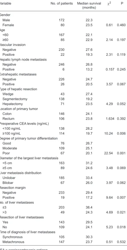
Univariate analysis of outcome
We analyzed the effects of 15 clinical pathologic factors on survival.
Preoperative CEA level of less or more than 100 ng/mL (P = 0.006), degree of primary tumor differentiation (P =
0.001), positive or negative resection
margin (P = 0.007), more or less than 3 liver metastases (P = 0.021), resection of liver metastases or not (P = 0.018) showed a significant prognostic value
for survival (Table 1).
Multivariate analysis of outcome
The prognostic factors in univariate analysis were entered into a multi-variate model to identify independent predictors of long-term survival.
Among the five significant variables, preoperative CEA level, number of
liver metastases, and resection of
liver metastases were identified as
independent prognostic factors and used to calculate the PI (Figure 2). Of
these, preoperative CEA levels were clearly the most influential, with an
increase in the likelihood of death of
1.746 times if preoperative CEA levels
were higher than 100 ng/mL, followed by number of liver metastases of 3 or
more (relative risk, RR = 1.432), and
resection of liver metastases or not
represented a favorable factor (RR =
0.406) (Table 2, Figure 2).
Table 1. Univariate analysis of prognostic factors after resection of liver metastases from colorectal cancer.
Variable No. of patients Median survival
(months)
c2 P
Gender
Male 172 22.3
Female 80 23.5 0.61 0.460
Age
<60 167 22.1
≥60 85 22.9 2.14 0.197
Vascular invasion
Negative 230 27.6
Positive 22 19.3 2.31 0.119
Hepatic lymph node metastasis
Negative 246 26.8
Positive 6 13.2 0.157 0.245
Extrahepatic metastases
Negative 226 24.7
Positive 26 20.5 3.57 0.067
Type of hepatic resection
Wedge 43 27.4
Segmentectomy 138 19.2
Hepatectomy 71 23.5 4.29 0.052
Location of primary tumor
Colon 146 24.1
Rectum 106 23.8 1.634 0.392
Preoperative CEA levels (ng/mL)
<100 ng/mL 138 28.2
≥100 ng/mL 114 19.7 10.24 0.006
Degree of primary tumor differentiation
Good 76 26.7
Moderate 109 25.1
Poor 67 20.1 22.54 0.001
Diameter of the largest liver metastasis
<5 cm 163 31.2
≥5 cm 89 24.6 3.48 0.069
Liver metastasis distribution
Unilobar 185 33.4
Bilobar 67 26.0 3.97 0.062
Resection margin
Negative 233 29.4
Positive 19 17.2 9.64 0.007
No. of liver metastases
≤3 203 38.4
>3 49 24.3 4.69 0.021
Resection of liver metastases
Yes 143 29.5
No 109 24.1 5.23 0.018
Time of diagnosis of liver metastases
Synchronous 105 30.3
Metachronous 147 23.7 0.51 0.532
Colorectal cancer prognosis 1119
Calculation of PI
The PI equation was constructed including all significant variables and coefficients from Table 2 as follows: PI = (0.349 x number of liver metastases) + (0.751 x preopera
-tive CEA levels) - (0.817 x resection of liver metastases).
In this equation the value of each variable was scored as 1 or 2: liver metastasis resection, 1; no liver metastasis resection, 2; less than 3 liver metastases, 1; more than 3
liver metastases, 2; preoperative CEA level of less than 100 ng/mL, 1; preoperative CEA level of more than 100 ng/mL,
2. The maximum PI value is 1.383, and the minimum value is -0.534. With the upper and lower quartile of PI, patients were divided into three risk groups and the differences in
survival between groups were significant by the
Kruskal-Wallis H test (Table 3, Figure 1B).
Calculation of individual survival
To estimate the expected survival rate of each individual,
we first calculated the average level of the t-year survival rate
S(t). In this study, we used the median prognostic index M
as a benchmark, and individual’s RR = exp(Pl-M). Thus, the
individual t-year survival rate is S(t)RR. Next, we randomly selected four individuals to calculate the expected survival rate compared to the benchmark (Table 4).
Discussion
The liver is the most common site of distant metastases from CRC. Resection remains the only chance of cure for patients with hepatic colorectal metastases, resulting in pro-longed survival compared to patients treated with palliative
Table 2. Multivariate analysis of prognostic factors after resection of synchronous liver metastases from colorectal cancer.
Variable β Wald c2 P Relative risk
(RR)
RR (95%CI)
Lower Upper
Number of liver metastases 0.349 5.542 0.019 1.432 1.062 1.931
Preoperative CEA levels 0.751 4.149 0.002 1.746 1.021 2.985
Resection of liver metastases -0.817 4.526 0.033 0.406 0.177 0.934
Gender 0.126 0.096 0.757 1.134 0.512 2.515
Age -0.425 1.578 0.209 0.654 0.337 1.269
Vascular invasion 0.284 0.458 0.498 1.328 0.584 3.020
Hepatic lymph node metastasis 0.384 1.094 0.296 1.468 0.715 3.015
Extrahepatic metastases 0.080 0.021 0.886 1.084 0.362 3.247
Type of hepatic resection wedge 0.994 1.989 0.158 2.701 0.679 10.744
Location of primary tumor 0.036 0.094 0.759 1.037 0.824 1.305
Degree of primary tumor differentiation -0.185 0.746 0.388 0.831 0.547 1.264
Diameter of the largest liver metastasis 0.329 0.252 0.616 1.389 0.385 5.016
Liver metastasis distribution 0.125 0.017 0.895 1.133 0.177 7.236
Resection margin 0.188 0.128 0.720 1.206 0.432 3.372
Time of diagnosis of liver metastases 0.092 0.035 0.852 1.096 0.418 2.875
CEA = carcinoembryonic antigen.
Table 3. Survival rate of the different risk groups (Kruskal-Wallis H test).
PI classification Median PI Number of cases Survival rate (%) c2 P
1 year 3 years 5 years
Low risk 31 P ≤ 0.016 63 74.9 34.5 20.8 27.3 <0.00
Moderate risk 15 0.016 < PI < 1.383 123 55.7 24.3 10.3
High risk 6 PI ≥ 1.383 66 32.3 1.7 0
Figure 2. Survival curve according to five significant variables. + = censored.
chemotherapy (7-9). The safety of major hepatic resection has been demonstrated in many institutions, encouraging surgeons to pursue more extensive resections (10-13). This has resulted in increasing complexity of the surgical approach. Resections involving the removal of more than two-thirds of the liver parenchyma are common, as are resections of more than four hepatic metastases (14-16).
Advances in surgical technique and perioperative care, such
as portal vein embolization, have enabled a more radical approach to the treatment of CRC liver metastases and extended the indications for surgical therapy. Thus, there is an ongoing need for a system to be used for the scrutiny of such invasive therapy.
In the 1980’s, two staging systems were developed by Gennari et al. (17,18). These systems were based on the
Table 4. t-year survival rate of four selected individuals.
Random individual
PI RR Expected t-year
survival rate (%)
1 year 3 years 5 years
1 1.383 2.61 24.5 3.9 0.5
2 0.632 1.29 49.9 20.3 7.7
Benchmark 0.425 1.0 58.3 29.1 13.7
3 0.217 0.81 64.5 36.7 19.9
4 -0.534 0.38 81.3 62.3 46.7
PI = prognostic index; RR = relative risk.
Colorectal cancer prognosis 1121
in some institutions because a favorable prognosis can be anticipated if the tumors are removed completely (23,24). Many ablative methods have been proposed for the treat-ment of liver tumors that are not suitable for resection. For instance, cryoablation and radiofrequency ablation show the greatest promise as treatment for metastatic colorectal
cancer. Although unlikely to be curative for large lesions,
it is possible that these ablative techniques may produce
complete destruction of smaller metastases. An
ever-more-aggressive approach is being undertaken based on the improving safety of hepatic resection and as a result of the inability of other therapies to produce long-term survival. It is also clear that systemic chemotherapy may have the greatest impact in the adjuvant setting where residual disease is minimal and microscopic. In a recent study of combined regional and systemic adjuvant chemotherapy,
patients in the high risk group benefited most from such
aggressive adjuvant chemotherapy.
The application of the Cox model to select the variables
that influence long-term survival, to calculate the individual
PI and then to stratify the patients into different groups is widely used in clinical research. Liu et al. (25) stated that
the Cox model has practical significance only when the entire PI is stratified into different groups according to the
estimated survival rate. Some studies have applied the PI to nasopharyngeal and endometrial cancer, showing that the PI is of good practical use (26).
The present study was based on Cox regression
analysis of the β value and significant prognostic factors
to calculate the PI for each patient. With PI of the upper and lower quartiles for the sector, patients were divided into three groups. The results showed that there is a large difference in survival rates, suggesting that PI is of clinical and practical value. Obviously, PI could predict long-term patient survival, facilitating the choice of treatment for clinical reference.
Acknowledgments
The authors would like to thank Song Wang (Shen Yang Environmental Protection Bureau) for assistance with the statistical analysis.
References
1. Khatri VP, Petrelli NJ, Belghiti J. Extending the frontiers of surgical therapy for hepatic colorectal metastases: is there a limit? J Clin Oncol 2005; 23: 8490-8499.
2. Benson AB III. Epidemiology, disease progression, and
economic burden of colorectal cancer. J Manag Care Pharm 2007; 13: S5-S18.
3. Folprecht G, Grothey A, Alberts S, Raab HR, Kohne CH.
Neoadjuvant treatment of unresectable colorectal liver me-tastases: correlation between tumour response and
resec-tion rates. Ann Oncol 2005; 16: 1311-1319.
4. Chung KY, Saltz LB. Antibody-based therapies for colorectal
cancer. Oncologist 2005; 10: 701-709.
5. Choti MA, Sitzmann JV, Tiburi MF, Sumetchotimetha W,
Rangsin R, Schulick RD, et al. Trends in long-term survival following liver resection for hepatic colorectal metastases. Ann Surg 2002; 235: 759-766.
6. Abdalla EK, Vauthey JN, Ellis LM, Ellis V, Pollock R, Broglio
KR, et al. Recurrence and outcomes following hepatic
re-related to primary CRC (17,18). These staging systems
were based on a multivariate survival analysis reflecting
prognosis but using 7 factors. Thus, all of the factors must be explored to determine the stage, which may make it
difficult to use these staging systems. Thus, a prognostic
system might be helpful to grade patients’ risk and to be used to tailor their further management. We have chosen a novel approach to develop a prognostic system based
on variables that were derived from significant independent
prognostic factors for consideration of liver surgery. The prognostic index model presented here can improve the accuracy of patient selection for surgery and be used to allocate patients to combined treatment modalities. More-over, the P values for high risk vs moderate risk groups and moderate risk vs low risk groups are significant.
The PI was calculated from an equation based on
variables and their correlation coefficients and related to
high, moderate and poor risk groups. Three groups with
different prognoses were identified among all candidates
for liver surgery.
Although liver resection improved survival prospects in
all patients regardless of their prognosis, none of the patients assigned to the high risk group were alive at 5-year follow-up. From our results, it is obvious that patients in the high risk group have very poor outcomes, and resection without additional effective adjuvant therapy is highly questionable. Patients in the moderate risk group have a much more guarded prognosis, and resection should be planned in the context of adjuvant therapies. Patients in the low risk group have a highly favorable outcome, and surgical resection is undoubtedly a rational therapy. Moreover, extensive pre-operative staging involving positron emission tomography and magnetic resonance imaging might be performed in patients who are in the high risk group, but are otherwise suitable for major liver resection (19-22).
section, radiofrequency ablation, and combined resection/ ablation for colorectal liver metastases. Ann Surg 2004; 239: 818-825.
7. Kerr DJ, McArdle CS, Ledermann J, Taylor I, Sherlock DJ,
Schlag PM, et al. Intrahepatic arterial versus intravenous
fluorouracil and folinic acid for colorectal cancer liver me -tastases: a multicentre randomised trial. Lancet 2003; 361: 368-373.
8. Yuste AL, Aparicio J, Segura A, Lopez-Tendero P, Girones R, Perez-Fidalgo JA, et al. Analysis of clinical prognostic factors
for survival and time to progression in patients with
meta-static colorectal cancer treated with 5-fluorouracil-based
chemotherapy. Clin Colorectal Cancer 2003; 2: 231-234. 9. Lorenz M, Muller HH. Randomized, multicenter trial of
fluorouracil plus leucovorin administered either via hepatic arterial or intravenous infusion versus fluorodeoxyuridine
administered via hepatic arterial infusion in patients with nonresectable liver metastases from colorectal carcinoma. J Clin Oncol 2000; 18: 243-254.
10. Yamada H, Katoh H, Kondo S, Okushiba S, Morikawa T.
Mesenteric lymph nodes status influencing survival and
recurrence pattern after hepatectomy for colorectal liver me-tastases. Hepatogastroenterology 2002; 49: 1265-1268.
11. Ercolani G, Grazi GL, Ravaioli M, Cescon M, Gardini A,
Varotti G, et al. Liver resection for multiple colorectal
metas-tases: influence of parenchymal involvement and total tumor
volume, vs number or location, on long-term survival. Arch Surg 2002; 137: 1187-1192.
12. Weber SM, Jarnagin WR, DeMatteo RP, Blumgart LH, Fong Y. Survival after resection of multiple hepatic colorectal me-tastases. Ann Surg Oncol 2000; 7: 643-650.
13. Jarnagin WR, Gonen M, Fong Y, DeMatteo RP, Ben-Porat L, Little S, et al. Improvement in perioperative outcome after hepatic resection: analysis of 1,803 consecutive cases over the past decade. Ann Surg 2002; 236: 397-406.
14. Scheele J, Stang R, Altendorf-Hofmann A, Paul M. Resec -tion of colorectal liver metastases. World J Surg 1995; 19: 59-71.
15. Gayowski TJ, Iwatsuki S, Madariaga JR, Selby R, Todo S, Irish W, et al. Experience in hepatic resection for metastatic colorectal cancer: analysis of clinical and pathologic risk factors. Surgery 1994; 116: 703-710.
16. Nordlinger B, Guiguet M, Vaillant JC, Balladur P, Boudjema
K, Bachellier P, et al. Surgical resection of colorectal
carci-noma metastases to the liver. A prognostic scoring system to improve case selection, based on 1568 patients. Association
Francaise de Chirurgie. Cancer 1996; 77: 1254-1262. 17. Gennari L, Doci R, Bignami P, Bozzetti F. Surgical treatment
of hepatic metastases from colorectal cancer. Ann Surg 1986; 203: 49-54.
18. Gennari L, Doci R, Bozzetti F, Bignami P. Proposal for stag-ing liver metastases. Recent Results Cancer Res 1986; 100: 80-84.
19. Strasberg SM, Dehdashti F, Siegel BA, Drebin JA, Linehan
D. Survival of patients evaluated by FDG-PET before hepatic resection for metastatic colorectal carcinoma: a prospective database study. Ann Surg 2001; 233: 293-299.
20. Zealley IA, Skehan SJ, Rawlinson J, Coates G, Nahmias
C, Somers S. Selection of patients for resection of hepatic metastases: improved detection of extrahepatic disease with FDG pet. Radiographics 2001; 21 (Spec. No.): S55-S69.
21. Strasberg SM, Siegal BA. Survival of patients staged by
FDG-PET before resection of hepatic metastases from colorectal cancer [letter]. Ann Surg 2002; 235: 308-310. 22. Rohren EM, Paulson EK, Hagge R, Wong TZ, Killius J,
Clavien PA, et al. The role of F-18 FDG positron emission
tomography in preoperative assessment of the liver in patients being considered for curative resection of hepatic metastases from colorectal cancer. Clin Nucl Med 2002; 27: 550-555.
23. Headrick JR, Miller DL, Nagorney DM, Allen MS, Des -champs C, Trastek VF, et al. Surgical treatment of hepatic and pulmonary metastases from colon cancer. Ann Thorac Surg 2001; 71: 975-979.
24. Elias D, Ouellet JF, Bellon N, Pignon JP, Pocard M, Lasser P. Extrahepatic disease does not contraindicate hepatectomy for colorectal liver metastases. Br J Surg 2003; 90: 567-574.
25. Liu Q, Jin PH, Da-Wei HE. A study of estimating the survival
rate of Cox’s model classed by PV. Chinese Health Stat 1997; 14: 4-6.