Antiviral activity of shikonin ester derivative PMM-034
against enterovirus 71
in vitro
Y. Zhang
1,2*, H. Han
1*, L. Sun
3*, H. Qiu
1, H. Lin
1, L. Yu
2, W. Zhu
2, J. Qi
1, R. Yang
1, Y. Pang
1,
X. Wang
1, G. Lu
1and Y. Yang
1 1State Key Laboratory of Pharmaceutical Biotechnology, NJU-NJFU Joint Institute of Plant Molecular Biology,Nanjing University, Nanjing, China 2Suzhou Industrial Park Center for Disease Control and Prevention, Suzhou, China 3College of Agriculture, Nanjing Agricultural University, Nanjing, China
Abstract
Human enterovirus 71 (EV71) is the major causative agent of hand, foot, and mouth disease (HFMD), particularly in infants and children below 4 years of age. Shikonin is a bioactive compound with anti-inflammatory, antiviral, and antibacterial activities derived from the roots of the Chinese medicinal herbLithospermum erythrorhizon. This study aimed to examine the antiviral activity of PMM-034, a shikonin ester derivative, against EV71 in rhabdomyosarcoma (RD) cells. Cytotoxicity of PMM-034 on RD cells was determined using WST-1 assay. Dose- and time-dependent effects of PMM-034 on EV71 replication in RD cells were determined using plaque reduction assay. mRNA expression levels of EV71/VP1 and pro-inflammatory cytokines (IL-1b, IL-6, IL-8, and TNF-a) were determined by real-time RT-PCR, and EV71/VP1 and phospho-p65 protein expressions were determined by western blot analysis. PMM-034 exhibited only weak cytotoxicity against RD cells. However, PMM-034 exhibited significant antiviral activity against EV71 in RD cells with 50% inhibitory concentration of 2.31mg/mL. The VP1 mRNA and protein levels were significantly reduced in cells treated with PMM-034. Furthermore, relative mRNA expression levels of IL-1b, IL-6, IL-8, and TNF-asignificantly decreased in the cells treated with PMM-034, while the phospho-p65 protein expression was also significantly lower in the treated cells. These results indicated that PMM-034 suppressed the expressions of pro-inflammatory cytokines in RD cells, exhibiting antiviral activity against EV71, as evidenced by the reduced VP1 mRNA and protein levels in PMM-034-treated cells. Thus, PMM-034 is a promising candidate for further development as an EV71 inhibitor.
Key words: EV71; VP1; Shikonin ester derivatives PMM-034; Rhabdomyosarcoma cells; NF-kB
Introduction
Hand, foot, and mouth disease (HFMD) is a common viral infection that commonly affects children below the age of 4 years (1). Human enterovirus 71 (EV71) belongs to the Enterovirus genus of the family Picornaviridae and causes sporadic outbreaks of HFMD. EV71 infection targets the central nervous system (CNS) (2). Once EV71 infects the CNS, a patient can die rapidly from severe complications including encephalitis and pulmonary edema (3). Recently, several consecutive EV71 epidemics have been reported in the Asia-Pacific region and no effective treatment for this infection exists. Selection of resistant viruses after treatment with antiviral drugs is inevitable (4), and while resistant viral strains have increased in recent years, only a few antiviral drugs have been approved for clinical use (5). The pace of pathogen resistance has exceeded the development of new antiviral
drugs. Therefore, there is an urgent need to develop new effective antiviral compounds (4) to meet the grow-ing need and combat resistance to existgrow-ing antiviral drugs.
Signal transduction pathways are essential for normal viral immunity and for virus replication. Viral infections induce the production of pro-inflammatory cytokines and chemokines. A previous study has shown that excessive pro-inflammatory cytokine and chemokine responses were induced in human monocyte-derived macrophages, and interleukin (IL)-1b, IL-6, IL-8, and TNF-a levels, but not IFN-a and g levels, were increased in EV71-infected patients (6). The NF-kB complex is activated in response to viral and bacterial infections, and the binding of the virion to its receptor can trigger membrane-proximal sig-naling cascades to activate NF-kB (7). Sometimes, viral
Correspondence: Y. Yang:<yangyh@nju.edu.cn>| G. Lu:<guihua.lu@nju.edu.cn>| X. Wang:<wangxm07@nju.edu.cn>
*These authors contributed equally to this study.
products such as dsRNA and viral proteins can trigger NF-kB activation (8).
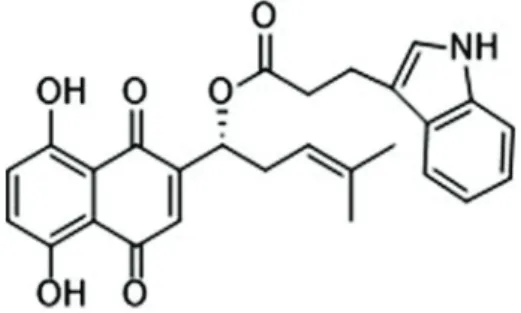
Several herbal prescriptions of traditional Chinese medicine (TCM) have been used to manage endemic infections due to their antiviral activity, for example, directly inhibiting or inactivating viruses, or improving the host immune system (9–11). Historically, root extracts obtained from the Chinese medicinal herb L. erythrorhizon have been used to treat burns, inflammation, trauma, and ulcers (12). Several studies have demonstrated that L. erythro-rhizonhas strong inhibitory activity towards neuraminidase and exhibits antiviral effects [i.e., against influenza A (H1N1) virus] by decreasing the viral reproduction and cytopathic effects (CPE) on virus-infected cells (13). Shikonin is an important naphthoquinone natural product derived from the roots of L. erythrorhizon (14). Shikonin exhibits a broad range of pharmacological activities includ-ing oxidant, cancer, inflammatory and anti-bacterial effects (15,16). Shikonin may also exert antiviral activity, and it has been found to exhibit anti-human immunodeficiency virus-1 (HIV-1) and adenovirus activ-ities (7,17). Recent research on shikonin has shifted to its derivatives (18), of which PMM-034 is one type (Figure 1). However, the antiviral effect of PMM-034 on EV71 infection remains unclear.
Here, we evaluated the extent to which PMM-034 could inhibit EV71 replication, and then assessed its effect on EV71/VP1 mRNA and protein expressions in rhabdomyo-sarcoma (RD) cellsin vitro. Furthermore, we explored the stage in the replication cycle at which PMM-034 inhibits EV71 and assessed the anti-inflammatory activity of PMM-034 on EV71-induced inflammationin vitro.
Material and Methods
Cells and viruses
RD cells were purchased from the American Type Cul-ture Collection (USA) and culCul-tured in Dulbecco’s modified Eagle’s medium (DMEM; Gibco, USA) with 10% fetal bovine serum (FBS; Gibco). The cell culture plates were incubated in a humidified incubator containing 5% CO2at 37°C.
EV71 was provided by the Suzhou Centre for Disease Prevention and Control (China). Finally, virus particles
were amplified using RD cells, and the virus titer was determined by a plaque reduction assay using RD cells.
Compounds
Shikonin ester derivatives were synthesized by our lab (19–21), and PMM-034 was dissolved in dimethyl sulf-oxide (DMSO) forin vitrostudies.
Cytotoxicity assay
Cytotoxicity of PMM-034 against RD cells was determined using cell viability assay in 96-well plates with cells cultured to 80–90% confluence (approximately 5104 cells/well). The cells were treated with various concentrations of PMM-034 dissolved in DMSO (0, 2.5, 5, 10, and 20 mg/mL) for 48 h, and then cell viability was assayed using WST-1 (Beyotime, China). Briefly, 10mL of WST-1 solution was added to the cells and the suspension was incubated at 37°C for 2 h. Medium without cells was used as the blank control. Absorbance was measured at a wavelength of 450 nm using an AMR-100 Microplate Reader (ThermoRui-OS, USA). Cell viability was calculated using the following formula: Cell viability (%) = (absorbance (Ab)experimental group – Abblank control) / (Abnegative control –Abblank control) 100%. The half-maximal cytotoxic concentration (CC50) was defined as the con-centration that reduced the Ab of PMM-034-treated cells to 50% the Ab of negative control (untreated cells) (22).
Plaque reduction assay
RD cells (3105 cells/well) were seeded into 6-well plates and cultured to 80% confluence. After EV71 [80 plaque-forming units (pfu)/well] were incubated with RD cells to allow absorption for one hour at 37°C, the culture supernatants were replaced with fresh DMEM containing 1–3% FBS and 1.5% methyl cellulose, and then cultured for an additional 96 h at 37°C in the presence of 5% CO2.
The cells were then washed with phosphate-buffered saline, fixed with 4% paraformaldehyde, stained with 5% crystal violet for 15 min, and then washed in running water. The plaques were counted under an inverted microscope (Nikon, TS100, Japan), and the virus titer (pfu/mL) was determined. Inhibition rate (%) was calcu-lated as follows: [(mean number of plaques in the untreated group)–(mean number of plaques in the experi-mental group)] / (mean number of plaques in the untreated group) 100 (23).
Time-dependent effect of PMM-034 on EV71 repli-cation in RD cells. RD cells (3105 cells/well) were seeded and incubated in the wells of 6-well plates. RD cell monolayers were infected with EV71 at 80 pfu/well. After incubation for 1 h to facilitate absorption, culture supernatants were replaced with fresh DMEM contain-ing 1–3% FBS. Then, 10 mg/mL PMM-034 was added to the cells at 0, 4, 8, 12, 16, and 24 h post-infection. Cell supernatants were collected for the viral plaque reduction assay.
Dose-dependent effect of PMM-034 on EV71 replica-tion in RD cells. RD cells (3105cells/well) were plated
into 6-well culture plates and incubated for 24 h. The medium was then removed and cells were infected with EV71 at 80 pfu/well. After virus absorption for 1 h, the medium was aspirated from the wells to remove unab-sorbed virus, and the cells were washed thrice with serum-free DMEM, and treated with different PMM-034 concentrations to test for antiviral activity. Finally, EV71-infected cells and culture supernatants were collected 48 h after infection for the plaque reduction assay.
Quantitative real-time reverse transcription (RT)-PCR
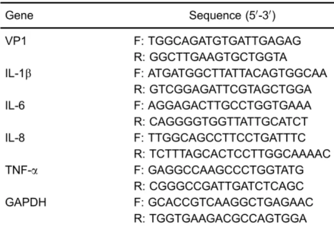
Total RNA was extracted using Trizol (Invitrogen, USA) according to the manufacturer’s instructions. The RNA purity and concentration were determined by spectroscopy [Ab260/Ab280] to be 1.8–2.0. RNA was reverse tran-scribed using a Reverse Transcription System (Promega, USA). Primers forIL-1,IL-6,IL-8,TNF-a,VP1andGAPDH were designed using Beacon Designer 7.0 and synthe-sized by Sangon Biotech Company (China; Table 1). Specific mRNAs were quantified by real-time PCR using Fast SYBR Green Master Mix (Invitrogen) in a Bio Rad CFX96 Real-Time System (Bio Rad, USA). Real-time PCR reactions were performed in triplicate in 96-well plates under the following conditions; 15 s at 95°C followed by 40 cycles of 15 s at 95°C, 30 s at 65°C, and 30 s at 72°C. GAPDH was used as the internal control. The relative expression levels of the genes were compared with those of GAPDH by the 2–DDCtmethod.
Western blot analysis
Total protein was extracted using radio-immunopreci-pitation assay plus phenylmethylsulfonyl fluoride (Solar-bio, China) according to the manufacturer’s instructions. Protein concentration was determined by the BCA Protein Assay Kit (Thermo, USA). Equal amounts of protein
(60 mg) were subjected to sodium dodecyl sulfate-polyacrylamide gel electrophoresis using 10% gels. Pro-teins were transferred to nitrocellulose membrane (GE Healthcare, USA). After the transfer, the membranes were blocked with 5% (w/v) nonfat dry milk in TBS-T (10 mM Tris-HCl, pH 8.0, 150 mM NaCl containing 0.1% v/v Tween-20) for at least 1 h. The membranes were then incubated with the following primary antibodies: Anti-Norovirus capsid protein VP1 antibodies (ab92976, 1:500; Abcam, UK); phospho-NF-kB/p65 (S536) rabbit anti-bodies (3013, 1:1000; CST, USA); b-actin mouse mono-clonal antibodies (A1978, 1:3000; Sigma Aldrich, USA) in blocking buffer overnight at 4°C. After washing thrice with TBST, membranes were incubated with secondary anti-bodies for 30 min and detected using an ECL PLUS detection kit (Thermo).
Statistical analysis
Statistical analyses were conducted using SPSS 17.0 software. Data are reported as means±SD from at least three independent experiments performed in triplicate. Statistical significance was evaluated by the Student’s t-test. Po0.05 was considered to be statistically significant.
Results
Cytotoxicity of PMM-034 on RD cells
The results of the WST-1 assay revealed that PMM-034 did not significantly affect cell viability at concentra-tions ofp100mg/mL, and its CC50was 137.9mg/mL. This
finding suggested that PMM-034 was only weakly cyto-toxic to RD cells (Figure 2).
Table 1.Primers used for real-time PCR.
Gene Sequence (50-30)
VP1 F: TGGCAGATGTGATTGAGAG
R: GGCTTGAAGTGCTGGTA IL-1b F: ATGATGGCTTATTACAGTGGCAA
R: GTCGGAGATTCGTAGCTGGA
IL-6 F: AGGAGACTTGCCTGGTGAAA
R: CAGGGGTGGTTATTGCATCT
IL-8 F: TTGGCAGCCTTCCTGATTTC
R: TCTTTAGCACTCCTTGGCAAAAC
TNF-a F: GAGGCCAAGCCCTGGTATG
R: CGGGCCGATTGATCTCAGC
GAPDH F: GCACCGTCAAGGCTGAGAAC
R: TGGTGAAGACGCCAGTGGA
F: forward; R: reverse.
Figure 2.Cytotoxicity of shikonin ester derivative PMM-034 on rhabdomyosarcoma cell viability. PMM-034 was serially diluted to 0, 2.5, 5, 10, 20, 50, 100, 200, and 400 mg/mL in DMEM containing 2.5% FBS. Subsequently, the cytotoxicity of PMM-034 on RD cells was determined by the WST-1 assay. Data are reported as means±standard deviation (SD) from at least three
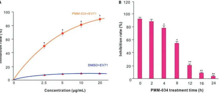
Dose- and time-dependent effects of PMM-034 on EV71 replication in RD cells
The plaque reduction assay using culture super-natants showed inhibition rates of 54.9±1.4, 67.9±1.7, 79.8±3.1, and 91.3±1.9%, respectively, for the tested concentrations. The 50% inhibitory concentration (IC50)
of PMM-034 for EV71 replication was 2.31mg/mL. EV71 replication was not inhibited in EV71-infected RD cells treated with the same concentration of DMSO (Figure 3A). The result showed that PMM-034 exhibited strong antiviral activity against EV71 in a dose-dependent manner.
A time course assay was performed to explore the stages of the EV71 viral replication cycle that were affected by PMM-034. EV71 replication in RD cells was
suppressed from 0–8 h, whereas only a partial inhibitory effect was observed at 12–24 h (Figure 3B). Therefore, anti-EV71 activity of PMM-034 was most prominent when it was applied early in the EV71 replication cycle.
PMM-034 reduced the mRNA and protein expressions of EV71 VP1
To study how PMM-034 affected the mRNA and protein levels of EV71/VP1, total RNA was extracted from EV71-infected cells treated with or without PMM-034 for 24 h. The results of the qRT-PCR revealed that EV71 VP1 mRNA level was significantly reduced in the PMM-034-treated cells (Figure 4A). Consistent with these results, PMM-034 decreased the expression of VP1 protein (Figure 4B).
Figure 3.Dose- and time-dependent effects of PMM-034 on human enterovirus 71 (EV71) replication in rhabdomyosarcoma (RD) cells. A, RD cells were infected with EV71 for 1 h, then treated with PMM-034 or DMSO for 23 h. Infected cells and culture supernatants were collected 48 h post-infection.B, RD cells were infected with EV71 for 1 h, and 10mg/mL PMM-034 was added at 0, 2, 4, 8, 12, 16, and 24 h after infection. Virus titers were measured by the plaque reduction assay. Data are reported as means±SD from at least 3
independent experiments. *Po0.05; **Po0.01, Student’st-test.
Figure 4.Effects of PMM-034 on human enterovirus 71 (EV71/VP1) mRNA and protein expressions in rhabdomyosarcoma (RD) cells. RD cells were infected with EV71 for 1 h and then treated with 10mg/mL PMM-034 or DMSO for 23 h.A, The EV71/VP1 mRNA level was detected by real-time RT-PCR. GAPDH was used as the internal control. Data are reported as means±SD of at least 3 independent
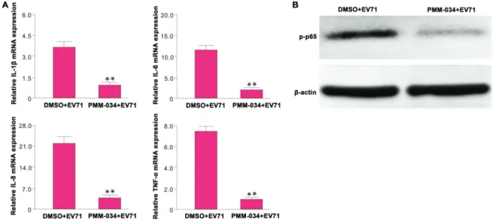
PMM-034 reduced the severity of EV71-induced inflammation
The TNF-a, IL-1b, IL-6, and IL-8 expression levels were significantly lower in EV71-infected RD cells treated with PMM-034 compared to DMSO-treated EV71-infected cells (Figure 5A).
Furthermore, PMM-034 decreased the phospho-p65 protein levels, which might account for the reduction of pro-inflammatory cytokines in EV71-infected RD cells treated with PMM-034 (Figure 5B).
Discussion
Certain TCMs have shown antiviral activity (24), and often function via multiple mechanisms of action and are associated with few side effects and no drug resistance (25). Lariciresinol-4-O-b-d-glucopyranoside, which is one of lignin compounds extracted from the root of Isatis indigotica, has been found to be effective against influ-enza A virus-induced CPE in MDCK cells. These studies also revealed that the lignin glycoside suppressed H1N1-induced expression of the proinflammatory molecules IL-6, TNF-a, IL-8, MCP- 1, IP-10, and IFN-a(26). Baicalin, a flavonoid compound extracted from Scutellaria roots, also exhibits potent antiviral effects on EV71 infection through inhibition of EV71/3D polymerase expression and Fas/FasL signaling pathways (27). Shikonin also pos-sesses anti-AdV3 capabilities and the potential antiviral mechanism might involve inhibition of the degree of apop-tosis and hexon protein expression of AdV (7). Recent
research on shikonin has shifted to its derivatives, of which PMM-034 is one type. To date, it is still poorly understood whether shikonin can inhibit EV71 infection, let alone the antiviral activity of PMM-034 on EV71. In the present study, we found thatp100mg/mL PMM-034 was not cytotoxic to RD cells. We found that PMM-034 exhib-ited a strong antiviral effect against EV71, and could inhibit 54.9–91.3% of the virus in a concentration-dependent manner. Furthermore, the IC50 of this compound against
EV71 was 2.31mg/mL. PMM-034 significantly suppressed EV71 replication in RD cells at 0–8 h after infection, but weakly inhibited replication at 12–24 h after infection. This indicates that the antiviral effects of PMM-034 occurred mainly at the early stage of EV71 infection.
The EV71 genome consists of a positive-sense single-stranded 7400-bp RNA (28). Upon infection, the internal ribosome entry site element in the 50 -untrans-lated region drives the translation of the viral processor polyprotein (29). This polyprotein, which is encoded as NH2-VP4-VP2-VP3-VP1-2A-2B-2C-3A-3B-3C-3D-COOH,
is then proteolytically cleaved by viral 2A and 3C pro-teases to form a range of viral structural proteins (VP1, VP2, VP3, and VP4) and 7 non-structural viral proteins (2A, 2B, 2C, 3A, 3B, 3C, and 3D) (30). Thefirst three viral proteins (VP1–VP3) are present on the outer surface of the virus, and the shorter VP4 is located completely on the inner surface of the capsid. The capsid proteins initiate infection by binding to a receptor on the host membrane (31). VP1 is the most external and immunodominant of the picornavirus capsid proteins, and the VP1 sequence may
be applied to classify enteroviruses and to analyze the phylogenetic relationships among human enteroviruses (32). VP1 has often been used for EV71 molecular geno-typing and epidemiological monitoring (33). Here, we found that VP1 protein expression was significantly blocked by PMM-034 at the early stage of EV71 infection. Further-more, EV71 mRNA abundance was also reduced after treatment with PMM-034, which was consistent with the results of the western blot analysis.
Previous studies have shown that the severity of clinical manifestations associated with EV71 infection pos-sibly depend on the host immune inflammatory response, including pro-inflammatory and anti-inflammatory cyto-kines and chemokine storms in the blood and cerebro-spinalfluid (34). Systemic inflammation caused by EV71 infection further deteriorated CNS disease, resulting in disease progression to the critical illness stage (35). NLRP3 inflammasomes have been shown to play a crucial role in the pathogenesis of coxsackievirus B3-induced myocar-ditis (36). Infection with CBV3, an enterovirus of the Pico-naviridae family, induced production of IL-1b in cardiac tissues of VMC mice, which was positively correlated with the severity of myocarditis (37). Excessive host immune responses may play a critical role in the course of CVB3-induced myocarditis (37). In addition, IAV-elicited NF-kB activity helps viral replication and spreading, and inhibition of IKK activity by the small molecule inhibitors BAY11-7085 and BAY11-7082 severely impaired IAV infection in human lung carcinoma cell lines (38). It is well known that active NF-kB is a transcription factor that induces hundreds of genes as part of an adjustment program to cope with the danger and stress signals leading to NF-kB activation. The target genes of NF-kB include regulators of inflammatory cytokines (e.g., IL-8) and cell survival, proliferation, and cell surface proteins (38). In this study, EV71 infection enhanced the expression of pro-inflamma-tory cytokines such as TNF-a, IL-1b, IL-6, and IL-8. We further showed that PMM-034 could suppress the
phosphorylation of NF-kB/p65 induced by EV71 infec-tion. Therefore, we propose that PMM-034 might reduce the severity of the inflammatory response involved in the inhibition of NF-kB activation. Researchers usually choose non-human primates like green monkeys, cyno-molgus, rhesus, and mouse models to investigate the pathogenesis of EV71 infection. Arita and his group reported that neonatal mice are susceptible to EV71 infection, and the mice exhibited an age-dependent susceptibility to EV71 infection (39). Mice older than 14 days were resistant to infection with EV71 clinical isolates. Yu et al. reported that EV71 infected immuno-competent-ICR mice developed rear limb paralysis and neuropathologies in the brain stem and spinal cord before death. For 1 to 7 days, MP-26 M infections in BALB/c mice can lead to limb paralysis, and the viruses can be isolated from skeletal muscle, blood, brains, livers, spleens and hearts (40). In the next step, we may also use neonatal mice as animal models to further investigate the inhibition effect of PMM-034 on EV71 infection and the underlying mechanism.
In this study, we found that PMM-034 could effectively suppress the expression of pro-inflammatory cytokines in EV71-infected RD cells, and exhibit antiviral activity against EV71, as evidenced by the reduced VP1 mRNA and protein levels in PMM-034-treated cells. Additionally, PMM-034 was not found to exert significant cytotoxicity against RD cells, making it a promising candidate for further development as an EV71 inhibitor.
Acknowledgments
This research was supported by the Program for Changjiang Scholars and Innovative Research Team in University (IRT_14R27), the National Natural Science Foundation of China (#31470384, #31171161, and #31670298), and the Fundamental Research Funds for the Central Universities (#020814380002).
References
1. Chen S, Yang Y, Yan X, Chen J, Yu H, Wang W. Influence of vitamin A status on the antiviral immunity of children with hand, foot and mouth disease.Clin Nutr2012; 31: 543–548, doi: 10.1016/j.clnu.2011.12.005.
2. He Y, Ong KC, Gao Z, Zhao X, Anderson VM, McNutt MA, et al. Tonsillar crypt epithelium is an important extra-central nervous system site for viral replication in EV71 encepha-lomyelitis.Am J Pathol2014; 184: 714–720, doi: 10.1016/ j.ajpath.2013.11.009.
3. Yu ZL, Xu JL. Research progress on the pathogenesis of EV71-induced neurogenic pulmonary edema.Hn Jornal of Mroology2013; 1: R725.721.
4. Everts M, Cihlar T, Bostwick JR, Whitley RJ. Accelerating drug development: antiviral therapies for emerging viruses as a model.Annu Rev Pharmacol Toxicol2017; 57: 155– 169, doi: 10.1146/annurev-pharmtox-010716-104533.
5. Razonable RR. Antiviral drugs for viruses other than human immunodeficiency virus. Mayo Clin Proc2011; 86: 1009– 1026, doi: 10.4065/mcp.2011.0309.
6. Gong X, Zhou J, Zhu W, Liu N, Li J, Li L, et al. Excessive proinflammatory cytokine and chemokine responses of human monocyte-derived macrophages to enterovirus 71 infection.BMC Infect Dis2012; 12: 224, doi: 10.1186/1471-2334-12-224.
7. Wang C, Gao L, Jin Y, Cardona CJ, Xing Z. Regulation of host responses and viral replication by the mitogen-activated protein kinases in intestinal epithelial cells infected with Enterovirus 71.Virus Res2015; 197: 75–84, doi: 10.1016/ j.virusres.2014.12.016.
through TLR4 signaling. PLoS One 2014; 9: e111496, doi: 10.1371/journal.pone.0111496.
9. Hsieh YJ, Yen MH, Chiang YW, Yeh CF, Chiang LC, Shieh DE, et al. Gan-Lu-Siao-Du-yin, a prescription of traditional Chinese medicine, inhibited enterovirus 71 replication, translation, and virus-induced cell apoptosis. J Ethnophar-macol2016; 185: 132–139, doi: 10.1016/j.jep.2016.03.034. 10. Esposito F, Carli I, Del Vecchio C, Xu L, Corona A, Grandi N, et al. Sennoside A, derived from the traditional chinese medicine plant Rheum L., is a new dual HIV-1 inhibitor effective on HIV-1 replication. Phytomedicine 2016; 23: 1383–1391, doi: 10.1016/j.phymed.2016.08.001.
11. Ji P, Chen C, Hu Y, Zhan Z, Pan W, Li R, et al. Antiviral activity of Paulownia tomentosa against enterovirus 71 of hand, foot, and mouth disease.Biol Pharm Bull2015; 38: 1–6, doi: 10.1248/bpb.b14-00357.
12. Lin HY, Chen W, Shi J, Kong WY, Qi JL, Wang XM, et al. Design, synthesis and biological evaluation of cinnamic acyl shikonin derivatives.Chem Biol Drug Des2013; 81: 275– 283, doi: 10.1111/cbdd.12077.
13. Zhang M, Zhao H, Zhao Z, Yan H, Lv R, Cui L, et al. Rapid screening, identification, and purification of neuraminidase inhibitors from Lithospermum erythrorhizon Sieb.et Zucc. by ultrafiltration with HPLC-ESI-TOF-MS combined with semipreparative HPLC. J Sep Sci2016; 39: 2097–2104, doi: 10.1002/jssc.201600087.
14. Baloch SK, Ma L, Xu GH, Bai LF, Zhao H, Tang CY, et al. A potent anticancer agent of shikonin derivative target-ing tubulin. Chirality 2015; 27: 274–280, doi: 10.1002/ chir.22425.
15. Andujar I, Rios JL, Giner RM, Recio MC. Pharmacological properties of shikonin - a review of literature since 2002. Planta Med 2013; 79: 1685–1697, doi: 10.1055/s-0033-1350934.
16. Kourounakis AP, Assimopoulou AN, Papageorgiou VP, Gavalas A, Kourounakis PN. Alkannin and shikonin: effect on free radical processes and on inflammation - a preliminary pharmacochemical investigation. Arch Pharm 2002; 335: 262–266, doi: 10.1002/1521-4184(200208)335:6o 262::AID-ARDP26243.0.CO;2-Y.
17. Chen X, Yang L, Zhang N, Turpin JA, Buckheit RW, Osterling C, et al. Shikonin, a component of chinese herbal medicine, inhibits chemokine receptor function and sup-presses human immunodeficiency virus type 1.Antimicrob Agents Chemother 2003; 47: 2810–2816, doi: 10.1128/ AAC.47.9.2810-2816.2003.
18. Wang R, Yin R, Zhou W, Xu D, Li S. Shikonin and its derivatives: a patent review.Expert Opin Ther Pat2012; 22: 977–997, doi: 10.1517/13543776.2012.709237.
19. Lin HY, Li ZK, Bai LF, Baloch SK, Wang F, Qiu HY, et al. Synthesis of aryl dihydrothiazol acyl shikonin ester derivatives as anticancer agents through microtubule stabi-lization.Biochem Pharmacol2015; 96: 93–106, doi: 10.1016/ j.bcp.2015.04.021.
20. Lin H-Y, Han H-W, Bai L-F, Qiu H-Y, Yin D-Z, Qi J-L, et al. Design, synthesis and biological evaluation of shikonin thio-glycoside derivatives: new anti-tubulin agents. RSC Adv 2014; 4: 49796–49805, doi: 10.1039/C4RA08810G. 21. Lu L, Qin A, Huang H, Zhou P, Zhang C, Liu N, et al.
Shikonin extracted from medicinal Chinese herbs exerts
anti-inflammatory effect via proteasome inhibition. Eur J Pharmacol2011; 658: 242–247, doi: 10.1016/j.ejphar.2011. 02.043.
22. Lin CW, Wu CF, Hsiao NW, Chang CY, Li SW, Wan L, et al. Aloe-emodin is an interferon-inducing agent with antiviral activity against Japanese encephalitis virus and enterovirus 71.Int J Antimicrob Agents2008; 32: 355–359, doi: 10.1016/ j.ijantimicag.2008.04.018.
23. Zhu W, Chiu LC, Ooi VE, Chan PK, Ang PO, Jr. Antiviral property and mode of action of a sulphated polysaccharide from Sargassum patens against herpes simplex virus type 2. Int J Antimicrob Agents2004; 24: 279–283, doi: 10.1016/ j.ijantimicag.2004.02.022.
24. Li Y, Ooi LS, Wang H, But PP, Ooi VE. Antiviral activities of medicinal herbs traditionally used in southern mainland China. Phytother Res 2004; 18: 718–722, doi: 10.1002/ ptr.1518.
25. Li T, Peng T. Traditional Chinese herbal medicine as a source of molecules with antiviral activity. Antiviral Res 2013; 97: 1–9, doi: 10.1016/j.antiviral.2012.10.006. 26. Li J, Zhou B, Li C, Chen Q, Wang Y, Li Z, et al.
Lariciresinol-4-O-beta-D-glucopyranoside from the root of Isatis indigotica inhibits influenza A virus-induced pro-inflammatory response. J Ethnopharmacol2015; 174: 379–386, doi: 10.1016/j.jep. 2015.08.037.
27. Li X, Liu Y, Wu T, Jin Y, Cheng J, Wan C, et al. The antiviral effect of baicalin on enterovirus 71in vitro.Viruses2015; 7: 4756–4771, doi: 10.3390/v7082841.
28. Han JF, Cao RY, Tian X, Yu M, Qin ED, Qin CF. Producing infectious enterovirus type 71 in a rapid strategy. Virol J 2010; 7: 116, doi: 10.1186/1743-422X-7-116.
29. Li X, Liu Y, Hou X, Peng H, Zhang L, Jiang Q, et al. Chlorogenic acid inhibits the replication and viability of enterovirus 71 in vitro. PLoS One 2013; 8: e76007, doi: 10.1371/journal.pone.0076007.
30. Hung HC, Chen TC, Fang MY, Yen KJ, Shih SR, Hsu JT, et al. Inhibition of enterovirus 71 replication and the viral 3D polymerase by aurintricarboxylic acid. J Antimicrob Che-mother2010; 65: 676–683, doi: 10.1093/jac/dkp502. 31. Lin JY, Chen TC, Weng KF, Chang SC, Chen LL, Shih SR.
Viral and host proteins involved in picornavirus life cycle. J Biomed Sci2009; 16: 103, doi: 10.1186/1423-0127-16-103. 32. Oberste MS, Maher K, Kilpatrick DR, Pallansch MA. Molec-ular evolution of the human enteroviruses: correlation of serotype with VP1 sequence and application to picornavirus classification.J Virol1999; 73: 1941–1948.
33. Chong P, Hsieh SY, Liu CC, Chou AH, Chang JY, Wu SC, et al. Production of EV71 vaccine candidates.Hum Vaccin Immunother2012; 8: 1775–1783, doi: 10.4161/hv.21739. 34. Huang HI, Weng KF, Shih SR. Viral and host factors that
contribute to pathogenicity of enterovirus 71.Future Micro-biol2012; 7: 467–479, doi: 10.2217/fmb.12.22.
35. Ye N, Gong X, Pang LL, Gao WJ, Zhang YT, Li XL, et al. Cytokine responses and correlations thereof with clinical profiles in children with enterovirus 71 infections.BMC Infect Dis2015; 15: 225, doi: 10.1186/s12879-015-0965-1. 36. Wang Y, Gao B, Xiong S. Involvement of NLRP3 infl
37. Mason JW. Myocarditis and dilated cardiomyopathy: an inflammatory link. Cardiovasc Res 2003; 60: 5–10, doi: 10.1016/S0008-6363(03)00437-1.
38. Schmitz ML, Kracht M, Saul VV. The intricate interplay between RNA viruses and NF-kappaB.Biochim Biophys Acta 2014; 1843: 2754–2764, doi: 10.1016/j.bbamcr.2014.08.004. 39. Arita M. Cooperative effect of the attenuation determinants derived from poliovirus sabin 1 strain is essential for
attenuation of enterovirus 71 in the NOD/SCID mouse infection model.J Virol2008; 82: 1787–1797, doi: 10.1128/ JVI.01798-07.