Article
*e-mail: [email protected], [email protected]
In Vitro Evaluation of Triazenes: DNA Cleavage, Antibacterial Activity and
Cytotoxicity against Acute Myeloid Leukemia Cells
Vanessa O. Domingues,a Rosmari Hörner,*,a Luiz G. B. Reetz,a Fábio Kuhn,a
Virgínia M. Coser,b Jacqueline N. Rodrigues,b Rita Bauchspiess,b
Waldir V. Pereira,b Gustavo L. Paraginski,c Aline Locatelli,c
Juliana de O. Fank,c Vinícius F. Giglioc and Manfredo Hörner*,c
aUniversidade Federal de Santa Maria, Departamento de Análises Clínicas e Toxicológicas,
97105-970 Santa Maria-RS, Brazil
bHospital Universitário de Santa Maria, Departmento de Hematologia-Oncologia,
97105-970 Santa Maria-RS, Brazil
cUniversidade Federal de Santa Maria, Departamento de Química, CP 5031,
97105-970 Santa Maria-RS, Brazil
As diazoaminas assimétricas 1-(2-clorofenil)-3-(4-carboxifenil)triazeno (1),
1-(2-luorofenil)-3-(4-carboxifenil)triazeno (2) e 1-(2-luorofenil)-3-(4-amidofenil)triazeno (3) foram avaliadas quanto à capacidade de clivagem do DNA plasmidial pUC18 e pBSKII, atividade antibacteriana e citotoxicidade in vitro frente a células de leucemia mielóide aguda e leucócitos normais utilizando
o bioensaio da redução do sal brometo de 3-(4,5-dimetiltiazol-2-il)-2,5-difeniltetrazólio (MTT). Os triazenos analisados demonstraram capacidade de clivagem dos dois tipos de DNA plasmidial: triazeno 1 em pH 8,0 e 50 °C; triazeno 2 em pH 6,5 a 37 e 50 °C; triazeno 3 em pH 6,5 e 37 °C. Os compostos apresentaram atividade citotóxica frente a células leucêmicas. O composto 1 mostrou
alta atividade frente a B. cereus (CIM = 32 mg mL-1). A associação molecular através de ligações
de hidrogênio no estado sólido do composto 3 com base na análise estrutural por difratometria de
raios X em monocristal, bem como os resultados das análises espectroscópicas nas regiões do IV e UV-Vis dos compostos 1, 2 e 3 são discutidos no presente trabalho.
The asymmetric diazoamines 1-(2-chlorophenyl)-3-(4-carboxyphenyl)triazene (1),
1-(2-luorophenyl)-3-(4-carboxyphenyl)triazene (2) and1-(2-luorophenyl)-3-(4-amidophenyl)
triazene (3) were evaluated for their ability to cleave pUC18 and pBSKII plasmid DNA, antibacterial
activity and in vitro cytotoxicity against acute myeloid leukemia cells and normal leukocytes using
the bioassay of reduction of 3-(4,5-dimethylthiazol-2-yl)-2,5-diphenyltetrazolium bromide (MTT). The triazenes showed ability to cleave the two types of plasmid DNA: triazene 1 at pH 8.0 and
50 °C; triazene 2 at pH 6.5 and 37 and 50 °C; triazene 3 at pH 6.5 and 37 °C. The compounds presented cytotoxic activity against myeloid leukemia cells. Compound 1 showed high activity
against B. cereus (MIC = 32 mg mL-1). The observation of intermolecular hydrogen bonding in
the solid state of compound 3, based on the structural analysis by X-ray crystallography, as well
as the results of IR and UV-Vis spectroscopic analyses of compounds 1, 2 and 3 are discussed in
the present work.
Keywords: triazenes, DNA cleavage, antibacterial activity, cytotoxicity
Introduction
Triazenes or diazoamines constitute a class of compounds containing three consecutive nitrogen atoms
in an acyclic arrangement.1 Diazoamino compounds can be obtained by diverse synthetic ways, the classical method including the coupling reaction of a diazonium salt, [R-N≡N]+X-, with an amine (R'-NH
symmetric triazenes; R ≠ R' for asymmetric compounds) by stepwise neutralization of the reaction system. They have diverse applications as DNA alkylating agents in tumor therapy, protecting groups in natural product synthesis and combinatorial chemistry, as well as precursors incorporated into polymer and oligomer synthesis.2-8
Hydrolysis of DNA or RNA catalyzed by relevant enzymes is an important subject in biotechnology, medicine and drug development.9,10 Synthetic nucleases which rapidly cleave nucleic acids under mild conditions have many important potential applications ranging from the synthesis of custom-designed artiicial restriction enzymes to the development of new antitumor agents.11 To mimic the biological function of enzymes, triazene compounds that cleave DNA are interesting for the development of artiicial nucleases. In this context, N-(3-hydroxypropyl) berenil represents one example of a triazene capable to promote DNA cleavage.12
Triazenes also act as chemotherapeutic agents for many tumors, such as brain, leukemia, melanoma, lymphoma and sarcoma.13-17 Among all chemotherapeutic agents tested against metastatic melanoma, DTIC (5-(3,3-dimethyltriazene)imidazol-4-carboxamide) - an FDA-approved pro-drug activated by N-demethylation in liver microssomes (microsomal enzyme CYP450) - is the most active single agent and thus considered a reference drug.18,19
Temozolomide (TMZ), i . e.,
8-carbamoyl-3-methylimidazol[5,1-d]-1,2,3,5-tetrazin-4(3H)-one, is another example of a methylating triazene like DTIC, which acts under physiological conditions against primary and metastatic tumors in the brain.20 Despite the need for more clinical experiments for the use of triazene compounds in acute leukemia and myelodysplasia treatments, some studies have shown that both DTIC and TMZ have antineoplastic activity in patients with leukemia.21-23
Recently the antimicrobial susceptibility proile of ifteen triazenes was evaluated by our group based on the reference broth microdilution method according to CLS guidelines.24
The mechanism of cytotoxicity of triazenes involves mainly methylation of the O6 position of guanine in DNA.25 On the other hand, it is well known that molecular association plays an important role in biological systems. Strategic modiications of electronic and geometric characteristics applied in molecular design determine the dimensionality of molecular association by diverse types of intermolecular secondary non-bonding interactions such as the large palette of hydrogen bonding. Hydrogen bonding is an important non-covalent interaction in structural investigations relative to molecular recognition and reactivity of molecules with
biological importance. From this point of view, diverse biological reaction mechanisms can be related to hydrogen bonding due to its energy range situated between van der Waals interactions and covalent bonds.26,27
The present study deals with the synthesis and characterization of 1-(2-chlorophenyl)-3-(4-carboxyphenyl) triazene (1),1-(2-luorophenyl)-3-(4-carboxyphenyl)triazene
(2) and1-(2-luorophenyl)-3-(4-amidophenyl)triazene (3)
(Figure 1). The study also demonstrates that the compounds are able to cleave pUC18 and pBSKII plasmid DNA through a possible hydrolytic mechanism. The antibacterial activity and in vitro cytotoxicity of the three triazenes against acute myeloid leukemia (AML) cell lines and control cell (without AML) using the bioassay of reduction of 3-(4,5-dimethylthiazol-2-yl)-2,5-diphenyltetrazolium bromide (MTT) are also analyzed. The single crystal structural analysis of 1-(2-luorophenyl)-3-(4-amidophenyl) triazene (3), including its self-assembly in the solid state
through hydrogen bonding, and the vibrational and electronic spectra of compounds 1, 2 and 3 are also presented.
Experimental
Materials
All reagents and solvents used in the study were reagent grade and were used without further puriication. For biological assays, water was puriied by passage through a Millipore water puriication system with a resistance of 18 MW and sterilized by autoclave. Reaction solutions for DNA cleavage studies, determination of minimum inhibitory concentrations and cytotoxicity essays were prepared according to standard sterile techniques. The DNA bands were stained, visualized on a UV transilluminator at 254 nm, photographed using a digital photographic camera and quantiied using Scion Image Beta 4.03 software (Scion Corp.).
1H and 13C NMR spectra were recorded on a Bruker
DPX 200 instrument. IR spectra were recorded on a Bruker FT-IR Spectrometer TENSOR 27 instrument with samples embedded in KBr pellets. UV-Vis spectra were measured on a Shimadzu UV-1650-PC spectrophotometer.
Synthesis of 1-(2-chlorophenyl)-3-(4-carboxyphenyl)
triazene (1)
The synthesis of triazene 1 was formerly reported
nitrite (0.60 g, 8.70 mmol) in water (10 mL). The reaction mixture was kept at -2 °C and a solution of 2-chloroaniline (0.93 g, 7.30 mmol) in 6 mol L-1 HCl (5 mL) was slowly added under continuous stirring. Stirring was continued for 20 min at a temperature between -2 and -5 °C. A cold sodium acetate solution (10%) was then added in small amounts until pH 7.0 was reached while a yellow solid precipitated. The crude product, separated by iltration, was washed with small amounts of cold water and puriied by column chromatography after its adsorption onto silica gel (70/230 mesh) using 5% acetone in hexane as eluent. The yellow product, in form of ine microcrystalline needles, was dried under vacuum. Yield 53% (0.95 g, 3.84 mmol) based on 4-aminobenzoic acid, mp 172 °C; IR nmax/cm-1, KBr pellets: 3174 (N−H), 2986 (O−H), 1682 (C=O), 1597 (C=C), 1510 (d(N−H), deformation), 1430 (d(O−H), deformation in plane), 1414 (N=N), 1312 (C-O), 1209 (N-N), 756 (d(C−H), deformation). 1H NMR (200 MHz, dmso-d6/tms) d12.33(br, s, 1H, OH), 7.16-7.70 (m, 8H, Ph); 13C NMR (200 MHz, dmso-d
6) d146.6 (C=O), 130.6, 129.4, 129.2, 129.0, 128.9, 128.5, 125.4, 121.4, 121.3, 120.1, 119.4, 119.2 (Ph); lmax/nm (MeOH) 203, 242, 361 (e/dm3 mol-1 cm-1 18.6 × 10³).
Synthesis of 1-(2-fluorophenyl)-3-(4-carboxyphenyl)
triazene (2)
To a solution of 4-aminobenzoic acid (1.00 g, 7.30 mmol) in 10 mL 6 mol L-1 HCl previously cooled to -2 °C, a
sodium nitrite solution (0.60 g, 8.70 mmol) in water (10 mL) was added under continuous stirring throughout 10 min. The reaction mixture was kept at -2 °C and a solution of 2-luoroaniline (0.81 g, 7.30 mmol) in 6 mol L-1 HCl (5 mL) was slowly added under continuous stirring. Stirring was continued for 20 min at a temperature between -2 and
-5 °C. A cold sodium acetate solution (10%) was then added in small amounts until pH 7.0 was reached while a yellow solid precipitated. The crude product, separated by iltration, was washed with small amounts of cold water and puriied by column chromatography after its adsorption onto silica gel (70/230 mesh) using diethyl ether as eluent. The yellow product in form of ine microcrystalline needles was dried under vacuum. Yield 64% (1.21 g, 4.70 mmol) based on 4-aminobenzoic acid, mp 175 °C. IR nmax/cm-1, KBr pellets: 3175 (N−H), 2985 (O−H), 1680 (C=O), 1607 (C=C), 1491 (d(N−H), deformation), 1431 (d(O−H), deformation in plane), 1404 (N=N), 1312 (C-O), 1201 (N-N), 756 (d(C−H), deformation). 1H NMR (200 MHz, dmso-d
6/tms) d13.06 (br, s, 1H, OH), 7.21-7.96 (m, 8H, Ph); 13C NMR (200 MHz, dmso-d6) d 166.9 (C=O), 131.1, 128.5, 128.4, 124.9, 124.8, 119.1, 116.8, 116.4, 114.0 (Ph); lmax/nm (MeOH) 202, 237, 361 (e/dm3 mol-1 cm-1 21.9 × 10³).
Synthesis of 1-(2-luorophenyl)-3-(4-amidophenyl)triazene
(3)
To a solution of 2-luoroaniline (0.89 g, 8.01 mmol) in 10 mL of 6 mol L-1 HCl previously cooled to -2 °C, a
Figure 1. Molecular structure of 1-(2-chlorophenyl)-3-(4-carboxyphenyl)-triazene (1), 1-(2-fluorophenyl)-3-(4-carboxyphenyl)triazene (2),
1-(2-luorophenyl)-3-(4-amidophenyl)triazene (3), 1,3-bis(4-carboxyphenyl)triazene (4), 1,3-bis(4-luorophenyl)triazene (5) and 1,3-bis(4-acetamidophenyl)
sodium nitrite solution (0.58 g, 8.41 mmol) in water (5 mL) was added under continuous stirring throughout 10 min. The reaction mixture was kept at -2 °C and a solution of 4-aminobenzamide (1.09 g, 8.00 mmol) in acetone (10 mL) was slowly added under continuous stirring. Stirring was continued for 20 min at a temperature between -2 and
-5 °C. A cold concentrated sodium carbonate solution was then added in small amounts until pH 7.0 was reached while a yellow solid precipitated. The crude product, separated by iltration, was washed with small amounts of cold water, followed by small amounts of cold ethanol and puriied by column chromatography after its adsorption onto silica gel (70/230 mesh) using hexane:ethyl acetate (3:7) as eluent. The microcrystalline product was recrystallized from a hexane:ethyl acetate mixture (1:3). Yellow vitreous bar-shaped crystals of 3 suitable for X-ray diffraction analysis
were obtained by slow evaporation of the solvent mixture within ive days. Yield 86% (1.48 g, 5.7 mmol) based on 2-luroaniline, mp 200 °C. IR nmax/cm-1, KBr pellets: 3493 (N−H, amide), 3404 (N−H, amide), 3167 (N−H, amine), 1665 (C=O), 1611 (C=C), 1489 (d(N−H), deformation), 1427 (d(O-H), deformation in plane), 1391 (N=N), 1312 (C-O), 1202 (N-N), 752 (d(C-H), deformation). 1H NMR (200 MHz, dmso-d6/tms) d 13.05 (br, s, 1H, OH), 7.22-7.97 (m, 8H, Ph); 13C NMR (200 MHz, dmso-d
6) d 167.9 (C=O), 144.4, 129.6, 129.1, 128.8, 125.4, 125.3, 119.6, 117.2, 116.9, 114.0 (Ph);lmax/nm (MeOH) 202, 233, 361 (e/dm3 mol-1 cm-1 26.7 × 10³).
DNA cleavage studies
DNA cleavage was measured by the conversion of supercoiled pUC18 and pBSKII plasmid DNA (Stratagene) to nicked circular and linear DNA forms. The screening assay to achieve nucleic acids cleavage was performed in a inal volume of 20 mL where the following reagents were present: 1 mg of plasmid DNA and different concentrations of tested compounds (3.75, 1.875, 1.25, 0.75 and 0.375 mmol L-1) at pH 6.5 (200 mmol L-1 PIPES buffer) and 8.0 (200 mmol L-1 Tris-HCl buffer) at 37 and 50 °C for 24 h; all assays were made twice. Samples were submitted to agarose gel (0.8%) electrophoresis stained with ethidium bromide. The resulting gels were digitalized with a photodocumentation system.
In order to verify the probable mechanism of DNA strand scission, the assays were repeated in the presence of free radical scavengers (thiourea 40 mmol L-1, glycerol 0.1% and 1.0% and dimethyl sulfoxide 0.04 mol L-1) in the same cleavage conditions (pUC18 and pBSKII plasmids at pH 6.5 and 8.0, 37 and 50 °C for 24 h). Cleavage assays were also performed in argon atmosphere in order to verify
the cleavage ability in absence of atmospheric oxygen according to the methodology described.29
Bacterial strains
In vitro antimicrobial activities of the triazene
compounds were evaluated against 30 test organisms, including 14 Gram-positive and 16 Gram-negative bacterial strains. The groups included nine bacteria of the American Type Culture Collection (ATCC): Pseudomonas aeruginosa ATCC 27853, Escherichia coli ATCC 25922, Escherichia coli ATCC 35218, Salmonella choleraesuis ATCC 10708,
Klebsiella pneumoniae ATCC 700603, Staphylococcus
aureus ATCC 25923, Staphylococcus epidermidis ATCC
12228, Enterococcus faecalis ATCC 29212 and Bacillus
cereus ATCC 10987. Twenty one clinical bacteria
from the Microbiology Laboratory of the Hospital Universitário de Santa Maria, Brazil, were identiied by a Siemens MicroScan automated system: Shigella sonnei,
Enterobacter cloacae, Klebsiella oxytoca, Bacillus cereus,
Micrococcus sp., Streptococcus pyogenes, Acintobacter
baumannii, Ralstonia pickettii, Serratia marcescens,
Enterococcus sp., Listeria monocytogenes, Aeromonas
hidrophyla, Enterobacter aerogenes, Streptococcus
agalactiae, Salmonella tiphymurium, Corynebacterium sp.,
Salmonella sp., Staphylococcus epidermidis, Acinetobacter
lwofii, Klebsiella pneumoniae, Staphylococcus hominis
subsp. hominis.
Determination of minimum inhibitory concentrations (MICs)
of the triazene compounds that inhibited visible growth of microorganisms. All tests were made three times.
Cell lines
Leukemic cells were obtained from three acute myeloid leukemia patients without treatment diagnosed at the Hematology-Oncology Laboratory of the Hospital Universitário de Santa Maria, Brazil, by light microscopic examination of the bone marrow as well as low cytometry to diagnose the presence of leukemia, to differentiate AML from other types of leukemia and to classify the subtype of disease. Cell lines of control patient (without AML) was obtained by collecting peripheral blood with EDTA. The mononuclear cells from bone marrow (1 mL) and peripheral blood (6 mL) were isolated by Ficoll-Paque PLUS (Sigma) density separation (centrifuged at 4000 rpm for 20 min). After isolation, the cells were washed three times in RPMI-1640 (Sigma) and the erythrocytes were lysed with hemolytic buffer. The cells were grown in RPMI with 20% fetal bovine serum (Sigma), penicillin 100 U mL-1, streptomycin 100 mg mL-1, HEPES buffer and sodium bicarbonate during 24 h at 37 °C, with 5% CO2.
MTT assay
MTT was dissolved in phosphate buffered saline (PBS) at 5 mg mL-1 and iltered to sterilize and remove a small amount of insoluble residue. Trypan blue (1% in distilled water) and cell suspensions were mixed in 1:6 quantities (50 mL of cells + 10 mL trypan blue). The quantity of cells was estimated by hemocytometry and (blue) stained cells were scored as dead. About 3 × 10⁶ cells mL-1 were grown with compounds for 24 h at 37 °C and 5% CO2. The triazene doses varied from 12.5, 50 and 100 mmol mL-1; the compounds were dissolved in dimethyl sulfoxide, which did not exceed 0.25% in the cell cultures. The DTIC solution was prepared in water. The microculture plates included 90 mL cell suspension and 10 mL of the compound solution. Control cultures received cells in absence of assayed compounds. After 20 h the MTT solution was added to all wells and plates were incubated at 37 °C for 4 h. At the end of the incubation period, the formazan product was dissolved in 100 mL of dimethyl sulfoxide. The optical density (OD) of each well was measured using a Fisher Bio-Tek BT2000 MicroKinetics reader (l = 570 nm). Each assay was carried out three times. The cell survival fraction was calculated by the following equation:
Cell survival = (OD of the treated well / mean OD of the control wells) × 100%
The IC50 value, deined as the drug concentration causing a 50% reduction in cellular viability, was calculated for each compound. The experimental data were transformed to sigmoid dose-response curves using nonlinear regression analysis (GraphPad Prizm 5), which enabled the calculation of the corresponding IC50.
Crystal structure determination
A single crystal on a glass iber was used for the X-ray data collection. Data were collected at -123 °C with a Bruker APEX II CCD area-detector diffractometer and graphite monochromatized Mo-Ka radiation using COSMO program.32 No crystal decay was observed. Crystal system and space group were unequivocally determined from the data sets and reciprocal space images. Cell reinement, data reduction and absorption correction were performed using SAINT and SADABS programs, respectively.32 The structure of 3 was solved by direct methods and reined
on F2 with anisotropic temperature parameters for all non-hydrogen atoms.33,34 The H atoms in the structure of 3 were identiied in Fourier difference maps and reined as individual atoms with isotropic displacement parameters. Table 1 shows the crystallographic data and details of data collection and reinement, and Table 2 the selected bond lengths and angles. Graphical representations were drawn with the DIAMOND software.35
Results and Discussion
Spectroscopic characterization
Infrared spectra
The infrared spectra of 1, 2 and 3 were analyzed in the
spectral region 4000-400 cm-1. The main absorptions and vibrational assignments were compared with literature data.36-38 Amide derivatives are characterized by two absorptions in sequence with medium intensities and 120-200 cm-1 spacing in the 3550-3150 cm-1 region. In compound 3 these absorptions were assigned at 3493 and
3404 cm-1.36,37 The shift to lower frequency can be attributed to electronic delocalization towards the terminal aryl substituents of the [-N=N-N(H)-] diazoamino group. The characteristic N–H stretching at 3181 cm-1 for the [-N=N-N(H)-] triazene group was observed as weak to medium signals at 3167-3175 cm-1 in 1, 2 and 3. Compared with 1,3-bis(phenyl) triazene the [-N=N-N(H)-] diazoamino group in 1, 2 and 3 was undoubtedly characterized by strong absorptions
at 1489-1510 cm-1, 1391-1414 cm-1 and 1201-1208 cm-1 corresponding to N-H in plane deformation, nas(N=N) and
Compounds 1 and 2 could be analyzed as R-C(=O)OH carboxylic derivatives, usually characterized in the solid state by a strong, very broad O-H stretching near 3000 cm-1.36 In compounds 1 and 2 this broad band was observed at 2986 and 2985 cm-1, respectively. In compound 3 this absorption was absent. Carboxylic acids
and derivatives usually exist as (R-C=O…H-O-C-R)
2 hydrogen-bonded dimers. If the dimer unit is considered, two carbonyl stretching frequencies, symmetric and asymmetric, are observed. The former is Raman active, and the asymmetric will be IR active only, assigned for most carboxyl dimers at 1720-1680 cm-1.36 Such observation agrees with the absorptions observed at 1665-1682 cm-1 assigned to n
as(C=O) for 1, 2 and 3. Further, the carboxyl dimer presents a characteristic infrared band at 960-875 cm-1 which corresponds to out-of-plane OH…O hydrogen deformation.36 In the case of 1 and 2, this characteristic absorption is observed near 850 cm-1 probably due to electronic delocalization towards the terminal aryl substituents of the [-N=N-N(H)-] diazoamino group. In the case of 3 the crystal structure shows intermolecular
associations promoted by C-O…H-N hydrogen bonding
between the carbonyl group of the amido substituent and the [-N=N-N(H)-] diazoamino group (Figure 3), which can be associated to the out-of-plane OH…N hydrogen
deformation in analogy with the out-of-plane OH…O
hydrogen deformation of R-C(=O)OH derivatives. This explains the well-deined absorption observed at 843 cm-1 in the spectra of 3. Besides the characteristic out-of-plane
OH…O hydrogen deformation, (R-C=O…H-O-C-R)
2 carboxyl dimers are characterized by O−H in plane deformation and C-O stretching in the spectral region of 1440-1395 cm-1 and 1315-1280 cm-1, respectively.36 Compounds 1 and 2 show the d(O−H) vibration at 1430 and 1431 cm-1, respectively, while in compound 3 the strong absorption at 1427 cm-1 was assigned to a N−H deformation in plane correspondent to the C-O…H-N intermolecular
hydrogen bonding (Figure 3). All compounds showed the C-O stretching at 1312 cm-1.
Strong bands observed for 1, 2 and 3 near 750 cm-1 were attributed to the C-H out-of-plane deformation and the very strong absorptions observed at 1597-1611 cm-1 were assigned to the C=C axial deformation, both characteristic of aromatic compounds.38
UV-Vis spectra
The UV-Vis spectra of 1, 2 and 3 dissolved in methanol
were analyzed in the 190-440 nm spectral region. The assignments made for the main absorptions are based on literature data.39,40 Aryl and/or phenyl-substituted triazenes show maximum absorptions at 190-440 nm
demonstrating their efficient chromophoric behavior. The electronic spectra of 1, 2 and 3 were compared with
those recorded in methanol for the compounds 1,3-bis (4-carboxyphenyl)triazene (4), 1,3-bis(4-fluorophenyl) triazene (5) and 1,3-bis(4-acetamidophenyl)triazene (6) respectively (Figure 1). The electronic spectra of
compounds 4, 5 and 6 dissolved in solvents with different
polarities showed solvatochromism related to the n→p* absorption due to the electron-withdrawing or donating property of fragments attached to terminal aryl substituents of the [-N=N-N(H)-] diazoamino group. Dissolved in methanol, 4, 5 and 6 showed a lmax value of 348, 351 and 369 nm assigned to this n →p* absorption, respectively.38 These values agree with the maximum observed of 361 nm for 1, 2 and 3. Further, the band observed at 242, 237 and
233 nm in the spectra of 1, 2 and 3 respectively is attributed
to the p →p* transition for aromatic rings, which were observed for 4 and 5 at 221 and 231 nm, respectively.38 A third intense band observed near 200 nm for 1, 2 and 3 was
attributed to the n →s* transition of the carbonyl group.40
X-ray crystallography
Since the molecular association promoted by hydrogen bonding plays an important role in biological systems, especially for molecular recognition and reactivity of molecules with biological importance, the crystal structure analysis of one of the triazene derivatives was carried out to help understand the hydrogen bond network established by the terminal aryl group substituents of the [-N=N-N(H)-] diazoamino moiety, as well as the inluence of this non-covalent interaction on the planarity of the molecule in the solid state. Attempts to obtain single crystals suitable for structural analysis of 1 and 2 are in progress.
Crystal data and experimental parameters of the structural analysis of 3 are presented in Table 1. Selected
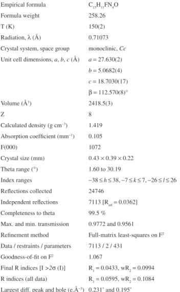
bond distances and angles are listed in Table 2. Figure 2 shows a projection of the molecular structure of FC6H4N=N(H)C6H4C(=O)NH2 (3), while Figure 3
represents a section of the one-dimensional assembly of the molecules via secondary N-H…O, N-H…F, N-H…N
and C-H…O hydrogen bonds, in a projection slightly tilted
toward the [001] crystallographic direction.
Compound 3 crystallizes in the monoclinic crystal
system, the non-centrosymmetric polar space group Cc. To test if a higher symmetry was eventually missed, an attempt was made to solve the structure in the centrosymmetric C2/c group, with negative results. Since the molecule of 3 is built
Table 1. Crystal and structure reinement data for 3
Empirical formula C13H11FN4O Formula weight 258.26
T(K) 150(2)
Radiation, l (Å) 0.71073 Crystal system, space group monoclinic, Cc
Unit cell dimensions, a, b, c (Å) a = 27.630(2)
b= 5.0682(4)
c= 18.7030(17)
b=112.570(8)° Volume (Å3) 2418.5(3)
Z 8
Calculated density (g cm-3) 1.419
Absorption coeficient (mm-1) 0.105
F(000) 1072
Crystal size (mm) 0.43 × 0.39 × 0.22 Theta range (°) 1.60 to 30.19
Index ranges -38 ≤h≤ 38, -7 ≤k≤ 7, -26 ≤l ≤ 26 Relections collected 24746
Independent relections 7113 [Rint= 0.0362] Completeness to theta 99.5 %
Max. and min. transmission 0.9772 and 0.9561
Reinement method Full-matrix least-squares on F2
Data / restraints / parameters 7113 / 2 / 431 Goodness-of-it on F2 1.067
Final R indices [I >2s (I)] R1= 0.0433, wR2= 0.0994 R indices (all data) R1= 0.0595, wR2= 0.1084 Largest diff. peak and hole (e.Å-3) 0.231* and 0.195*
*Highest peak:0.231 (e.Å-3) at 0.1935, 0.6264, 0.0511 [0.77 Å from H4].
*Deepest hole: -0.195 (e.Å-3) at 0.0306, 0.3744, 0.3387 [0.68 Å from
N12].
Table 2. Selected bond lengths [Å] and angles [°] for 3
Bond lengths [Å] Bond lengths [Å] C1-O1 1.248(2) C2-O2 1.234(2) C1-N1 1.327(2) C2-N2 1.336(3) C1-C24 1.485(2) C2-C44 1.490(2) C12-F1 1.361(2) C32-F2 1.349(2)
N1-H3 0.81(3) N2-H5 0.83(3)
N1-H4 1.02(3) N2-H6 1.06(3)
N13-H1 1.01(3) N23-H2 0.88(3) N11-N12 1.280(2) N21-N22 1.271(2) N12-N13 1.327(2) N22-N23 1.335(2) Bond angles [°] Bond angles [°] O1-C1-N1 121.29(17) O2-C2-N2 121.04(17) O1-C1-C24 120.06(15) O2-C2-C44 119.87(17) N1-C1-C24 118.60(14) N2-C2-C44 119.08(16) F1-C12-C13 118.12(18) F2-C32-C33 118.48(16) F1-C12-C11 118.44(17) F2-C32-C31 118.54(15) C25-C24-C1 119.31(14) C45-C44-C2 118.40(16) C23-C24-C1 121.82(15) C43-C44-C2 123.14(16) C1-N1-H3 117.8(17) C2-N2-H5 120.0(19) C1-N1-H4 113.2(14) C2-N2-H6 121.9(16) H3-N1-H4 129(2) H5-N2-H6 118(2) N11-N12-N13 112.22(14) N21-N22-N23 111.02(14) N12-N13-H1 118(2) N22-N23-H2 120.9(19) C21-N13-H1 122(2) C41-N23-H2 118.2(19)
Figure 2. Molecular structure with atom-labeling scheme of the two
symmetrically independent FC6H4N=N(H)C6H4C(=O)NH2 molecules of
3 in the asymmetric unit of the space group Cc. Displacement ellipsoids are drawn at the 50% level.
about the orientation of the molecules relative to the polar axes in the unit cell of 3. Two symmetrically independent
molecules of 3 were observed in the asymmetric unit of the
unit cell (Figure 2). The observed intermolecular hydrogen bonding involving the amide group made the independent molecules deviate from planarity, becoming chiral. The lack of planarity can be observed by the interplanar angles O1C1N1/C21-C26 [22.5(3)°; whole molecule r.m.s. deviation = 0.1610 Å] and O2C2N2/C41-C46 [2.7(3)°; whole molecule r.m.s. deviation = 0.0310 Å].
The molecular structure corresponds to the expected
trans stereochemistry about the N11=N12 and N21=N22
and Caryl-N bond lengths. Accordingly, the N11=N12 [1.280(2) Å] and N21=N22 [1.271(2) Å] bond lengths are longer than the characteristic value for a double bond (1.236 Å), whereas N12-N13 [1.327(2) Å] and N22-N23 [1.335(2) Å] bond distances are shorter than the characteristic value for a single bond (1.404 Å).41 On the other hand, C11-N11 [1.414(2) Å] and C21-N13 [1.393(2) Å], as well as C31-N21 [1.416(2) Å] and C41-N23 [1.398(2) Å] bonds are shorter than the characteristic N-Caryl single bonds (secondary amines, R2NH, R = Csp2; 1.452 Å).42 These values agree with those observed in similar compounds: 1,3-bis(2-triluoromethylphenyl)triazene [N=N 1.281(2) and N-N 1.302(2) Å],40 1,3-bis(2,4-dibromophenyl) triazene [N=N 1.267(7) and N-N 1.332(7) Å],44 and 1-(4-bromophenyl)-3-(4-nitrophenyl)triazene [N=N 1.257(8) and N-N 1.326(10) Å].43 The bond angles of the N=N-N triad [N11-N12-N13 112.22(14)°] and [N21-N22-N33 111.02(14)°] are very close to the angles found in 1,3-bis(2-triluoromethylphenyl)triazene [111.89(14)°] and 1-(4-bromophenyl)-3-(4-nitrophenyl) triazene [110.9(7)°].43,45
The PLATON software was used to analyze the intermolecular hydrogen bonding observed in the crystal of 3, as shown in Figure 3.46
The crystal structure shows that the symmetrically independent FC6H4N=N(H)C6H4C(=O)NH2 molecules are individually related to each other by translation into
chains along the crystallographic direction [010]. Table 3 lists the geometric parameters of the intermolecular hydrogen-bonding in 3. The molecules are associated
through hydrogen bonds including a bifurcated acceptor geometry (D1-H1, D2-H2)…A (D = donor, A = acceptor
atom), (N13-H1, C22-H22)…O2', and a bifurcated
donor geometry D-H…(A
1, A2), N2-H6…(F1''', N11'''); symmetry codes: (') -1/2+x, -3/2-y, -1/2+z, (''') 1/2+x,
-3/2-y, 1/2+z. The angle F1'''…H6…N11''' is 70.4(8)° and the elevation of H6 from the plane deined by the atoms F1''', N2, N11''' is 0.02(3) Å. Atom O2' deviates 0.054(2) Å from the best plane deined by the atoms C22, C2', N13, while the angle H1…O2'…H22 is 63(1)°. The one-dimensional
arrangement of each symmetrically independent molecule of 3 along the crystallographic direction [010] results from
the classical N1-H3…O1'' hydrogen bond, symmetry code:
('') x, -1+y, z. Due to the symmetry independence of each molecule of 3, which originates the 1-D arrangement, both
chains running in opposite directions to each other along the crystallographic b axis are also symmetrically independent, as indicated in Figure 3.
Biological activity
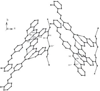
The ability of triazenes 1 and 2 to cleave supercoiled
pUC 18 and pBSKII plasmids in pH 8.0 (200 mmol L-1 Tris-HCl buffer) upon incubation at 50 °C for 24 h is evidenced in Figure 4, while Figure S1 (see Supplementary Information) shows that triazene 3 promotes DNA cleavage
in pH 6.5 (200 mmol L-1 PIPES buffer) after 24 h at 37 and 50 °C. Compounds 1 and 2 are able to cleave plasmid
DNA by transforming the supercoiled form (FI) into the
circular relaxed form II by single strand scission, while
compound 3 was not able to show cleavage activity in the
same conditions employed for 1 and 2. However, compound 3 has demonstrated more cleavage eficiency in pH 6.5 after
24 h at 37 and 50 °C, due to additional cleavage of form II
into III (linear form) as shown in Figure S1. The shape of the
linear form can be attributed to an increase in the number of random cleavage events indicating low selectivity.47
Figure 3. Section of the one-dimensional (1-D) assembling of the molecules
in the crystal structure of FC6H4N=N(H)C6H4C(=O)NH2 (3). Intermolecular hydrogen bonds including bifurcated acceptor geometry (D1-H1, D2-H2)∙∙∙A (D = donor, A = acceptor atom), (N13-H1, C22-H22)∙∙∙O2’, bifurcated donor geometry D-H∙∙∙(A1, A2), N2-H6∙∙∙(F1’’’, N11’’’), and classical N1-H3∙∙∙O1’’ hydrogen bonding along the crystallographic direction [010] are drawn with dashed lines. Symmetry transformations used to generate equivalent atoms: (') -1/2+x, -3/2-y, -1/2+z,
('') x, -1+y, z, (''') 1/2+x, -3/2-y, 1/2+z.
Table 3. Hydrogen-bonding geometric parameters (Å, °) for 3
D-H∙∙∙A D-H H∙∙∙A D∙∙∙A ∠D-H∙∙∙A
N13-H1∙∙∙O2' 1.01(3) 1.78(3) 2.758(2) 162(3) C22-H22∙∙∙O2' 0.97(3) 2.52(2) 3.295(2) 136(2) N2-H6∙∙∙F1''' 1.06(3) 2.51(3) 3.223(2) 123(2) N2-H6∙∙∙N11''' 1.06(3) 2.10(3) 3.142(2) 166(2) N1-H3∙∙∙O1'' 0.81(3) 2.31(3) 2.963(2) 139(2) (D= donor atom, A= acceptor atom). Symmetry codes: (') -1/2+x, -3/2-y,
In order to verify the plasmid DNA cleavage mechanism for 1, 2 and 3, assays were carried out in the presence of
0.04 mol L-1 dmso or 0.1-1.0% glycerol as hydroxyl radical scavengers. The experiments showed that these conditions did not affect the cleavage activity of 1, 2 and 3, suggesting
that free hydroxyl radicals do not participate in the cleavage reaction.48,49 To conirm that molecular oxygen did not participate in the reaction, other assays were conducted under anaerobic conditions, showing that the cleavage ability of 1, 2 and 3 is not based on an oxidative mechanism.
In conclusion, the plasmid DNA cleavage activity of 1, 2
and 3 is probably based on a hydrolytic mechanism. It is
important to point out that all natural nucleases act through a hydrolytic mechanism.47
The MIC values of 1, 2 and 3 showed weak activity
against Gram-positive and Gram-negative bacteria. Two susceptibility endpoints were recorded for each isolate. The MIC is the lowest concentration of the compound in which the microorganism does not demonstrate visible growth. Minimum bactericidal concentration (MBC), in its turn, is deined as the lowest concentration yielding negative subculture or only one colony. The results show that 1 is the most active triazene, inhibiting the
growth of some strains with MIC values of 128 mg mL-1
(Enterococcus faecalis ATCC 29212, Klebsiella oxytoca
and Aeromonashidrophyla) and 32 mg mL-1 for Bacillus
cereus.Compound 2 revealed a MIC value of 64 mg mL-1
for Streptococcus agalactiae, while triazene 3 did not
show promising antimicrobial activity (MIC values higher than 128 mg mL-1 against all bacteria assessed). The MBC values for 1, 2 and 3 were higher than 128 mg mL-1 for all organisms investigated. The observed MICs of 1, 2 and 3
against the tested organisms are comparable to those of the similar compounds 1-(2-luorophenyl)-3-(4-acetylphenyl) triazene, 1-(4-ethoxyphenyl)-3-(4-carboxyphenyl)triazene, 1-(4-nitrophenyl)-3-(4-carboxyphenyl)triazene and 1-(4-acetylaminophenyl)-3-(4-carboxyphenyl)triazene.25 Other studies have shown that triazenes have better activity against gram-positive than gram-negative bacteria,24 given the fact that the gram-negative bacterial membrane is more complex than the gram-positive one and presents a barrier to the penetration of numerous antimicrobial molecules. Also, the periplasmic space contains large concentrations of enzymes which are able to decompose exogenous molecules. However, the mode of action of the reported compounds is still undetailed.
Most anticancer agents act on DNA or its precursors, inhibiting their synthesis or causing irreparable damage to their molecules. Examples are alkylating agents, cisplatin and antibiotics with antitumor activity, which are able to act at various cell cycle phases and are called non phase-speciic. In this context, the bleomycins A2 and B2 form an important group of antibiotics able to cleave DNA through an oxidative mechanism.50 The cytotoxic action of bleomycin transformed it into an anticancer drug with significant activity against squamous cell carcinoma of the uterine cervix, lymphomas and testicular tumors. Other examples of antibiotics used in anticancer therapy are dactinomycin, daunorubicin, doxorubicin, epirubicin, mitoxantrone, idarubicin and doxorubicin (or adriamycin).50,51 The default schema for the Hodgkin’s disease treatment consists of the combination chemotherapy of adriamycin, bleomycin, vinblastine and dacarbazine (ABVD).52
Figure 4. Totalcleavage (%) of pUC18 and pBSKII plasmid DNA by triazenes 1 and 2. Lane 1: control plasmid DNA (untreated pUC18 and pBSKII);
The ability of the triazenes 1, 2 and 3 to kill human
cancer cells was investigated using marrow bone cultures of leukemia patients and peripheral blood of control patient with a standard bioassay, MTT. Cells were exposed to 1, 2 and 3 during 24 h in order to evaluate the effects of the
investigated triazenes as described in the experimental section. For comparison purposes, DTIC cytotoxicity was evaluated under the same conditions. Based on the concentration-response curve, the cell survival did not depend on the triazene concentration for compounds 1, 2
and 3 (Figure 5).
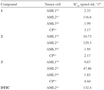
Table 4 shows the results of the antiproliferative effect of 1, 2 and 3 on acute myeloid leukemia cells
and control patient cells. IC50 values in the range of 1.83-129.30 mmol mL-1 were observed for the leukemia cells. The compounds 1, 2 and 3 demonstrated cytotoxicity
very similar against AML3, while compound 1 appeared
to be more cytotoxic than 2 and 3 against AML1. These
results suggest that the replacement of luorine by chlorine in compound 1 increased the antileukemic activity of the
triazenes in all leukemic cells tested. Against AML3 cells the compounds presented the best anti-leukemic activity and against AML2 the worst. However, the three compounds were more active against AML2 cells compared to DTIC. The correct dose is one of the most important aspects to achieve the best antitumor effect without increasing toxicity too much, considering that the therapeutic window of most antineoplastic agents is narrow. The fact that the IC50 of the three compounds for AML3 is half the CP allows us to suggest the possibility of administering a lower dosage, reducing toxicity. Data also showed that the compounds evaluated were highly toxic to normal leukocytes. The results of the MTT-dye reduction assay indicated that 1, 2
and 3 had a signiicant cytotoxic/antiproliferative effect in
comparison with other triazenes.53,54
The three triazene compounds described in this work belong to the alkylating agents group with similar chemical, physical, antimicrobial, nuclease and antitumor properties. DTIC and TMZ also belong to this group, showing an activity mechanism mainly related to the methylation of
Figure 5. Concentration dependence of the effect of the 1, 2 and 3
triazenes on cell proliferation of acute myeloid leukemia (AML1, AML2, AML3) and normal leukocytes (CP). Cells were exposed and viability determined after 24 h. Each data point represents the arithmetic mean ±
standard deviation.
Table 4. Inhibition of cell growth by triazenes 1, 2 and 3 and DTIC
assessed after 24 h exposure (MTT-dye reduction assay)
Compound Tumor cell IC50 (mmol mL-1)(ª)
1 AML1(b) 2.33
AML2(c) 116.6
AML3(d) 1.99
CP(e) 3.17
2 AML1(b) 10.73
AML2(c) 129.3
AML3(d) 1.95
CP(e) 2.17
3 AML1(b) 9.67
AML2(c) 47.86
AML3(d) 1.83
CP(e) 4.44
DTIC AML2(c) 132.4
(ª) IC
50 (dose required to achieve 50% decrease in cell growth). (b)AML1 =
acute myeloid leukemia cells of patient number 1. (c)AML2 = acute
myeloid leukemia cells of patient 2. (d)AML3 = acute myeloid leukemia
O6-guanine, mediated by the methyldiazonium ion, a highly reactive derivative of these chemotherapeutics agents. The antitumor effects of alkylating agents and cell resistance to them depend on at least three DNA repair systems: a) increase in O6-methyl-guanine methyl-tranferase (MGMT), also called O6-alkylguanine-DNA-alkyltranferase; b) loss of mismatch repair (the MMR system recognizes and repairs base-base mismatches and small insertion-deletions loops, IDLs); and c) base excision repair (BER) deiciency.
Conclusions
The 1-(2-chlorophenyl)-3-(4-carboxyphenyl)triazene (1), 1-(2-luorophenyl)-3-(4-carboxyphenyl)triazene (2)
and 1-(2-fluorophenyl)-3-(4-amidophenyl)triazene (3)
compounds assessed in the present study are capable to cleave plasmid DNA under deined pH, temperature and time conditions. Since the eficiency of plasmid DNA cleavage by the investigated triazenes was not inhibited in the presence of free hydroxyl scavengers and anaerobic atmosphere conditions, the hydrolytic mechanism for the DNA reaction is suggested, a mechanism similar to the one promoted by natural nucleases. The experimental results indicated that triazenes 1 and 2 had considerable action against
gram-positive bacteria. All compounds tested had pronounced cytotoxic activity against acute myeloid leukemia cells and normal leukocytes. Further studies to establish whether methylated triazene compounds in human leukemia could offer a new approach to control this disease are required. The crystal and molecular structure of one of the triazene derivatives (3) revealed intramolecular and intermolecular
interactions promoted by hydrogen bonding, which establish a signiicant inluence on the molecular planarity in the solid state. This observation suggests that 3 is probably not
susceptible to DNA binding by intercalation. The presence of luorine and amide substituents favor hydrogen bonding, indicating that the mode of action of 3 may involve an
hydrolytic mechanism initiated by an external approach directed to the major or minor grooves of the DNA molecule.
Supplementary Information
Crystallographic data for the structural analysis have been deposited with the Cambridge Crystallographic Data Centre (CCDC no. 751661). Further details of the crystal structure investigation are available free of charge via www. ccdc.cam.ac.uk/conts/retrieving.html (or from the CCDC, 12 Union Road, Cambridge CB2 1EZ, UK; fax: +44 1223 336033; e-mail: [email protected]).
Table S1 and Figure S1 are available free of charge at http://jbcs.sbq.org.br, as pdf ile.
Acknowledgements
M. H. thanks CNPq (proc. 305254/2009-0), F. K. thanks CNPq/PIBIC, A. L. thanks CNPq and V. F. G. thanks CAPES for grants. The diffractometer was supported by the Financiadora de Estudos e Projetos (FINEP, CT-INFRA 03/2001). The authors are grateful to PRPGP/UFSM/Pró-Publicações Internacionais for the language review of the manuscript.
References
1. Khramov, D. M.; Belawski, C. W.; J. Org. Chem.2007, 72, 9407.
2. Rouzer, C. A.; Sabourin, M.; Skinner, T. L.; Thompson, E. J.; Wood, T. O.; Chmurny, G. N.; Klose, J. R.; Roman, J. M.; Smith, R. H. Jr.; Michejda, C. J.; Chem. Res. Toxicol. 1996, 9,172.
3. Nicolaou, K. C.; Boddy, C. N. C.; Li, H.; Koumbis, A. E.; Hughes, R.; Natarajan, S.; Jain, N. F.; Ramanjulu, J. M.; Bräse, S.; Solomon, E.; Chem. Eur. J.1999, 5, 2602.
4. Bräse, S.; Dahmen, S.; Pfefferkorn, M.; J. Comb. Chem.2000,
2, 710.
5. Jones II, L.; Schumm, J. S.; Tour, J. M.; J. Org. Chem.1997,
62, 1388.
6. Moore, J. S.; Acc. Chem. Res.1997, 30, 402.
7. Jean-Claude, B. J.; Mustafa, A.; Damian, Z.; De Marte, J.; Vasillescu, D. E.; Yen, R.; Chan, T. H.; Leyland-Jones, B.;
Biochem. Pharmacol. 1999, 57, 753.
8. Doucet, K. G.; Pye, C. C.; Vaughan, K.; Enright, T. G.;
THEOCHEM2006, 801, 21.
9. An, Y.; Liu, S.; Deng, S.; Ji, L.; Mao, Z.; J. Inorg. Biochem.
2006, 100, 1586.
10. Shao, Y.; Ding, Y.; Jia, Z.; Lu, X.; Ke, Z.; Xu, W.; Lu, G.; Bioorg. Med.Chem. 2009, 17, 4274.
11. Takasaki, B. K.; Jik Chin, J.; J. Am. Chem. Soc. 1994, 116, 1122.
12. Burr, S. J.; Mselati, A.; Thomas, E. W.; Tetrahedron Lett.2003,
44, 7307.
13. O’ Reilly, S. M.; Newlands, E. S.; Glaser, M. G.; Brampton, M.; Rice-Edwards, J. M.; Illingworth, R. D.; Richards, P. G.; Kennard, C.; Colquohoun, I. R.; Lewis, P.; Stevens, M. F. G.;
Eur. J. Cancer1993, 29A, 940.
14. Bleehen, N. M.; Newlands, E. S.; Lee, S. M.; Thatcher, N.; Selby, P.; Calvert, A. H.; Rustin, G. J.; Brampton, M.; Stevens, M. F.; J. Clin. Oncol.1995, 13, 910.
15. Hoang-Xuan, K.; Camilleri-Broet, S.; Soussain, C.; Curr. Opin. Oncol. 2004, 16, 601.
16. Tani, M.; Fina, M.; Alinari, L.; Stefoni, V.; Baccarani, M.; Zinzani, P. L.; Haematologica2005, 90, 1283.
18. D’Incalci, M.; Souteyrand, P.; Ann. Dermatol. Venereol.2001,
128, 517.
19. Perry, M. J.; Mendes, E.; Simplício, A. L.; Coelho, A.; Soares, R. V.; Iley, J.; Moreira, R.; Francisco, A. P.; Eur. J. Med. Chem.
2009, 44, 3228.
20. Rijn, J. V.; Heimans, J. J.; Berg, J. V. D.; Valk, P. V. D.; Slotman, B. J.; J. Rad. Oncol. Biol. Phys. 2000, 47, 779.
21. Franchi, A.; Papa, G.; D’ATri, S.; Piccioni, D.; Masi, M.; Bonmassar, E.; Haematologica1992, 77, 146.
22. Seiter, K.; Liu, D.; Loughran, T.; Siddiqui, A.; Baskind, P.; Ahmed, T.; J. Oncol.2002, 20, 3249.
23. Seiter, K.; Liu, D.; Siddiqui, A. D.; Lerner, R.; Nelson, J.; Ahmed, T.; Leuk. Lymph.2004, 45, 1209.
24. Hörner, M.; Giglio, V. F.; Santos, A. J. R. W.; Westphalen, A. B.; Iglesias, B. A.; Martins, P. R.; Amaral, C. H.; Michelot, T. M.; Reetz, L. G. B.; Bertoncheli, C. M.; Paraginski, G. L.; Hörner, R.; Braz. J. Pharm. Sci.2008, 44, 441.
25. Marchesi, F.; Turriziani, M.; Tortorelli, G.; Avvisati, G.; Torino, F.; De Vecchis, L.; Pharm. Res.2007, 56, 275.
26. Desiraju, G. R.; Steiner, T.; The Weak Hydrogen Bond in Structural Chemistry and Biology, IUCr Monographs on Crystallography 9, Oxford University Press Inc: New York, 1999.
27. Steiner, T.; Angew. Chem., Int. Ed.2002, 41, 48.
28. Grammaticakis, P.; Bull. Soc. Chim. Fr.1956, 139.
29. Horn Jr., A.; Vencato, I.; Bortoluzzi, A. J.; Hörner, R.; Silva, R. A. N.; Spoganicz, B.; Drago, V.; Terenzi, H.; Oliveira, M. C. B.; Werner, R.; Haase, W.; Neves, A.; Inorg. Chim. Acta2005,
358, 339.
30. Clinical and Laboratory Standards Institute; Methods for Dilution AntimicrobialSusceptibility Tests for Bacteria that
Grow Aerobically. Approved Standard; 6th ed.;Wayne: NCCLS,
2003; p.53. (NCCLS document M7-A6).
31. Beloti, V.; Barros, M. A. F.; Freitas, J. C.; Nero, L. A.; Souza, J. A.; Santana, E. H. W.; Franco, B. D. G. M.; Rev. Microbiol.
1999, 30, 137.
32. COSMO (Version 1.48), SAINT (Version 7.06°), SADABS
(Version 2.10); Bruker AXS Inc.; Madison: Wisconsin, USA, 2005.
33. Burla, M. C.; Caliandro, R.; Camalli, M.; Carrozzini, B.; Cascarano, G. L.; De Caro, L.; Giacovazzo, C.; Polidori, G.; Spagna, R.; (SIR2004) J. Appl. Crystallogr.2005, 38, 381.
34. Sheldrick, G. M.; SHELXL-97; Program for Crystal Structure Reinement; University of Göttingen, Germany, 1997.
35. Brandenburg, K.; DIAMOND 3.1a. 1997-2005; Crystal Impact GbR, Bonn, Germany, 1997.
36. Colthup, N. B.; Daly, L. H.; Wiberley, S. E.; Introduction to Infrared and Raman Spectroscopy, 3rd ed., Academic Press:
New York, 1990.
37. Kübler, R.; Lüttke, W.; Weckherlin, S.; Z. Elektrochem.1960,
64, 650.
38. Silverstein, R. M.; Webster, F. X.; Spectrometric Identiication of Organic Compounds, 6th ed., John Wiley & Sons: New York,
1998.
39. Masoud, M. S.; Ali, A. E.; Shaker, M. A.; Ghani, M. A.;
Spectrochim. Acta 2005, 61A, 3102.
40. Silverstein, R. M.; Bassler, G. C.; Morrill, T. C.; Spectrometric Identiication of Organic Compounds, John Wiley & Sons: New
York, 1981.
41. Allen, F. H.; Kennard, O.; Watson, D. G.; Brammer, L.; Orpen, A. G.; Taylor, R.; J. Chem. Soc., Perkin Trans. I1987, 2, S1; Teatum, E.; Gschneider, K.; Waber, J.; Report LA-2345. Los Alamos Scientiic Laboratory, New Mexico, USA, 1960. 42. Orpen, A. G.; Brammer, L.; Allen, F. H.; Kennard, O.; Watson,
D. G.; Taylor, R.; J. Chem. Soc., Dalton Trans.1989, S1-83. 43. Hörner, M.; Silva, A.; Fenner, H.; Anal. Sci.: X-ray Struct. Anal.
Online2006, 22, x295.
44. Hörner, M.; Casagrande, I. C.; Bordinhão, J.; Mössmer, C. M.;
Acta Crystallogr. 2002, C58, o193.
45. Hörner, M.; Visentin, L. C.; Behm, M. B.; Machado, F. C.; Bortoluzzi, A. J.; Anal. Sci.: X-ray Struct. Anal. Online2007,
23, x247.
46. Spek, A. L.; PLATON, A Multipurpose Crystallographic Tool, Utrecht University, Utrecht: The Netherlands, 2008.
47. Fernandes, C.; Parrilha, G. L.; Lessa, J. A.; Santiago, L. J. M.; Kanashiro, M. M.; Boniolo, F. S.; Bortoluzzi, A. J.; Vugman, N. V.; Herbst, M. H.; Horn Jr., A.; Inorg. Chim. Acta2006, 359,
3176.
48. Basile, L. A.; Barton, J. K.; J. Am. Chem. Soc. 1987, 109, 7548. 49. Cowan, J. A.; Curr. Opin. Chem. Biol. 2001, 5, 634.
50. Brunton, L. L.; Lazo, J. S.; Parkker, K. L; Goodman & Gilman,
McGraw-Hill Interamericana do Brasil: Rio de Janeiro, 2006. 51. Gewirtz, D. A. A.; Biochem. Pharmacol. 1999, 57, 727.
52. Duggan, D. B.; Petroni, G. R.; Johnson, J. L.; Glick, J. H.; Fisher, R. I.; Connors, J. M.; Canellos, G. P.; Peterson B. A.;
J. Clin. Oncol. 2003, 21, 607.
53. Smith, R. H.; Scudiero, D. A.; Michejda, C. J.; J. Med. Chem.
1990, 33, 2579.
54. Zheleva, A. M.; Gadjeva, V. G.; Int. J. Pharm.2001, 212, 257.
Submitted: October 25, 2009
Supplementary Information
*e-mail: [email protected], [email protected]
In Vitro Evaluation of Triazenes: DNA Cleavage, Antibacterial Activity and
Cytotoxicity against Acute Myeloid Leukemia Cells
Vanessa O. Domingues,a Rosmari Hörner,*,a Luiz G. B. Reetz,a Fábio Kuhn,a
Virgínia M. Coser,b Jacqueline N. Rodrigues,b Rita Bauchspiess,b
Waldir V. Pereira,b Gustavo L. Paraginski,c Aline Locatelli,c
Juliana de O. Fank,c Vinícius F. Giglioc and Manfredo Hörner*,c
a Universidade Federal de Santa Maria, Departamento de Análises Clínicas e Toxicológicas,
97105-970 Santa Maria-RS, Brazil
bHospital Universitário de Santa Maria, Departmento de Hematologia-Oncologia,
97105-970 Santa Maria-RS, Brazil
c Universidade Federal de Santa Maria, Departamento de Química, C.P. 5031,
97105-970 Santa Maria-RS, Brazil
Table S1. Minimum inhibitory concentration values (µg mL−1) determined for the triazenes 1, 2 and 3
Bacterial strain Compound 1 Compound 2 Compound 3
Pseudomonas aeruginosa ATCC 27853 > 128 > 128 > 128
Escherichia coli ATCC 25922 > 128 > 128 > 128
Escherichia coli ATCC 35218 > 128 > 128 > 128
Salmonella choleraesuis ATCC 10708 > 128 > 128 > 128
Klebsiella pneumoniae ATCC 700603 > 128 > 128 > 128
Staphylococcus aureus ATCC 25923 > 128 > 128 > 128
Staphylococcus epidermidis ATCC 12228 > 128 > 128 > 128
Enterococcus faecalis ATCC 29212 = 128 > 128 > 128
Bacillus cereus ATCC 10987 > 128 > 128 > 128
Shigella sonnei > 128 > 128 > 128
Enterobacter cloacae > 128 > 128 > 128
Klebsiella oxytoca = 128 > 128 > 128
Bacillus cereus = 32 > 128 > 128
Micrococcus sp. > 128 > 128 > 128
Streptococcus pyogenes > 128 > 128 > 128
Acintobacter baumannii > 128 > 128 > 128
Raustonia pickettii > 128 > 128 > 128
Serratia marcescens > 128 > 128 > 128
Enterococcus sp. > 128 > 128 > 128
Listeria monocytogenes > 128 > 128 > 128
Aeromonas hidrophyla = 128 > 128 > 128
Enterobacter aerogenes > 128 > 128 > 128
Streptococcus agalactiae > 128 = 64 > 128
Salmonella tiphymurium > 128 > 128 > 128
Corynebacterium sp. > 128 >128 > 128
Salmonella sp. > 128 >128 > 128
Staphylococcus epidermidis > 128 > 128 > 128
Acinetobacter lwofii > 128 > 128 > 128
Klebsiella pneumoniae > 128 > 128 > 128
Figure S1. Total cleavage (%) of pUC18 and pBSKII plasmid DNA by triazene 3. Lane 1: control plasmid DNA (untreated pUC18 and pBSKII); Lanes