ORIGINAL
ARTICLE
Detection and characterization of human rotavirus
in hospitalized patients in the cities of Ponta Grossa,
Londrina and Assai - Pr, Brazil
Authors
Carlos M Nozawa1
Gustavo Franthesco Kerntopf2 Érika da Silva Czernisz3 Daniele Albuquerque3 Priscila Romanin4 José Felipe Eliseu Freitas4 Norma Santos5 Fabrício José Benati6 Eduardo Pietruchinski7 Rosa Elisa Carvalho Linhares8 1PhD in Sciences (Virology)/ Universidade Estadual de Londrina, Londrina, Pr. 2Master’s Degree in Microbiology/Universidade Estadual de Londrina, Londrina, Pr.
3Bacheloring in Biological Sciences/ Universidade Estadual de Londrina, Londrina, Pr. 4Secondary School – Fellowship of IC Jr/ Universidade Estadual de Londrina, Londrina, Pr. 5PhD in Sciences (Microbiology)/ Universidade Federal do Rio de Janeiro, Rio de Janeiro, RJ. 6Master’s Degree in Microbiology/Universidade Federal do Rio de Janeiro, Rio de Janeiro, RJ.
7Master’s Degree in Microbiology/Centro de Ensino Superior de Campos Gerais, Ponta Grossa, Pr. 8PhD in Sciences (Microbiology)/ Universidade Estadual de Londrina, Londrina, Pr.
Submitted on: 3/2/2010 Approved on: 9/6/2010
Correspondence to:
Universidade Estadual de Londrina
Rodovia Celso Garcia Cid, (Pr 445) Km 380 Caixa Postal 6001 CEP: 86.051-990 - Londrina Paraná - Brasil
Phone: +55 (43) 3371-4000 E-mail: [email protected]
Financial Support: CNPq, Brazilian Ministry of Health, Capes, Fundação Araucária, Proppg/UEL
We declare no confl ict of interest.
ABSTRACT
Acute diarrheal disease is still one of the major public health problems worldwide. Rotaviruses (RV) are the most important viral etiologic agents and children under fi ve years of age are the target population. Objective: To investigate the rate of RV infection in hospitalized patients due to acute diarrhea in the cities of Ponta Grossa, Londrina and Assai - Paraná. Methods: Latex agglutination (LA); immunochromatography (ICG); polyacrylamide gel electrophoresis (PAGE) and negative staining electron microscopy (ME) tests were used to detect the virus. For the genotyping, RT-PCR and RT-PCR-ELISA were used, respectively, for NSP4 and VP4/VP7. Result: Out of 124 samples there were 69 positive stool samples for RV, for at least one of the used tests, 67 of them being RV group A (RV-A). Overall, most of the RV positive stool samples came from children under thirteen years of age. However, 12 positive cases occurred in patients aged 13 years or above, including an 81-year old patient. Conclusion: The data showed similar electropherotypes and genotypes G, P and NSP4 of the inland wild circulating strains of RV.
Keywords: rotavirus; determination; hospitalized patients; genotyping.
[Braz J Infect Dis 2010;14(6):553-557]©Elsevier Editora Ltda.
INTRODUCTION
Acute diarrheal disease is still one of the major public health problems worldwide. Rotaviruses (RV) are the most important etiologic agents and children under fi ve years of age are the preferred target population. Moreover, a variety of infant animals are equally affected by species-specifi c vi-rus strains. Worldwide, RV cause approximately 112 millions of domestic episodes of diarrhea, 25 millions of clinic visits, 2 millions of hospitaliza-tions and about 611,000 deaths of children under fi ve years of age, annually.1
RV are icosahedric virions, non-enveloped, and present a triple concentric layers of proteins.2,3 The inner layer is formed by virus protein 2 (VP2) that involves the genome, the VP1 (RNA polymerase dependent of RNA) e a VP3 (guani-lyltransferase and methylase). The intermediate layer is made up of VP6 associated with VP2 and confers the structure the so-called double-layered particles (DLP). The outer layer is constituted by trimeric structures of VP7 glycoprotein and the dimeric spikes of VP4 forming the triple-layered particles (TLP), the infectious form of the virus. Virus genome is represented by 11 segments of
double-stranded RNA that encode, respectively, six structural proteins (VP1-VP4, VP6 and VP7) and six nonstructural proteins (NSP1-NSP6).
Based on antigenic specifi city of VP6, RV are classifi ed into seven groups (A-G), prevailing the infections by group A strains (RV-A).2 Moreover, VP6 epitopes allow the differentiation of group A strains into sub-groups (SG-I, SG-II, SG-I/II, and non-SG-I/II), prevailing SG-II in human infec-tions.4 These strains are molecularly distinguished into genogroup I (SG-I) and genogroup II (SG-II, SG-I/II, and SG non-I,non II).5 G and P genotyp-ing is attributed, respectively, to VP7 and VP4.2 Twenty-three G and 31 P genotypes have been described.6,7 Due to the importance of NSP4 pro-tein in the virion morphogenesis, replication and in the pathogeny of the infection this protein was also defi ned molecularly into six genotypes (A-F).4
Genotypes G1-G4 and G9 combined with P[8] and P[4] are the most prevalent worldwide (approximately 90%).9,10 Concerning NSP4 gen-otyping, B type has been shown to be the most common in the world.11,12
By poliacrylamide gel electrophoresis (PAGE) RV are classi-fi ed into seven electropherotypes (e-type) from A-G, according to the migration pattern of the 11 RNA segments. Moreover, group A strains can be further classifi ed according to the mo-bility of the segments 10 and 11 into long (L), short (S) and super-short (SS) electropherotypes according to the migration pattern.13,14
Presently, two types of RV vaccine are commercially avail-able for human use (Rotarix and RotaTeq) and their safety and effi cacy to prevent and/or attenuate severe diarrheal episodes have been proved.15,16
Bearing in mind the genetic variability of the virus, either vaccine or host natural immunity to the virus or both may pose a selective pressure that may result in emergence of unusual genotypes. Crossing species barrier is also another possibility for the appearance of mutant strains. In fact, hitherto undescribed genotypes have been found.17 These selective events may repre-sent a signifi cant antigenic “shift” or “drift”, as has been shown for infl uenza virus with a real impact in the epidemiology of the disease. Therefore, it is important to monitor wild strains of the virus in order to evaluate all these consequences, and to accompany the evolution of the infection.
In this paper we evaluated circulating human RV strains in three locations of the State of Paraná, Brazil.
MATERIAL AND METHODS
Feces
One hundred and twenty four fecal samples were collected from April 2005 to March 2009 from hospitalized patients suffering from acute diarrhea, admitted to private and pub-lic hospitals in the cities of Assai (Hospital Climas), Lond-rina (Hospital Universitário Regional do Norte do Paraná)
and Ponta Grossa (Hospital Bom Jesus) - Paraná. The study protocol was approved by the Experimental Ethics Commit-tee of the Universidade Estadual de Londrina, under the nº 01840268000-07.
Negative staining electron microscopy
For transmission electron microscopy (EM) raw stool sam-ples were processed by super direct negative staining with 2% sodium phosphotungstate, pH 6.3, as described elsewhere.18
Immunochromatography and latex agglutination tests
Stool samples were also homogenized at 20% (vol/vol) in PBS, pH 7.3, and clarifi ed by centrifugation at 450xg/10 min. Clarifi ed homogenate were submitted to Vikia - Rota Adeno, Biomérieux SA, Fr. and/or Virotect Rota, Omega Diagnostic Ltd., UK., according to the manufacturers recommendations.
Polyacrilamide gel electrophoresis
Clarifi ed fecal homogenates were further submitted to virus RNA extraction, as described before,19 for PAGE.
RT-PCR and PCR-ELISA
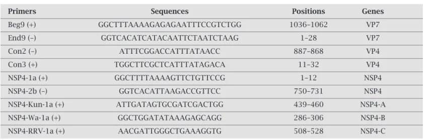
For RT-PCR virus RNA was extracted from stools using the TRIzol method (Invitrogen, Carlsbad, USA) and subjected to reverse transcription followed by PCR.20 The cDNA of VP7 or VP4 gene was synthesized by using primers labeled with biotin at their 5’ ends. For PCR-ELISA detection of labeled PCR products and identifi cation of the genotypes of posi-tive samples, briefl y, 96-well microplates (Nunc-Immuno module, Nunc, Roskilde, Denmark) coated with streptavi-din (Roche Diagnostic GmbH, Mannheim, Germany) were used. To each well the biotin-labeled PCR product was dis-tributed (one reaction per genotype) followed by the addi-tion of type-specifi c digoxigenin-labeled probe. Individual G- or P-type specifi c probe mixtures (three type-specifi c probes/genotype/mixture) were distributed onto the plates. This was followed by the addition of anti-digoxigenin per-oxidase conjugate (Roche Diagnostic GmbH); the substrate (TMB peroxidase substrate system; KPL, Gaithersburg, MD), and absorbance read at 450 nm within 10 min.21 For NSP4 typing, nested-PCR was used and primers are listed in Table 1, including those for VP7 and VP4 typing.
Table 1. VP7, VP4 and NSP4 primers used for amplification and genotyping
Primers Sequences Positions Genes
Beg9 (+) GGCTTTAAAAGAGAGAATTTCCGTCTGG 1036–1062 VP7
End9 (–) GGTCACATCATACAATTCTAATCTAAG 1–28 VP7
Con2 (–) ATTTCGGACCATTTATAACC 887–868 VP4
Con3 (+) TGGCTTCGCTCATTTATAGACA 11–32 VP4
NSP4-1a (+) GGCTTTTAAAAGTTCTGTTCCG 1–12 NSP4
NSP4-2b (–) GGTCACATTAAGACCGTTCC 750–731 NSP4
NSP4-Kun-1a (+) ATTGATAGTGCGATCGACTGG 439–460 NSP4-A
NSP4-Wa-1a (+) GGCTGGATATAAAGAGCAGG 286–306 NSP4-B
RESULTS
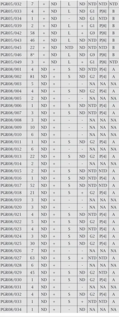
From 124 stool samples evaluated 69 were positive for RV (55.6%), for at least one of the methods used. Amongst these 69 positive samples, 63 (88.4%) were considered group A (RV-A), as demonstrated by serological methods, being positive either by ICG or LA. Additionally, four strains were also defi ned as RV-A by electrophoretic pattern by PAGE. Therefore, overwhelmingly, a total of 67 strains out of 69 (97.1%) were RV-A. Individually, the performance of the methods used accounted for the follow-ing results (Table 2): For LA, 66 samples were analyzed and 23 were positive (34.8%). For ICG, 40 samples were positive out of 64 (62.5%). For PAGE, out of 124 samples 47 were positive (37.9%), being 26 strains, 55.2% (26/47), with short pat-tern electropherotypes and 20, 42.5% (20/47), with long pattern. For EM, 47 samples were tested and 11 were positive (23.4%). As far as genotyping is concerned, out of the 124 samples, 44 samples (35.4%) were amplifi ed as following.
PGR05/032 7 + ND L ND NTD NTD NTD
PGR05/033 4 + ND L ND G1 P[8] B
PGR05/034 1 + ND - ND G1 NTD B
PGR05/039 2 + ND L + G1 P[8] B
PGR05/042 58 + ND L + G9 P[8] B
PGR05/043 46 + ND L ND NTD P[8] B
PGR05/045 22 + ND NTD ND NTD NTD B
PGR05/046 8* + ND L ND G9 P[8] B
PGR05/049 3 + ND L + G1 P[8] NTD
PGR08/001 4 ND + S ND NTD P[4] A
PGR08/002 81 ND + S ND G2 P[4] A
PGR08/003 5 ND + - - NA NA NA
PGR08/004 4 ND + S ND G2 P[4] A
PGR08/005 2 ND + - - NA NA NA
PGR08/006 1 ND + S ND NTD P[4] A
PGR08/007 3 ND + S ND NTD P[4] A
PGR08/008 3 ND + - - NA NA NA
PGR08/009 10 ND + - - NA NA NA
PGR08/010 6 ND + - - NA NA NA
PGR08/011 1 ND + S ND G2 P[4] A
PGR08/012 6 ND + - - NA NA NA
PGR08/013 22 ND + S ND G2 P[4] A
PGR08/014 2 ND + - - NA NA NA
PGR08/015 2 ND + S ND NTD NTD A
PGR08/016 1 ND + S ND NTD P[4] A
PGR08/017 52 ND + S ND NTD NTD A
PGR08/018 21 ND + S + G2 P[4] A
PGR08/019 3 ND + - - NA NA NA
PGR08/020 3 ND + - - NA NA NA
PGR08/021 4 ND + S ND NTD P[4] A
PGR08/022 5 ND + S ND G2 P[4] A
PGR08/023 4 ND + S ND NTD P[4] A
PGR08/024 3 ND + S ND G2 P[4] A
PGR08/025 30 ND + S ND G2 P[4] A
PGR08/026 7 ND + - - NA NA NA
PGR08/027 63 ND + S + NTD NTD A
PGR08/028 6 ND + - - NA NA NA
PGR08/029 45 ND + S ND G2 NTD A
PGR08/030 1 ND + S ND G2 P[4] A
PGR08/031 4 ND + - - NA NA NA
PGR08/032 4 ND + S ND G2 P[4] A
PGR08/033 1 ND + S + NTD NTD A
PGR08/034 1 ND + - ND NA NA NA
aEletropherotype: L - long, S - short.
bAntigenic specificity carried by indicated proteins.
cND, not done.
dNA, not amplified.
eNTD, not determined.
+ positive/- negative. Table 2. Rotavirus strains with the respective
methods of detection/typing
Strain Age LA ICG E-typea EM Genotypesb
(yrs./ mos.*)
VP7 VP4 NSP4
ASS08/001 1 NDc - - + NAd NA NA
ASS08/003 4 ND + L + NTDe NTD A
ASS08/004 2 ND - - + NA NA NA
LON08/016 2 + ND S ND G2 P[4] A
LON08/018 2 + ND S ND G2 P[4] A
LON08/019 3 - ND S ND NTD NTD A
LON08/022 7 + ND S + NTD NTD NTD
PGR05/001 1 - ND - - NTD NTD B
PGR05/002 3 + ND L ND G1 NTD B
PGR05/006 3 + ND - - NTD NTD NTD
PGR05/010 11 - ND L + G1 P[8] B
PGR05/011 6 + ND L ND NTD P[8] B
PGR05/012 1 + ND L ND NTD NTD NTD
PGR05/013 10 + ND - - NA NA NA
PGR05/015 1 + ND L ND G1 P[8] B
PGR05/016 12 + ND L ND G1 P[8] B
PGR05/019 14 - ND - - NTD NTD B
PGR05/020 2 + ND L ND NTD NTD B
PGR05/021 9 + ND - - NA NA NA
PGR05/023 4* + ND L ND NTD NTD NTD
PGR05/024 8* - ND L ND NTD NTD B
PGR05/025 1 + ND - ND NTD NTD NTD
PGR05/026 14 + ND S ND NTD P[8] B
PGR05/027 9* + ND L ND G1 P[8] B
PGR05/030 1 - ND L ND NTD NTD NTD
Eleven samples amplifi ed, individually, either for NSP4-A (6) or NSP4-B (5). Thirteen samples amplifi ed for the dou-ble combinations G1/NSP4-B (2), G1P[8] (1), G2/NSP4-A (1), P[4]/NSP4-A (6), and P[8]/NSP4-B (3). Twenty samples amplifi ed for the triple combinations G1P[8]/NSP4-B (6), G2P[4]/NSP4-A (12) and G9P[8]/NSP4-B (2). Genotypes G1, G2 and G9 accounted for 37.5% (9/24), 54.1% (13/24), and 8.3% (2/24) of the strains detected, respectively. In our study genotyping VP4 demonstrated that P[4] was prevalent in 56.2% (18/32) in comparison to 43.7% (14/32) of P[8]. Genotyping of NSP4 accounted for 56.8% (25/44) for type A and 43.2% for type B.
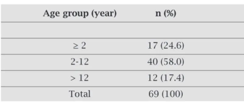
According to the ages of the patients, out of 124 stool samples 57 came from children and young children (≤ twelve years of age) (82.6%, 57/69). Positive cases in adults were detected in twelve patients over 12 years, includ-ing an 81-year-old male patient, overall representinclud-ing 17.4% (12/69) (Table 3).
DISCUSSION AND CONCLUSION
This study demonstrated the outstanding importance of RV as the causative of acute diarrheal disease, account-ing for a positivity of 55.6%, particularly because all the subjects studied were hospitalized patients. Overwhelm-ingly, most of the RV positive stool samples were from children under six years of age. This epidemiological feature has been taken for granted, but, increasingly, adults have been affected by the disease. Infection of RV in adults seems to be common and has been reported mainly among geriatric patients, disabled individuals, health attendants and those living in households with diseased children.22-25 In the present work, we found that 17.4% of RV positive samples were detected in adults from 12 years of age, including an 81-year old patient. It is suggested that the number of adults infected by RV would be greater, and studies involving them should be encouraged. It has been suggested that symptomatic or asymptomatic adults could serve as source of infection Table 3. Distribution of rotavirus positive cases according to the age of the patients
Age group (year) n (%)
≥ 2 17 (24.6)
2-12 40 (58.0)
> 12 12 (17.4)
Total 69 (100)
to children.26,27 In this study RV-A, as a rule, account-ed for 97.1% of the cases, as most of the world reports have demonstrated.28-30 The electropherotypes of these strains of RV-A showed a slight predominance of short pattern in comparison to the long-pattern strains, al-beit, unusual, this has been reported in India.31 In gen-eral, the predominance of long e-types has been re-ported throughout the world,32-36 however, variation has been shown according to some genotypes. As far as VP7 genotyping is concerned, our study revealed greater incidence of G2 secondly by G1. Although in general, greater incidence has been attributed to G1 worldwide, in Brazil, however, temporally/seasonally there has been a change of prevalence between G1 and G2, as demon-strated elsewhere.29,37 In our study, the greater incidence of G2 may be explained by geographical features or due to the small sampling. Nevertheless, these results dem-onstrated the occurrence of G types common in most of the countries, including Brazil9,10,37 combined with P[4] or P[8]. For VP4 genotyping, similar prevalence profile has been found in other countries and in Brazil, and the analysis underlying varied epidemiological features is also possible, between the two major genotypes presently found, P[4] and P[8]. As for NSP4 genotyping, we found that type A accounted for the most strains detected. B type NSP4 has been shown the most common in the world.11,12 However, in Brazil, this is true for the south-eastern area,38 but not for the northern region, where A type is prevalent.39 The data reported in our work dem-onstrated similar profile of the wild circulating strains of RV, as compared to data obtained elsewhere in the world and in our country. Molecular and serological nu-ances are mainly attributed to factors, such as RV geno-typic and phenogeno-typic variability; interspecies barriers crossing; host immunity pressure - naturally or artifi-cially acquired, as well as, geographical and seasonal fea-tures. The apparent increase of adult infection and the increasing number of untyped strains, albeit, positive by other tests, as we found in our work, may be a dem-onstration of a constant genetic variability. Therefore, the emergence of new RV strains should be expected. The effect of RV vaccination recently launched in Brazil is of major benefit for preventing the disease. However, changing in RV genotypes might also be expected as a result of a selective evolutionary process. In concluding, the threat posed by RV still represents a heavy sanitary and economical burden and has to be carefully treated.
ACKNOWLEDGMENTS
REFERENCES
1. Angel J, Franco MA, Greenberg HB. Rotavirus vaccines: recent developments and future considerations. Nat Rev Microbiol 2007;5:529-39.
2. Estes MK, Cohen J. Rotavirus gene structure and function. Microbiological Reviews 1989;53:410-49.
3. Yeager M, Dryden KA, Olson NH et al. Three-dimensional
structure of rhesus rotavirus by cryoelectron microscopy and image reconstruction. J Cell Biol 1990;110:2133-44.
4. Estes MK, Kapikian AZ. Rotaviruses. In: Knipe D.M., Howley PM, Griffi n DE et al. eds. Fields Virology. Philadelphia: Kluwer Healt Lippincott, Williams & Wilkins, 2007.
5. Iturriza-Gómara M, Wong C, Blome S, Desselberger U, Gray J. Molecular characterization of VP6 genes of human rotavirus isolates: correlation of genogroups with subgroups and evi-dence of independent segregation. J Virol 2002;76:6596-601. 6. Abe M, Ito N, Morikawa S et al. Molecular epidemiology of
rotaviruses health calves in Japan: Isolation of a novel bo-vine rotavirus bearing new P and G genotypes. Virus Res 2009;144:250-7.
7. Ursu K, Kisfali P, Rigó D et al. Molecular analysis of the VP7 gene of pheasant rotavirus identifi es a new genotype designat-ed G23. Arch Virol 2009;154:1365-9.
8. Matthijnssens J, Ciarlet M, Rahman M et al. Recommenda-tions for the classifi cation of group A rotaviruses using all 11 genomic RNA segments. Arch Virol 2007;153:1621-9. 9. Beards GM, Desselberger U, Flewett TH. Temporal and
geo-graphical distributions of human rotavirus serotypes, 1983 to 1988. J Clin Microbiol 1989;27:2827-33.
10. Santos N, Hoshino Y. Global distribution of rotavirus sero-types/genotypes and its implication for the development and implementation of an effective rotavirus vaccine. Rev Med Vi-rol 2005;15:29-56.
11. Iturriza-Gómara M, Anderton E, Kang G et al. Evidence for
ge-netic linkage between the gene segments encoding NSP4 and VP6 proteins in common and reassortant human rotavirus strains. J Clin Microbiol 2003;41:3566-73.
12. Cho SL, Ahn JH, Kim K et al. Genetic variation in the NSP4
gene of human rotavirus isolated in Seoul. J Bacteriol Virol 2006;36:79-87.
13. Espejo RT, Calderón E, González N et al. Presence of two
dis-tinct types of rotavirus in infants and young children hospital-ized with acute gastroenteritis in Mexico City, 1977. J Infect Dis 1979;139:474-7.
14. Matsuno S, Hasegawa A, Mukoyama A, Inouye S. A candidate for a new serotype of human rotavirus. J Virol 1985;54:623-4. 15. Vesikari T, Matson DO, Dennehy P et al. Safety and effi cacy
of a pentavalent human-bovine (WC3) reassortant rotavirus vaccine. N Engl J Med 2006;354:23-33.
16. Ruiz-Palacios GM, Pérez-Schael I, Velázquez FR et al. Safety and effi cacy of an attenuated vaccine against severe rotavirus gastroenteritis. N Engl J Med 2006;354:11-22.
17. Solberg OD, Hasing ME, Trueba G, Eisenberg JNS. Characteri-zation of novel VP7, VP4, and VP6 genotypes of a previously untypeable group A rotavirus. Virology 2009;385:58-67. 18. Santos N, Nozawa C. Comparação dos testes de aglutinação
do látex, microscopia eletrônica e eletroforese em gel de po-liacrilamida do RNA viral na detecção de rotavírus em fezes diarréicas de crianças. Revista Brasileira de Patologia Clínica 1989;25:117-20.
19. Herring AJ, Inglis NF, Ojeh CK et al. Rapid diagnosis of rotavi-rus infection by direct detection of viral nucleic acid in silver-stained polyacrylamide gels. J Clin Microbiol 1982;16:473-7.
20. Santos N, Soares CC, Volotão EM et al. Surveillance of rotavi-rus strains in Rio de Janeiro, Brazil, from 1997 to 1999. J Clin Microbiol 2003;41:3399-402
21. Santos N, Honma S, Timenetsky MCST et al. Development of a mi-crotiter plate hybridization-based PCR-enzyme-linked immuno-sorbent assay for identifi cation of clinically relevant human group A rotavirus G and P genotypes. J Clin Microbiol 2008;46:462-9. 22. Wenman WM, Hinde D, Feltham S, Gurwith M. Rotavirus
in-fection in adults. Results of a prospective family study. N Engl J Med 1979;301:303-6.
23. Anderson EJ, Weber SG. Rotavirus infection in adults. Lancet Infect Dis 2004;4:91-9.
24. Iijima Y, Iwamoto T, Nukuzuma S et al. An outbreak of
rotavi-rus infection among adults in an institution for rehabilitation: long-term residence in a closed community as a risk factor for rotavirus illness. Scand J Infect Dis 2006;38:490-6.
25. Carraro E, Perosa AHS, Siqueira I et al. Rotavirus infection in children and adult patients attending in a tertiary hospital of São Paulo, Brazil. Braz J Infect Dis 2008;12:44-6.
26. Barnes GL, Callaghan SL, Kirkwood CD et al. Excretion of
se-rotype G1 rotavirus strains by asymptomatic staff: a possible source of nosocomial infection. J Pediatr 2003;142:722-5. 27. Pietruchinski E, Benati F, Lauretti F et al. Rotavirus diarrhea
in children and adults in a southern city of Brazil in 2003: dis-tribution of G/P types and fi nding of a rare G12 strain. J Med Virol 2006;78:1241-9.
28. Parashar UD, Gibson CJ, Bresse JS, Glass RI. Rotavirus and se-vere childhood diarrhea. Emerg Infect Dis 2006;12:304-6.
29. Munford V, Souza EC, Caruzo TAR et al. Serological and
mo-lecular diversity of human rotavirus in São Paulo, Brazil. Braz J Microbiol 2007;38:459-66.
30. Surendran S. Rotavirus infection: molecular changes and pathophysiology. Experimental and Clinical Sciences Journal 2008;7:154-62.
31. Saravanan P, Ananthan S, Ananthasubramanian M. Rotavirus infection among infants and young children in Chennai, South India. Indian J Med Microbiol 2004;22:212-21.
32. Maunula L, von Bonsdorff C-H. Frequent reassortments may explain the genetic heterogeneity of rotaviruses: analysis of Finnish rotavirus strains. J Virol 2002;76:11793-800.
33. Urbina D, Rodríguez JG, Arzuza O et al. G and P genotypes
of rotavirus circulating among children with diarrhea in the Colombian northern coast. Int Microbiol 2004;7:113-20. 34. Modarres S, Rahbarimanesh AA, Karimi M et al.
Electro-phoretic RNA genomic profi les of rotavirus strains prevailing among hospitalized children with acute gastroenteritis in Te-hran, Iran. Arch Iran Med 2008;11:526-31.
35. Aminu M, Esona MD, Geyer A, Steele AD. Epidemiology of rotavirus and astrovirus infections in children in northwestern Nigeria. Ann Afr Med 2008;7:168-74.
36. Santos JS, Alfi eri AF, Leite JPG et al. Molecular epidemiology of the human group a rotavirus in the Paraná State, Brazil. Brazilian Archives of Biology and Technology 2008;51:287-94. 37. Leite JP, Carvalho-Costa FA, Linhares AC. Group A rotavirus genotypes and the ongoing Brazilian experience: a review. Mem Inst Oswaldo Cruz 2008;103:745-53.
38. Araújo IT, Heinemann MB, Mascarenhas JDP et al. Molecular
analysis of NSP4 and VP6 genes of rotavirus strains recovered from hospitalized children in Rio de Janeiro, Brazil. J Med Microbiol 2007;56:854-9.