SHORT COMMUNICATION
Episomal detection of
Banana streak OL virus
in single
and mixed infection with
Cucumber mosaic virus
in banana
‘Nanicão Jangada’
Patrícia R. Carnelossi1, Taise Bijora1, Cassiele U. Facco1, Jaqueline M. Silva1, Marcelo H. S. Picoli1, Eliezer R. Souto1 & Fernando T. de Oliveira2
1Departamento de Agronomia,Universidade Estadual de Maringá, Maringá, PR, 87020-900, Brazil; 2Instituto Paranaense de
Assistência Técnica e Extensão Rural, Andirá, PR, 86380-000, Brazil Author for correspondence: Eliezer R. Souto, e-mail: ersouto@uem.br
ABSTRACT
Banana streak virus (BSV) and Cucumber mosaic virus (CMV) are commonly found in all banana growing-areas of the world.
These viruses cause diseases that lead to yield reduction and constrain plant breeding and distribution of Musa germplasm. Most of the
diagnostic methods targeting BSV can generate dubious results because of the considerable genetic and serological diversity among BSV isolates and the presence of integrated BSV sequences in the banana plant. Both viruses are usually detected in single and mixed infections in banana plantations in the north region of Paraná state using DAS-ELISA and PCR. A rolling-circle amplification protocol tested in this study allowed specific detection and identification of an episomal BSV isolate infecting Nanicão Jangada cultivar, thus confirming the occurrence of Banana streak OL virus in Brazil.
Key words: Badnavirus, Cucumovirus, Musaceae, RT-PCR, rolling-circle amplification.
World banana (Musa spp.) production in 2011 was
102 million tons, primarily from plantations in India, Brazil, Ecuador, China and the Philippines (FAO, 2012). Banana cultivars originated from two wild species, Musa acuminata Colla (A genome) and M. balbisiana Colla
(B genome), resulting in a series of diploid, triploid and tetraploid genomes (AA, AB, AAA, AAB, ABB, AABB, AAAB, ABBB) (Simmonds, 1976). The worldwide movement of banana hybrids has been affected by the difficulty in obtaining virus-free plantlets through tissue culture due to the occurrence of mosaic disease caused by CMV (Cucumber mosaic virus), family Bromoviridae,
and streak disease caused by BSV (Banana streak virus).
Currently, BSV is considered a major constraint to banana improvement and poses a threat to Musa production
worldwide (James et al., 2011).
The most typical symptoms of BSV infection are broken or continuous streaks on the leaf lamina, which may vary from yellow, chlorotic, black or brown color. Mild symptoms include faint broken chlorotic lines or eyespots, while more severe symptoms consist of necrosis of various part of the plant, causing its death. Since BSV symptoms may be confounded with CMV symptoms, accurate diagnosis cannot rely on visual assessments (Lockhart & Jones, 2000). BSV is a member of the genus
Badnavirus in the family Caulimoviridae. Its genome
comprises noncovalently closed, double-stranded DNA of approximately 7.2 to 7.8 kbp. Four BSV species are currently
recognized: Banana streak Mysore virus (BSMyV), Banana streak GF virus (BSGFV), Banana streak OL virus (BSOLV), and Banana streak VN virus (BSVNV)
(Geering & Hull, 2012). Nevertheless, many other species of BSV may occur based on numerous reported partial sequences (Figueiredo et al., 2006). BSV can occur in two main forms within the host, firstly integrated into the host genome (endogenous BSV) and secondly in an episomal form (episomal BSV). Episomal infection may result due to the integrated form of the virus being expressed within the plant (Ndowora & Lockhart, 2000), or by introduction to the plant via mealybug vectors from an external source of viruses. Although incomplete integrants have been found in both A and B genomes, endogenous activatable forms of BSV have only been detected in the B genome of various banana accessions, particularly subjected to certain stress conditions, such as tissue culture (Geering et al., 2005).
A genetic mechanism of recombination may be responsible for triggering endogenous BSV to become episomal because of an inhibiting factor present with the homologous BB genome and absence of endogenous BSV in recombined genes such as AAB (Lheureux et al., 2003).
of these viruses. Hence, control strategies rely largely on the use of virus-free planting material (Brioso et al., 2000).
Leaf samples were taken from ‘Nanicão Jangada’ (AAA) showing leaf streaking symptoms, and from asymptomatic plants of ‘Grand Naine’ (AAA), ‘Nanicão comum’ (AAA), ‘IAC 2001’ (AAA), ‘Maçã’ and ‘Prata Anã’ (AAB).
Samples were tested through double-antibody sandwich (DAS)-ELISA, with a polyclonal SCBV
commercial kit, and with a CMV polyclonal antiserum following manufacturer's (Agdia) instructions. Samples of lyophilized healthy banana leaves acquired from Agdia were used as negative control. Each sample was tested in triplicate wells of a polystyrene microtiter plate. Tests were considered positive when the absorbance (A405) value of
each sample was at least two times greater than that of the healthy control.
For BSV detection, planttotal DNA extraction was performed according to James et al. (2011). Fresh (0.4 g) leaf tissue was ground in 3 mL of extraction buffer (100 mM Tris-HCl, pH 8.0; 50 mM EDTA; 1.4 M NaCl; 80 mM Na2SO3; 2% PVP-10; and 2% cetyltrimethylammonium
bromide) using a mortar and pestle. Samples were incubated at 65°C for 15 min and then centrifuged for 5 min at 12,000 rpm. The supernatant was subsequently mixed with an equal volume of chloroform: isoamylalcohol (24:1) and the mixture centrifuged at 12,000 rpmfor 5 min. After a second chloroform extraction, the supernatant was mixed with an equal volume of isopropanol and incubated at room temperature for 5 min. Nucleic acids were pelleted by centrifugation as described above, and pellets were washed with 70% ethanol, air dried, and resuspended in 50 μL of sterile distilled water.
The extraction of plant total RNA for CMV detection was made by combining the CTAB extraction protocol (James et al., 2011), and the Trizol reagent extraction method. Banana leaves (0,4g) ground in liquid nitrogen were homogenized in 3 mL of extraction buffer plus 2% cetyltrimethylammonium bromide and transferred to 1,5 mL microcentrifuge tubes. Samples were maintained at 65ºC for 30 min, shaken at 10 min intervals before adding 1 volume of Trizol reagent following manufacturer's protocol.
For BSV detection by PCR, reaction procedures were as described in Harper et al., (2002), with degenerate primers, forward 1A (5´-CTNTAYGARTGGYTGT NATGCCNTTYGG), and reverse primer 4’ (5´-TCCAYTTRCANAYNSCNCCC CANCC) to amplify a 600-bp fragment of the RT/RNaseH-coding region (ORF3). PCR cycle conditions were an initial denaturation at 94ºC for 10 min, 5 cycles of 94ºC for 30 s, 37ºC for 30 s and 72ºC for 90 s, followed by 30 cycles (94ºC for 30 s, 50ºC for 30 s, 72ºC for 1 min) and a final extension of 72ºC for 1 min.
For CMV detection, RT-PCR was performed with forward primer CMV-2 (5’-TATGATAAGAAGCTTGTTTCGCG), and reverse CMV-1 (5’-GCCGTAAGCTG GATGGACAA) (Wylie et
al., 1993), which amplify approximately a 500 bp fragment of the putative viral coat protein gene. For cDNA synthesis with M-MLV reverse transcriptase (Invitrogen) and reverse CMV-1 primer, total RNA was extracted from 1g of banana leaf, ground in liquid nitrogen, adding trizol reagent. PCR profile consisted of an initial denaturing step at 94ºC for 3 min; 35 cycles of 94ºC for 3min, 1 min at 60ºC and 90 s at 72ºC; with a final extension step at 72ºC for 10 min.
Rolling-circle amplification (RCA) using the Illustra TempliPhi 100 Amplification Kit (GE Healthcare), was essentially according to the manufacturer’s instructions with some modifications as proposed by James et al. (2011). Nucleic acid extract (1 μL) was mixed with 5 μL of kit sample buffer and 1 μL of a 50-μM stock solution (each primer at approximately 4.16 pmol/μL) of degenerate primers (1A/4’). The mixture was denatured at 95°C for 3 min and cooled on ice, and to this was added kit reaction buffer (5 μL) premixed with polymerase (0.2 μL). The mixtures were incubated at 30°C for 18 h and then the reactions were stopped by incubation at 65°C for 10 min. Products of RCA amplification of sample ‘Jangada’-13 were independently digested with KpnI, BamHI, EcoRI, and XbaI restriction endonucleases and then electrophoresed in
1% agarose gels in Tris-acetate-EDTA buffer at 100 V for 75 min. Gels were stained using 0.25× SYBR Safe DNA Gel Stain (Invitrogen) and visualized on a transilluminator. The approximately 1Kbp XbaI-digest fragment was excised
from gel, purified and ligated into appropriately digested pUC19 plasmid vector which was used to transform cells of Escherichia coli DH5α made competent by heat shock
treatment. Plasmid DNA containing inserts were purified and sent for sequencing with universal M13 forward and reverse primers, in a ABI 3730 DNA analyzer (Applied Biosystems), in a lab facility at Universidade de São Paulo.
Products of RT-PCR amplifications with primers (CMV-1/2) were excised from gels, purified and subsequently ligated into the pGem-T Easy Vector (Promega), transformed into competent Escherichia coli
DH5α and sequenced with CMV-1/2 primers following the previously described procedures.Sequences were compared with others from GenBank by the BLAST program of the National Center for Biotechnology Information (NCBI).
Banana producing areas were surveyed through DAS-ELISA with an antiserum for Sugarcane bacilliform virus (SCBV), a badnavirus serologically related to BSV
(Lockhart and Autrey, 1988), and BSV was found in single and in mixed infections with CMV (Table 1). DAS-ELISA was as effective as RCA for BSV detection (Table 1), and both tests would be recommended for episomal detection of BSV. However, the efficiency of serological based methods for the episomal detection of BSV has been questioned, considering the uneven BSV distribution within the Musa
the high diversity of BSV would result in serological variability among strains, likely to give false negatives during detection procedures such as ELISA (Ndowora & Lockhart, 2000).
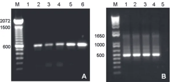
Likewise, PCR with primers 1A/4’ was successful in BSV detection in symptomatic ‘Jangada’ samples. However, it could not specifically discriminate infected from not infected plants, considering that many PCR positive samples as ‘Prata Ana’, ‘Maçã’, ‘Grand Naine’ and ‘Nanicão comum’ were not BSV infected as indicated by ELISA and RCA (Table 1, Figure 1). Several PCR based protocols directly on plant crude extracts or on purified DNA have been used to detect BSV strains, but they have the disadvantage of giving false positives, since PCR primers can detect BSV sequences integrated into the plant genome as episomal virus particles. Moreover, all Musa
spp. examined so far, harbor some sequence form of BSV in their DNA (Harper et al., 2002, James et al., 2011).
Results of this study confirmed previous works (James et al., 2011; Wambulwa et al., 2012) that RCA is very reliable to detect episomal BSV, as the sequence-independent nature of RCA can selectively amplify circular and not linear (integrated) DNA. RCA specifically amplified the genome of episomal BSV present in ‘Nanicão Jangada’ (Table 1, Figure 2). Subsequent sequence analysis of approximately 1.2-Kbp fragment of XbaI-digestion of RCA
product from DNA of ‘Nanicão Jangada’-13 revealed 99% similarity to part of the RT/RNaseH-coding region from
BSOLV. According to ICTV differences within ORF3 of more than 20% is a demarcation criterion of species in the
Badnavirus genus (Geering & Hull, 2012). Additionally,
the RCA polymorphic restriction fragment profile of 1 band obtained by KpnI and BamHI treatment, and the XbaI
profile of 3 bands are in agreement with the predicted for BSOLV, as reported by James et al., (2011), except for
EcoRI restriction profile of 3 bands instead of 4 (Figure 2),
an indication of a slight nucleotide sequence variability in the examined BSOLV strain.
In this study, detection of CMV and BSV through all tested procedures was sometimes inconsistent (Table 1), probably due to low titer and virus uneven distribution in some banana plants.
RCA has been used for detection of DNA genomes of viruses in the families Geminiviridae, Nanoviridae and Caulimoviridae (Inoue-Nagata et al., 2004; Johne et
al., 2009; James et al., 2011). Thus, in the case of BSV, it would be advisable to identify species using RCA amplification of episomal BSV to avoid misidentification by the amplification of virus endogenous sequences.
The episomal BSOLV isolate identified in this work could not come from an integrated form of the virus later expressed causing disease, as ‘Nanicão Jangada’ belongs to the Cavendish subgroup (AAA), and in A genomes the expression of functional viral transcripts is not found (Geering et al., 2005; James 2011). This emphasizes the Banana leaf samples BSV-DAS-ELISA
(1)
(405 nm)
(2)
BSV-PCR
(3)
BSV-RCA
(3)
CMV-DAS-ELISA (405 nm)
(2)
‘Nanicão Jangada’-1
-(0,019)
+ -
-(0,017)
‘Nanicão Jangada’-4
-(0,017)
+ - +
(0,108)
‘Nanicão Jangada’-10 +
(0,268)
+ + +
(0,295)
‘Nanicão Jangada’-12 +
(0,100)
+ + +
(0,042)
‘Nanicão Jangada’-13 +
(0,524)
+ + +
(0,225)
‘Prata Anã’
-(0,016)
+ -
-(0,013)
‘Maçã’
-(0,014)
+ - +
(0,216)
‘Grand Naine’
-(0,007)
+ - +
(0,218)
‘Nanicão comum’
-(0,019)
+ - +
(0,061)
‘IAC 2001’ +
(0,052)
+ -
-(0,016)
Banana leaf tissue (Agdia®),
-(0,014)
nt nt
-(0,014) negative control
TABLE 1 - Comparison ofdouble antibody sandwich (DAS)-ELISA, polymerase chain reaction (PCR) and rolling-circle amplification
(RCA) for detection of Banana streak virus (BSV)from Musa, and detection of Cucumber mosaic virus (CMV) by DAS-ELISA.
(1) Tested with Sugarcane bacilliform virus antiserum (Agdia), (2) Mean of three absorbance readings at 405 nm, (3) Tested with Badnavirus primers (1A/4’) (Harper et al., 2002), (+) positive, (-) negative, (nt) not tested.
FIGURE 2 - Analysis of rolling-circle
amplification (RCA) products with
Badnavirus primers 1A/4’ on DNA
from leaf extracts of ‘Nanicão Jangada’-13. M. 1Kbp plus DNA ladder; 1. RCA
product; 2. KpnI-digested, 3. Bam
HI-digested, 4. EcoRI-digested, 5. Xba
I-digested RCA product.
FIGURE 1 - Amplification of nucleic acids from banana leaf extracts. A. PCR with Badnavirus primers
1A/4’. M. 100 bp DNA ladder; 1. Negative control (no DNA); 2. ‘Maçã’; 3. ‘Grand Naine’; 4. ‘Prata Anã’; 5. ‘Nanicão Jangada’-10; 6. ‘Nanicão Jangada’-13. B. RT-PCR with CMV primers 1/5. M. 1Kbp plus DNA
ladder; 1. ‘Grand Naine’; 2. ‘Maçã’; 3. ‘Nanicão Jangada’-10; 4. ‘Nanicão Jangada’-13; 5. Healthy ‘Nanicão
comum’.
need of indexing banana planting material using the most reliable and sensitive detection tools to avoid the episomal BSV spread through infected planting material.
In Brazil, since the first report of CMV in banana (Ribeiro et al., 1975) there are reports of Musa infection by
distinct strains of BSV that are not recognized yet as species
in the Badnavirus genus (Brioso et al., 2012, Figueiredo et
al., 2006, Poltronieri et al., 2009). Also, there is a report of a PCR detection of a BSOLV isolate from banana ‘Terra Ana’ (AAB) from São Paulo state (Lombardi at al., 2010).
RCA-based methods can circumvent most of the drawbacks of these detection tools for banana BSV indexing. Future research should aim to optimize RCA protocols for more time and cost-effective BSV diagnostics.
ACKNOWLEDGMENTS
This work was supported by FINEP/SEBRAE and Fundação Araucária de Amparo à Pesquisa do Paraná. We also thank Conselho Nacional de Desenvolvimento Científico e Tecnológico - CNPq and Coordenação de Aperfeiçoamento de Pessoal de Nível Superior - CAPES for a fellowship grant.
REFERENCES
Brioso PST, Cordeiro ZJM, Rezende JAM, Kitajima EW, Pimentel JP, Figueiredo, AR (2000) Infecção mista em bananeiras pelos vírus do mosaico do pepino (“Cucumber mosaic virus” CMV) e da risca da bananeira (“Banana streak virus” BSV) no Brasil. Summa Phytopathologica 26:254-257.
Brioso PST (2012) Badnavirus e seu controle. In: 45o Congresso
Brasileiro de Fitopatologia.Tropical Plant Pathology (Suplemento) 37:1-19.
FAO. Food and Agriculture Organization of the United Nations. Food and Agricultural commodities production, 2012. Available at: http://faostat.fao.org/. Accessed on August 14th, 2013.
Figueiredo DV, Meissner Filho PE, Silva Neto SP, Brioso PST (2006) Detection and analysis of Banana streak virus (BSV)
sequences variability of banana from Brazil. Summa Phyto-pathologica 32:118-123.
Geering ADW, Olszewski NE, Harper G, Lockhart BEL, Hull R, Thomas, JE (2005) Banana contains a diverse array of endogenous badnaviruses. Journal of General Virology 86:511-520.
Geering ADW, Hull R (2012) Family Caulimoviridae. In: King
AMQ, Adams MJ, Carstens EB, Lefkowitz EJ (Eds.) Virus Taxonomy: Ninth Report of the International Committee on Taxonomy of Viruses. San Diego, CA, USA. Academic Press. Harper G, Hart D, Moult S, Hull R (2002). Detection of Banana streak virus in field samples of bananas from Uganda. Annals of
Applied Biology 141:247-257.
Inoue-Nagata AK, Albuquerque LC, Rocha WB, Nagata T (2004) A simple method for cloning the complete begomovirus genome using the bacteriophage phi29 DNA polymerase. Journal of
Virological Methods 116:209-211.
James AP, Geijskes RJ, Dale JL, Harding RM (2011) Development of a novel rolling-circle amplification technique to detect Banana streak virus that also discriminates between integrated and
episomal virus sequences. Plant Disease 45:57-62.
Johne R, Müller H, Rector A, van Ranst M, Stevens (2009) Rolling-circle amplification of viral DNA genomes using phi29 polymerase. Trends Microbiology 17:205-211.
Lheureux F, Carreel F, Jenny C, Lockhart BEL, Iskra-Caruana ML (2003) Identification of genetic markers linked to banana streak disease expression in inter-specific Musa hybrids. Theoretical and
Applied Genetics 106:594-598.
Lockhart BEL, Autrey JC (1988) Ocurrence in sugarcane of a bacilliform virus related serologically to banana streak virus. Plant Disease 72:230-233.
Lockhart BEL, Jones DR (2000) Banana streak. In: Jones D.R. (Ed.), Diseases of Banana Abaca and Ensete. Wallingford UK. CAB International. pp. 263-274.
Lombardi R, Harakava R, Colariccio A (2010) Clonagem e purificação de fragmento da proteína capsidial de Banana streak OL virus. Pesquisa Agropecuária Brasileira 45:811-817.
Ndowora T, Lockhart BEL (2000) Development of a serological assay detecting serologically diverse Banana streak virus isolates.
In: Craenen, K, Ortiz R, Karamura EB, Vuylsteke D (Eds.) Proceedings of the First International Conference on Banana and Plantain for Africa. Acta Horticulture 540:377-388.
Poltronieri LS, Figueiredo DV, Brioso PST, Verzignassi SSC (2009) Constatação do Banana streak Uganda B virus em Bananeiras no
Estado do Pará. Summa Phytopathologica 35:74.
Ribeiro MISD, Ribeiro RLD, Maiolino W, Robbs CF (1975) Nota sobre a ocorrência de uma forma severa de mosaico em bananais do Estado do Rio de Janeiro. Revista da Sociedade Brasileira de Fitopatologia 6, 7, 8:26-28.
Simmonds NW (1976) Banana. In: Simmonds NW (Ed.) Evolution of Crop Plants. London, UK. Longman. pp. 211-215.
Wambulwa MC, Francis N, Wachira FN, Karanha LS, Muturi SM (2012) Rolling circle amplification is more sensitive than PCR and serology-based methods in detection of Banana streak virus
in Musa germplasm. American Journal of Plant Sciences
3:1581-1587.
Wylie S, Wilson CR, Jones RAC, Jones MGK (1993) A polymerase chain reaction assay for Cucumber mosaic virus in lupin seeds.
Australian Journal of Agriculture Research 44:41-51.