RESUmo
O objeivo deste estudo foi avaliar o cres
-cimento microbiano em sondas para vi
-trectomia de uso único, reprocessadas na práica assistencial. Foram invesigadas nove sondas reusadas e reprocessadas por diferentes métodos. As sondas foram seg
-mentadas, individualmente, em porções de 3,5 cm, totalizando em 979 unidades amostrais (extensões, conectores e pon
-teiras) inoculadas em meio de cultura e in
-cubadas a 37°C, por 14 dias. Os resultados mostraram crescimento microbiano em 57 (5,8%) unidades amostrais, das quais, 25 foram esterilizadas por Óxido de Eileno, 16 por Plasma de Peróxido de Hidrogênio e 16 por Vapor à Baixa Temperatura e For
-maldeído. Foram ideniicadas 17 espécies microbianas, sendo as mais prevalentes o Micrococcus spp., Staphylococcus coagu
-lase negaiva, Pseudomonas spp. e Bacillus subilis. O reuso de sondas de uso único para vitrectomia não se mostrou seguro, portanto tal práica não é recomendada.
dEScRitoRES
Sonda Vitrectomia
Reuilização de equipamento Esterilização
Infecção da ferida operatória Endotalmite
Evaluation of microbial growth on
single-use vitrectomy probes reprocessed
in healthcare practice
O
riginal
a
r
ticle
AbStRAct
The aim of this study was to evaluate the microbial growth on single-use vitrectomy probes reprocessed in healthcare pracice. We invesigated nine vitrectomy probes that had been reused and reprocessed us
-ing diferent methods. The samples were secioned, individually, in porions of 3.5 cm, totaling 979 sampling units (exten
-sions, connectors and vitrectomy cuters), which were inoculated in culture me
-dium and incubated at 37°C for 14 days. The results showed microbial growth on 57 (5.8%) sample units, 25 of which had been sterilized using ethylene oxide, 16 by hydrogen peroxide plasma, and 16 by low-temperature steam and formalde
-hyde. Seventeen microbial species were ideniied. The most prevalent were: Mi
-crococcus spp., coagulase-negaive Staphy
-lococcus, Pseudomonas spp., and Bacillus subilis. The reuse of single-use vitrectomy probes was shown to be unsafe, therefore this pracice is not recommended.
dEScRiPtoRS
Probe Vitrectomy Equipment reuse Sterilizaion Surgical wound infecion Endophthalmiis
RESUmEn
Este estudio objeivó evaluar el crecimien
-to microbiano en sondas para vitrec-tomía de uso único recicladas en la prácica asis
-tencial. Se invesigaron nueve sondas reu
-ilizadas y recicladas mediante diferentes métodos. Las sondas fueron segmentadas individualmente en porciones de 3,5 cm, totalizándose 979 unidades de muestra (extensiones, conectores y punteras), ino
-culadas en medio de culivo e incubadas a 37°C por 14 días. Los resultados demostra
-ron crecimiento microbiano en 57 (5,8%) unidades de muestra, 25 de las cuales habían sido esterilizadas con óxido de ei
-leno, 16 con plasma de peróxido de hidró
-geno y 16 por vapor a baja temperatura y formaldehido. Se ideniicaron 17 especies microbianas, prevaleciendo el Micrococcus spp, Staphylococcus couagulasa negaivo, Pseudomonas spp y Bacillus subilis. La reuilización de sondas de uso único para vitrectomía no demostró seguridad, por lo que la prácica no es recomendable.
dEScRiPtoRES
Sonda Vitrectomía Equipo reuilizado Esterilización
Infección de herida operatória Endotalmiis
AvAliAção do crescimento microbiAno em sondAs de uso único pArA vitrectomiA reprocessAdAs nA práticA AssistenciAl
evAluAción de crecimiento microbiAno en sondAs de uso único pArA vitrectomíA reciclAdAs en lA prácticA AsistenciAl
1rn, ph.d. student, Graduate program – proesA, university of são paulo school of nursing and cApes grantee. são paulo, sp, brazil. fsmorais@usp. br 2rn, graduate from university of são paulo school of nursing. Guarujá, sp, brazil. [email protected] 3rn, ph.d. student, Graduate program – proesA, university of são paulo school of nursing and cnpq grantee. osasco, sp, brazil. [email protected] 4ph.d., Faculty, basic nursing department, universidade Federal de minas Gerais. belo Horizonte, mG, brazil. [email protected] 5rn, specialist in surgical center management. Head nurse, clínica oftalmocentro-ribeirão preto. president of brazilian ophthalmology society. ribeirão preto, sp, brazil. [email protected] 6rn, specialist nurse in surgical center and prevention and infection control related to Health Assistance. master’s student of department of collective Health of, university of são paulo school of nursing. são paulo, sp, brazil. [email protected] 7Full professor, medical-surgical nursing department, university
intRodUction
At Brazilian health insituions, the reprocessing of single-use materials is frequent, although the risks of this pracice has not been explored so as to guarantee safety in the use of these materials. The main jusiicaion for this pracice is the high cost of materials(1), like vitrectomy probes for example. In the Brazilian context, the reprocess -ing of s-ingle-use materials has been discussed and stud -ied in view of diferent aspects, which are: ethical, legal, technical and safety. Unil date, however, evidence does not permit a consensus(2). Besides going against manufac -turer indicaions for single use, the material traded as such is manufactured using raw materials that do not support aggressions inherent in cleaning and further sterilizaion, which can compromise funcionality during subsequent use. In addiion, there materials cannot be disassembled to permit the necessary cleaning, which is considered the main phase for the safety of the sterilized product.
In Brazil, the Ministry of Health, through the Naional Health Surveillance Agency (ANVISA), has been lead -ing discussions and regula-ing reprocess-ing since 1985(3). Despite the lack of oicial data or speciic studies on the actual dimensions of this pracice(4), it is known that oph -thalmology is one of the medical specialies that reuse single-use materials, including vitrectomy probes. Despite the gravity of infecions when sight is afected, the reuse of these materials is frequent and oten performed with -out any validaion as to the safety of their reprocessing.
Vitrectomy probes are used in vitrectomies, surgical procedures performed to remove the vitreous humor, a jelly substance that ills up most of the eye(5). This surgi -cal procedure is performed to treat advanced cases of reinal detachment, diabeic reinopathy, inlammaions and traumas leading to vitreous turbidity. Post-vitrectomy endophthalmiis rates in the reported situaions are low, ranging between 0.02% and 0.05%(6-7), when compared with endophthalmiis ater cataract surgeries including vitrectomy, which range between 0.04% and 0.13%(8-10). Atenion is due to the risk when thesevitrectomy probes
are used in cataract extracion surgeries that include vit -rectomy, in which there is a possibility that the posterior capsule will break, entailing vitreous loss. These events are related with risk factors for endophthalmiis(5,11).
Based on these risks, the reprocessing of single-use materials should be based on strong evidences for the absence of related risks, not only infecion, but also the presence of endotoxins, toxic residues of cleaning prod -ucts, material funcionality and integrity. Despite these muliple potenial risk factors, this research was limited to the safety assessment involving the sterility of single-use vitrectomy probesreused in care pracice, reinforcing that these devices cannot be disassembled for cleaning and in -clude narrow, long lumens, which represent a challenge for safe processing.
obJEctiVE
To assess microbial growth in single-use vitrectomy probes reprocessed in care pracice.
mEtHod
Considering, a priori, that the reuse of single-use mate -rials implies legal issues for the healthcare establishments that pracice it, access to analysis material was obtained through the intenional raional method, requesing the help of the Chairwoman of the Brazilian Ophthalmology Nursing Society (SOBRENO). She intermediated the dona -ion of vitrectomy probes, together with a short descrip -ion of the cleaning and steriliza-ion rouine each device was submited to (Figure 1).
Nine reprocessed vitrectomy probes were donated, properly wrapped and sterilized, coming from four insitu -ions that consituted the experimental group. Single-use vitrectomy probes consist of two extensions measuring 215cm each, with a 1.5 mm diameters, linked by connec -tors and atached to a ip.
Chart 1 – Description of cleaning and sterilization procedures for vitrectomy probes by the institutions that donated samples - São Paulo, 2009
* i = donating institution
I* ROUTINES
CLEANING STERILIZATION
A Manual without disassembly, no internal friction, immersion in enzymatic detergent, rinsing, drying.
Hydrogen Peroxide Plasma (HPP) and Low-temperature Steam Formaldehyde (LTSF)
B Idem A Ethylene Oxide (ETO)
C Manual without disassembly, no internal friction, immersion in
enzymatic detergent, rinsing. ETO
A new vitrectomy probe, brand ACCURUS®, originally
sterilized by the manufacturer using Ethylene Oxide (ETO), was used as a negaive control. Extensions were secioned in the same way as in the experimental group, totaling 121 extensions, 4 connectors and 1 ip.
Each sampling unit was individually and directly inoc -ulated in Trypic Soy Broth (TSB) culture medium and in -cubated at 37oC for 14 days, with daily turbidity reading.
The microbiological ideniicaion of posiive cultures was performed at the Microbiology Laboratory of the Hospital Infecion Service at Irmandade Santa Casa de
Misericórdia de São Paulo, Brazil. Samples that displayed
turbidity were plated in blood Agar medium and incu -bated at 35ºC ± 2ºC for 7 days. Plates showing posiive growth were ideniied according to the morphology and inctorial property visualized in Gram coloring. To colo -nies of Gram-posiive cocci, catalase, coagulase (Staphy Test®, Probac® do Brasil) or esculin hydrolysis, growth in
the presence of bile (Bile esculin) and growth in the pres -ence of 6.5% NaCl 6,5% (Kit for Enterococci, Probac® do
Brasil) tests were applied. Gram-negaive bacilli were ideniied through the biochemical series, containing Triple Sugar Iron (TSI), moility, citrate, phenylalanine and indole. In this series, bacilli that revealed to be non-glucose fermenters were ideniied through the NF II Kit (Probac® do Brasil), applying the following tests: oxidase,
culture in MacConkey, fermentaion of O/F glucose, maltose and lactose, arginine and lysine decarboxylaion and gelain liquefacion.
RESULtS
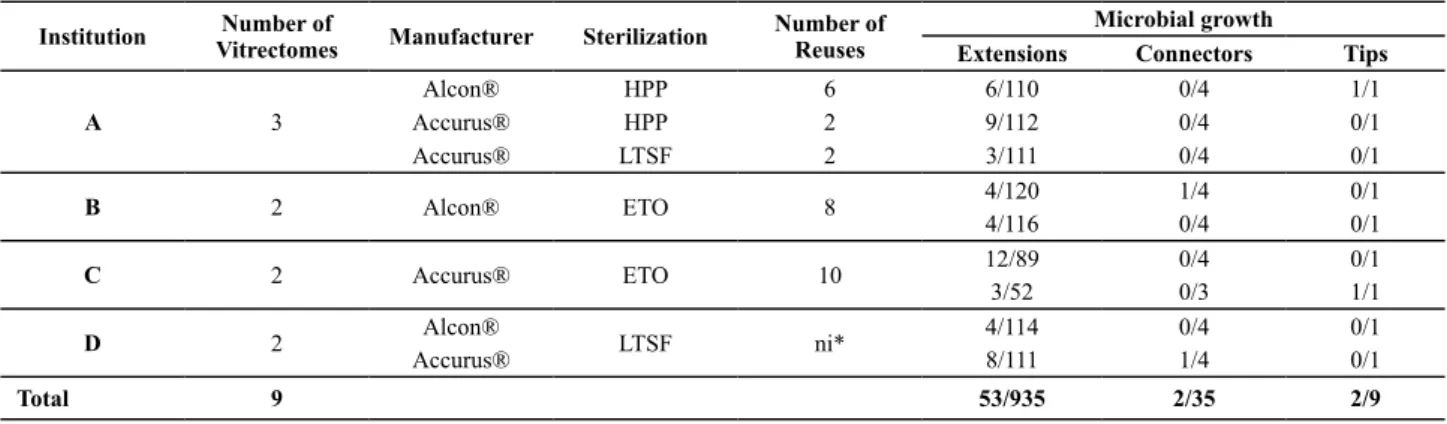
The nine reprocessed vitrectomy probes revealed microbial growth on some of their extensions, con -nectors and tips, according to the data displayed in Table 1.
Table 1 – Distribution of microbial growth in vitrectomy probes reused according to the donating institution, respective sterilization methods and number of reuses - São Paulo, 2009
* not informed
Institution VitrectomesNumber of Manufacturer Sterilization Number of Reuses Microbial growth
Extensions Connectors Tips
A 3
Alcon® HPP 6 6/110 0/4 1/1
Accurus® HPP 2 9/112 0/4 0/1
Accurus® LTSF 2 3/111 0/4 0/1
B 2 Alcon® ETO 8 4/120 1/4 0/1
4/116 0/4 0/1
C 2 Accurus® ETO 10 12/89 0/4 0/1
3/52 0/3 1/1
D 2 Alcon® LTSF ni* 4/114 0/4 0/1
Accurus® 8/111 1/4 0/1
Total 9 53/935 2/35 2/9
Fity-seven posiive samples were recovered from 979 sampling units. Among the 57 (5.8%) posiive cultures, 53 came from extension segments and two from connectors. Two out of nine ips were contaminated.
In the comparison of the sterilization methods used for reprocessing the vitrectomy probes, the chi-square test (χ2), whose result was 0.9951, proves that there was no
statistically signiicant difference among the three steril -ization methods used to reprocess the probes, which were: ETO, HPP, LTSF.
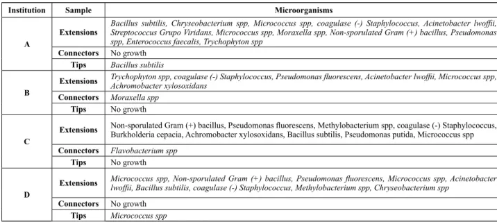
As for the recovered microorganisms, data are dis -played in Figure 2 and Table 2.
Seventeen microbial species were isolated. In some sample, more than one microorganism was found. Micro
-scopic analysis revealed 16 bacterial and one fungal spe -cies, with 7 Gram-posiive, 9 Gram-negaive bacteria and one fungus, according to the data displayed in Table 2.
No microbial cultures were found in negaive control sampling units (0/126).
diScUSSion
Most microorganisms found in this study belong to the skin and mucosa microbiota, such as Micrococcus and co -agulase negaive Staphylococcus, the later coinciding with
the ophthalmologic infecion agents appointed in literature.
as contaminaion sources and that adequate cleaning and sterilizaion methods should guarantee their eliminaion. Efecive internal channel cleaning of vitrectomy probes is very diicult, due to their long extension (215 cm) and narrow diameter (1.5 mm). Therefore, manual methods, without fricion of their surfaces, as found in pracice, are insuicient. Cleaning any health product with these char -acterisics represents the main challenge for professionals at processing units. The cleaning technique for this material shape should involve disassembly of the product, exposure to detergent, manual mechanic and complementary auto -mated acion (ultrasonic washer with retro-lux), rinsing, drying and visual inspecion(13). In this study, the devices were not submited to this cleaning standard. In all cases, only manual cleaning occurred, without disassembly of the device, as the material does permit this; without internal fricion, using brushes, immersion in enzymaic detergent, rinsing and drying, which one of the insituions that donat -ed the samples did not perform. In the processing of these devices, drying is a step that cannot be neglected, mainly in materials submited to sterilizaion in ETO. Both ETO and its sub-products ethylene chlorohydrin and ethylene glycol are extremely irritaing to issue. Ethylene glycol is a sub-prod -uct that slowly results from the reacion between ETO and water(14). Thus, in materials that are not dried adequately, the inal quanity of this substance could represent an ad -diional risk when reprocessing the devices.
In this study, risk severity was expressed by the recov -ery of vegetaive and not just sporulaing microorganisms
(Bacillus subilis), indicaing evident cleaning and steriliza
-ion laws. As opposed to the heat steriliza-ion method, in which the sterilizing agent is conducted, cold methods Chart 2 – Distribution of microorganisms identiied on the vitrectomy probes reused according to the donating institution and analyzed parts - São Paulo, 2009
Institution Sample Microorganisms
A
Extensions
Bacillus subtilis, Chryseobacterium spp, Micrococcus spp, coagulase (-) Staphylococcus, Acinetobacter lwofii,
Streptococcus Grupo Viridans, Micrococcus spp, Moraxella spp, Non-sporulated Gram (+) bacillus, Pseudomonas spp, Enterococcus faecalis, Trychophyton spp
Connectors No growth
Tips Bacillus subtilis
B
Extensions Trychophyton spp, coagulase (-) Staphylococcus, Pseudomonas luorescens, Acinetobacter lwofii, Micrococcus spp, Achromobacter xylosoxidans Connectors Moraxella spp
Tips No growth
C
Extensions Non-sporulated Gram (+) bacillus, Pseudomonas luorescens, Methylobacterium spp, coagulase (-) Staphylococcus, Burkholderia cepacia, Achromobacter xylosoxidans, Bacillus subtilis, Pseudomonas putida, Micrococcus spp
Connectors Flavobacterium spp
Tips No growth
D
Extensions Micrococcus spp, Non-sporulated Gram (+) bacillus, Pseudomonas luorescens, Micrococcus spp, Acinetobacter lwofii, Bacillus subtilis, coagulase (-) Staphylococcus, Methylobacterium spp, Chryseobacterium spp
Connectors No growth
Tips Micrococcus spp
Table 2 – Distribution of microorganisms identiied in vitrectomy probes reused in decreasing order of growth frequency per micro-bial group - São Paulo, 2009
Microorganism Number of
samples %
Gram Positive
Micrococcus spp 14 20.9
Coagulase negative Staphylococcus 8 11.9
Bacillussubtilis 6 9.0
Non-sporulated Gram positive bacilli 6 9.0
Streptococcus Grupo Viridans 4 6.0
Other Gram positive cocci 2 3.0
Enterococcusfaecalis 1 1.5
Gram Negative
Pseudomonas spp 8 12.0
Acinetobacter lwofii 3 4.5
Chryseobacterium spp 3 4.5
Achromobacterxylosoxidans 2 3.0
Methylobacterium spp 2 3.0
Moraxella spp 2 3.0
Burkholderia cepacia 1 1.4
Flavobacterium spp 1 1.4
Gram negative cocci 1 1.4
Fungi
Trychophyton spp 3 4.5
A study(15) that explored the presence of organic resi -dues on surgical instruments submited to immersion in enzymaic soluion for 60 minutes, mechanical cleaning during the immersion, ultrasonic washing for 10 minutes, rinsing three imes in tap water, lubricaion, inspecion of funcioning, drying with compressed air and verical posi -ioning for inal drying, detected the presence of residues in 84.3% (27/32), through visual microscope inspecion. Based on these study results, the authors conclude that, even when using cleaning protocols to reprocess the ma -terials, it is very hard to completely remove the residues. Considering the reprocessing of vitrectomy probes, which were submited to a cleaning process that does not com -ply with best-pracice recommendaions, it is highlighted that health establishments should take great cauion when making decisions on reprocessing and re-using dis -posable materials.
This research was limited to the assessment of po -tenial infecion risks, but other po-tenial risks are asso -ciated with the reuse of disposable materials, described in literature, which go beyond infecions, also evidencing pyrogenic reacions, adverse events deriving from toxic residues of processing products, funcional performance errors and damage to the material’s physical integrity(16).
One very relevant aspect in the processing of health -care materials is the quality of rinsing water, which can be a source of endotoxins (when contaminated with Gram-negaive microorganisms) and other organic and inorganic residues that can jeopardize processing safety(17). Oicial recommendaions indicate water treated through reverse osmosis, bacteria ilter, and disilled water for the inal rinsing. None of the insituions that donated the devices for this research menioned this care. Concerning mate -rial used in ophthalmologic surgeries, speciic recom -mendaions exist for rinsing, which should be performed abundantly to remove detergent residues, while sterilized disilled water should be used for the inal rinsing(18). This recommendaion is based on reports of post-surgical toxic eye syndromes, which the American Society of Cataract and Refracive Surgery – ASCRS - e a American Society of Ophthalmic Registered Nurses – ASORN(18) deined as TASS – Toxic Anterior Segment Syndrome.
The sterilizaion technologies that use low tempera -tures are indicated to sterilize thermosensiive materials. Since the 1950’s, ETO has been used, while HPP and LTSF are relaively new methods. Research on the later dates back to the 1990’s and methods difer in terms of difus -ibility power. In a way, the availability of these technolo -gies favored the choice to reuse disposable materials with
thermosensiive raw material, in view of the possibility to reduce hospital costs(19-20). The materials used in this study are thermosensiive, but include narrow and long lumens, which represent a challenge to difuse the steril -izing agent. One of the sterilizaion methods, used at one of the donaing insituions, HPP, is incompaible with the long extension of the material under analysis (215 cm).
These study results suggest the possibility of bioilm development on the internal surface of the devices. It is known that bioilm developed on health products can in -clude Gram-posiive (Enterococcus faecalis, Staphylococcus
aureus, Sthapylococcus epidermidis and Streptococcus
viri-dans), Gram-negaive bacteria (Escherichia coli, Klebsiella
pneumoniae, Proteus mirabilis and Pseudomonas
aerugi-nosa) or fungi(21). These microorganisms can come from
paients and health professionals’ skin, from tap water or other environmental sources(21). Many of the microorgan -isms described in bioilm composiions are in accordance with those ideniied in this study (Enterococcusfaecalis,
coagulase-negaive Staphylococcus, Streptococcusviridans
and Pseudomonasaeruginosa). As the vitrectomy probes
analyzed in this experiment were reused muliple imes, between two and ten imes, and without a validated clean -ing protocol, the recovered microorganisms may have origi -nated in bioilms developed in the narrow and long lumens of the device extensions, and also in connectors and ips.
The present study results leave no doubt as to the risk of reprocessing disposable vitrectomy probes, as per -formed by the insituions that donated the analyzed de -vices. The processing steps they adopted demonstrated deiciencies to guarantee material sterility, and no tests were performed to validate pracice, in compliance with current legislaion, Resoluion No. 2606(22). No studies were found in literature that assessed the technical feasi -bility of re-using vitrectomy probes and it is to be doubted whether favorable results would be achieved in view of the complexity of the device.
In the universe of disposable materials, some could be reused as their shape is simple, without internal spaces, funcioning like new ater reprocessing. According to the authors, the characterisics that would represent safe re -use do not apply to vitrectomy probes.
concLUSion
REFEREncES
1. Centro Cochrane do Brasil. Segurança e eicácia dos cateteres de eletroisiologia. São Paulo; 2005.
2. Denser CPAC, Lacerda RA. Reprocessamento e reuilização de material odonto-médico-hospitalar de uso único: busca de evidências pela revisão sistemáica de literatura cieníica. Acta Paul Enferm. 2006;19(3):316-22.
3. Brasil. Ministério da Saúde; Agência Nacional de Vigilância Sanitária (ANVISA). Portaria n. 4, de 7 de fevereiro de 1986. Dispõe sobre a normalização do uso e reuilização de mate
-riais médico-hospitalares descartáveis no país [Internet]. Bra
-sília; 1986 [citado 2003 ago. 4]. Disponível em: htp://www. anvisa.gov.br/legis/portarias/04_86.htm
4. Ribeiro SMCP. Cenário do reprocessamento de materiais de uso único: situação no Brasil e no mundo. In: Padoveze MC, Borrasca VL, organizadoras. Reprocessamento de materiais de uso único. São Paulo: APECIH; 2008. p. 1-6.
5. Hatch WV, Cernat G, Wong D, Devenyi R, Bell CM. Risk factors for acute endophthalmiis ater cataract surgery: a popula
-ion-based study. Ophthalmology. 2009;116(3):425-30.
6. Shimada H, Nakashizuka H, Hatori T, Mori R, Mizutani Y, Yu
-zawa M. Incidence of endophthalmiis ater 20- and 25-gauge vitrectomy. Ophthalmology. 2008;115(12): 2215-20.
7. Joondeph BC, Blanc JP, Polkinghorne PJ. Endophthalmiis af
-ter pars plana vitrectomy: a New Zeland experience. Reina. 2005;25(5):587-9.
8. Garcia-Sáenz MC, Arias-Puente A, Rodríguez-Caravaca G, Ba
-ñuelos JB. Efeciveness of intracameral cefuroxime in pre
-vening endophthalmiis ater cataract surgery Ten-year com
-paraive study. J Cataract Refract Surg. 2010;36(2):203-7.
9. Al-Mezaine HS, Kangave D, Al-Assiri A, Al-Rajhi AA. Acute-onset nosocomial endophthalmiis ater cataract surgery: incidence, clinical features, causaive organisms, and visual outcomes. J Cataract Refract Surg. 2009;35(4):643-9.
10. Wejde G, Samolov B, Seregard S, Koranyi G, Montan PG. Risk factors for endophthalmiis following cataract surgery: a ret
-rospecive case-control study. J Hosp Infect. 2005;61(3):251-6.
11. Endophthalmiis Study Group, European Society of Cataract & Refracive Surgeons. Prophylaxis of postoperaive endo
-phthalmiis following cataract surgery: results of the ESCRS mulicenter study and ideniicaion of risk factors. J Cata
-ract Ref-ract Surg. 2007;33(6):978-88.
12. Polit DF. Fundamentos da pesquisa em enfermagem: mé
-13. Graziano KU. Processo de limpeza, desinfecção e esteriliza
-ção de arigos odonto-médico-hospitalares e cuidados com o ambiente de centro cirúrgico. In: Lacerda RA, coordena
-dora. Controle de infecção em centro cirúrgico: fatos, mitos e controvérsias. São Paulo: Atheneu; 2003. p. 163-95.
14. Nogueira MH, Avelar MCQ, Barreto HHC, Cardoso DPA. Resíduos tóxicos em canulas e seringas esterilizadas e re
-esterilizadas com óxido de eileno. Rev Inst Adolfo Lutz. 1989;49(1):27-34.
15. DesCôteaux JG, Poulin EC, Julien M, Guidoin R. Residual organic debris on processed surgical instruments. AORN J. 1995;62(1):23-30.
16. Greene VW. Reuse of medical devices labeled for single-use. In: Mayhall CG. Hospital epidemiology and infecion control. 3ª ed. Philadelphia: Lippincot William & Wilkins; 2004. p. 1535-45.
17. Graziano KU, Lacerda RA, Turrini RNT, Bruna CQM, Silva CPR, Schmit C, et al. Indicators for evaluaion of process
-ing dental-medical-hospital supplies: elaboraion and vali
-daion. Rev Esc Enferm USP [Internet]. 2009 [cited 2010 jul. 22];43(n.esp 2)):1174-80. Available from: htp://www.sci
-elo.br/pdf/reeusp/v43nspe2/en_a05v43s2.pdf
18. Hellinger WC, Hasan SA, Bacalis LP, Thornblom DM, Beck
-mann SC. Outbreak of toxic anterior segment syndrome following cataract surgery associated with impuriies in autoclave steam moisture. Infect Control Hosp Epidemiol. 2006;27(3):294-8.
19. Goveia VR, Pinheiro SMC, Graziano KU. Métodos de ester
-ilização por baixa-temperatura e novas tecnologias. Rev La
-ino Am Enferm. 2007;15(3):373-6.
20. Neto SB, Graziano KU, Padoveze MC, Kawagoe JY. Eicácia da esterilização de canetas de bisturi elétrico de uso único re
-processadas. Rev Laino Am Enferm. 2010; 18(1):81-6.
21. Donlan RM. Bioilm formaion: a clinically relevant microbio
-logical process. Clin Infect Dis. 2001;33(8):1387-92.
22. Brasil. Ministério da Saúde; Agência Nacional de Vigilância Sanitária (ANVISA). Resolução n. 2.606, de 11 de agosto de 2006. Dispõe sobre as diretrizes para elaboração, validação e implantação de protocolos de reprocessamento de produ
-tos médicos e dá outras providências [Internet]. Brasília; 2006 [citado 2010 jul. 22]. Disponível em: htp://www.an