161
Original Article
Autoimmune and non-autoimmune thyroid dise ase s have
diffe re nt patte rns of ce llular HLA class II e xpre ssion
Service of Endocrinology and Department of Pathologic Anatomy,
School of Medical Sciences - State University of Campinas, Campinas, Brazil
Denise Engelbrecht Zantut-Wittmann, Luís Henrique Barbosa Boechat, Glauce Aparecida Pinto, Miriam Aparecida da Silva Trevisan, José VassalloINTRODUCTION
C la ss II a ntig e ns o f the huma n ma jo r histo co mpatibility co mplex (MHC), the human leuko cyte antig ens (HLA) DP, DQ and DR, are central elements in the presentatio n o f exo geno us a ntig e ns to T C D4 + lymp ho c yte s a nd the regulatio n o f immune respo nse. Expressio n o f these glyco pro teins o n the cell surface is restricted to B lymp ho c yte s, ma c ro p ha g e s, o the r a ntig e n-presenting cells and the capillary endo thelium.1 So me autho rs described aberrant HLA class II expressio n antig ens in fo llicular thyro id cells in G raves’ disease (G D)2 and Hashimo to thyro iditis (HT)3. Cell cultures fro m no rmal thyro ids o r G D cases generally express HLA-DR in fo llicles near areas infiltrated by lympho cytes, when stimulated by
γ
-interfero n.2 -4 These cells pro bably present self-antig ens to the immune system and perpetuate the auto immune pro cess.5 HLA class II expressio n has also been o bserved in so me no rmal thyro ids, a to xic multino d ula r g o ite r (A M G ), thyro id adeno mas and carcino mas.6 ,7 As there has been no repo rt o n the to po graphical pattern o f HLA-DR expressio n in thyro cytes, the aim o f the present stud y w a s to d e ta il this a sp e c t in no rma l, auto immune and no n-auto immune thyro id tissue, using the immuno pero xidase technique.ABSTRACT
Contex t: Surface HLA-DR antigen is usually o nly expressed by antigen-presenting cells (APC). In auto immune thyro id disease, fo llicle cells functio n as APC, thus expressing HLA-DR. Ho wever, no n-auto immune thyro id diseases may also express surface class II antigens.
Objective: To evaluate the presence and pattern o f HLA class II expressio n in auto immune and no n-auto immune thyro id diso rders.
Design: Retro spective: histo patho lo gical and immuno histo chemical analysis.
Loca tion: Referral center, university ho spital.
Sa mple: Ten histo lo g ically no rmal thyro ids, 1 1 G raves’ disease, 7 Hashimo to ’s thyro iditis, 1 0 ato xic multino dular g o iter and 3 to xic adeno mas were analyzed by
immuno histo chemistry, using a mo no clo nal antibo dy anti-HLA-DR.
M ain M easurements: The presence of these antigens in thyroid follicular cells and their relation to inflammatory infiltrate was evaluated. The pattern of HLA-DR expression in thyroid follicular cells was analyzed: membrane, cytoplasmic or both.
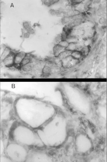
Results: Altho ugh HLA-DR antigens were sparsely present in o ne o f the 8 no rmal thyro ids, in 6 o f the 9 ato xic multino dular go iter and in 2 o f the 3 to xic adeno mas a net po sitivity co uld be seen in large areas. In all 5 Hashimo to ’s thyro iditis and in 7 o f the 1 0 G raves’ disease cases. This expressio n o ccurred in fo llicle cells either in co ntact with inflammato ry cells o r no t. In no n-auto immune thyro id disease, HLA-DR po sitivity was essentially cyto plasmic, whereas in G raves’ disease and Hashimo to thyro iditis it was mainly in cell membranes.
Conclusions: It is suggested that the HLA class II expressio n o n the surface o f fo llicle cells co uld be related to auto -antigen presentatio n to the immune system by these cells, leading to inflammatio n.
Key-w ords: G raves’ disease. Auto immune thyro iditis. HLA-DR antigens. Immuno histo chemistry
162
METHODS
Thyro id surgical specimens fro m 8 no rmal individuals, 1 0 with G D, 5 with HT, 9 with AMG and 3 with to xic adeno ma (TA) were fixed in 1 0 % fo rma lin, emb edded in pa ra ffin, sta ined with he ma to xylin a nd e o sin a nd se le c te d fo r immuno histo c he mic a l te c hniq ue s. Sp e c ific immuno staining was perfo rmed to identify HLA-DR using a mo dified streptavidin-bio tin-pero xidase te c hniq ue .8 Mo no c lo na l prima ry a nti-HLA-DR antibo dy (Dako ), was diluted at 1 :1 0 in 0 .1 % BSA. Detectio n o f the streptavidin-bio tin-pero xidase co mplex was achieved with diamino benzidine and slight co unterstaining with hemato xylin. At the end o f the reactio n, slides were mo unted with Entellan (Merck). Immuno stained sectio ns were sco red fo r the expressio n o f HLA-DR using the fo llo wing criteria: a) presence o f HLA-DR po sitive fo llicular cells in co ntact with inflammato ry infiltrate; b) presence of DR positive follicular cells in the absence o f inflammato ry infiltrate; c) presence o f DR po sitive inflammato ry infiltrate and d) presence o f DR po sitive inflammato ry infiltrate in the absence o f DR po sitive fo llicular cells.
The p a tte rn o f HLA -DR e xp re ssio n in fo llicular cells was classified as: 1 ) restricted to the cell membrane; 2 ) restricted to the cyto plasm and 3 ) in bo th, cell membrane and cyto plasm.
RESULTS
HLA-DR expressio n in o ne o f the 8 no rmal thyro id tissue cases, 6 o f the 9 AMG and 2 o f the
3 TA was restricted to small scattered gro ups o f fo llicular cells. Ho wever, in 7 o f the 1 0 G D cases and especially in all 5 HT cases, expressio n was mo re diffuse and extensive, generally o bserved in clo se relatio n to a lympho plasmacytic infiltrate. In additio n, in 6 G D and 4 HT cases, po sitive fo llicles were also fo und o utside these areas.
The p a tte rn o f HLA -DR e xp re ssio n in fo llicular cells in AMG (5 o f 6 ) and in TA (2 o f 2 ) co uld o nly be verified restricted to the cyto plasm. In o ne no rma l thyro id tha t presented a sma ll gro up o f DR po sitive fo llicular cells, the expressio n was in the membrane. In G D, HLA-DR was always expressed in the membrane (7 o f 7 ) and in 2 cases it was also o bserved in the cyto plasm. In 5 o f the 5 HT c a ses, the po sitive memb ra ne staining was mo re intense and was acco mpanied by cyto plasmic expressio n in 4 o f the 5 . (Fig ure 1 , Table 1 )
DISCUSSION
This study has sho wn that vario us benig n thyro id diseases may express HLA class II, in a g reement with o ther a utho rs.6 Ho wever, the p a tte rn o f e xp re ssio n w a s d iffe re nt w he n a uto immune a nd no n-a uto immune thyro id diseases were co mpared. O nly in auto immune p ro c e sse s w a s the re sig nific a nt, e xte nsive membrane expressio n o f HLA-DR whereas in no n-auto immune pro cesses it was g enerally limited to the cyto plasm.
The genes o f the human MHC situated in the HLA regio n are related to the immune respo nse
Ta ble 1 - Pa tterns of HLA-DR ex pression in thyrocytes of norma l tissue a nd of tissue from pa tients w ith
Gra ves’ disea se, Ha shimoto’s thyroiditis, a tox ic multinodula r goiter a nd tox ic a denoma
Diag no sis N umber HLA-DR HLA-DR expressio n type
o f cases expressio n Membrane Cyto plasmic Bo th
N o rmal 8 1 1 0 0
G raves’ disease 1 0 7 5 0 2
Hashimo to ’s thyro iditis 5 5 1 0 4 Multino dular g o iter 9 6 1 5 0
To xic adeno ma 3 2 0 2 0
Tota l 3 5 2 1 8 7 6
163
to pro te in a ntig e ns. Thus, C D4 + o r C D8 + T lympho cytes o nly reco gnize peptides pro cessed by antigen-presenting cells, the macro phages, B lympho cytes, capillary endo thelium and dendritic cells, when asso ciated with pro ducts o f the MHC genes expressed o n the cell surface.9 In the 1 9 8 0 ’s the ina deq ua te expressio n o f HLA c la ss II in fo llic ula r c e lls o f G D a nd HT c a se s w a s demo nstrated, mo st frequently in areas clo se to the lympho plasmacytic infiltrate. This suggests that the se thyro c yte s c o uld p la y a ro le in the develo pment and perpetuatio n o f the auto immune disturbance.6 ,7 ,1 0 In ag reement with these data, in o ur study a significant number o f po sitive cases (1 2 o ut o f 2 1 ) exhibited areas o f fo llicular cells expressing HLA-DR in the presence o f inflammato ry cells. In 1 9 o f these 2 1 cases there was also HLA-DR e xp re ssio n in a re a s d ista nt fro m the inflammato ry infiltrate.
Sub se q ue nt re p o rts ha ve sho w n tha t fo llicular cells in AMG , TA and carcino mas also express HLA-DR, especially in relatio n to fo ci o f infla mma to ry infiltra te. Studies using prima ry cultures o f no rmal thyro id cells, G D and o ther thyro id d ise a se s, ha ve d e mo nstra te d tha t thyro cytes in auto immune diseases co uld functio n a s a ntig en-presenting c ells, whic h a ppa rently do es no t o ccur in no n-auto immune pro cesses.6 ,1 1 In o ther investig atio ns using cultures o f thyro id murine cells o r FRTL-5 , it was o bserved that these cells can express the class II MHC mo lecules, but were unable to present antig ens.1 2 ,1 3 These co nflicting results may reflect co ntaminatio n o f the thyro id cell cultures by dendritic cells, which are abundant in this tissue and are po werful antig en-presenting cells.1 4
O ur find ing s ind ic a te a fund a me nta l difference in the pattern o f HLA class II expressio n in the fo llicular cells. Thus, in no n-auto immune thyro id disea ses, suc h a s AMG a nd TA, this expressio n was almo st exclusively o bserved in cell c yto pla sm, a nd in G D a nd HT, HLA-DR wa s expressed o n the cell memb ra ne in a ll ca ses irre sp e c tive o f the p re se nc e o f c yto p la smic expressio n. The pattern o f HLA-DR expressio n o n the cell membrane in auto immune thyro id diseases c o nfirms tha t the se thyro c yte s pla y a ro le in
antigenic presentatio n. O n the o ther hand, in cells o f no n-a uto immune thyro id d ise a se s suc h expressio n may reflect a general activatio n o f the mechanisms of cell proliferation and such activation may no t result in ho rmo ne o verpro ductio n, since it was also o bserved in AMG .5 ,6 ,1 2 ,1 3 ,1 5
O ur re sults p ro vid e e vid e nc e tha t, independent o f the thyro id functio n, class II MHC p ro d uc ts a re e xp re sse d o n the surfa c e o f thyro cytes in G D and HT, and that there are clear-c ut d iffe re nclear-c e s in the p a tte rns o f HLA -DR expressio n in auto immune and no n-auto immune thyro id diseases.
Sao Paulo Med J/Rev Paul Med 1999; 117(4):161-4.
164
Sao Paulo Med J/Rev Paul Med 1999; 117(4):161-4.
REFERENCES
1- Meuer SC, Scho ssman SF, Reinherz EL. Clo nal analysis o f human
cyto to xic T Lympho cytes: T4+ and T8+ effecto r T cells reco gnize pro ducts o f different majo r histo co mpatibility co mplex regio n. Pro c Nat Acad Sci USA 1982;79:4395-9.
2- Hanafusa T, Pujo l-Bo rrel R, Chio vato L, Russel RCG, Do niach D,
Bo ttazzo GF. Aberrant expressio n o f HLA-DR antigen o n thyro cytes in Graves’ disease: relevance fo r auto immunity. Lancet 1983;2:1111-5.
3- Aichinger G, Fill H, Wick G. In situ immune co mplexes, lympho cyte subpo pulatio ns, and HLA-DR-po sitive epithelial cells in Hashimo to thyro iditis. Lab Invest1985;52:132-40.
4- Iwatani Y, Gerstein HC, ltaka M, Ro w W, Vo lpé R. Thyro cyte HLA-DR
e xp re ssio n and inte rfe ro n-gam m a p ro d uc tio n in auto im m une thyro id disease. J Clin Endo crino l Metab 1986;63:695-708.
5- Faure GC, Benso ussan-Lyzero wicz D, Bene MC, Aubert V, Leclere J.
Co expressio n o f CD40 and class II antigen HLA-DR in Graves’ disease thyro id epithelial cells. Clin Immuno l Immuno patho l 1997;84:212-5.
6- Grubeck-Lo ebenstein B, Lo ndei M, Greenall C, Pirich K,Kassai H,
Waldhäusi W, Feldmann M. Patho genetic relevance o f HLA class II expressing thyro id fo llicular cells in no nto xic go iter and Graves’ disease. J Clin Invest 1988;81:1608-14.
7- Kno ll MR, Schwab M, Oestreich K, Rumstadt B, Hagmuller E. HLA
c las s II e xp re s s io n in we ll- d iffe re ntiate d thyro id c arc ino m a: co rrelatio n with clinico -patho lo gical features. J Exp Clin Cancer Res 1997;16:177-82.
8- Hsu SM, Raine L, Fanger H. Use o f avidin-bio tin-pero xidase co mplex (ABC) in immuno pero xidase techniques: A co mpariso n between ABC and unlabelled antibo dy (PAP) pro cedure. J Histo chem Cyto chem 1981;29:557-80.
9- Abbas AK, Lichtman AH, Po ber JS. The majo r histo co mpatibility
co mplex. In: Saunders WB (ed). Cellular and Mo lecular Immuno lo gy. Philadelphia; 1994:96.
10- Margo lik JB, We e tm an AP, Burm an KD. Im m uno histo c he m ic al analysis o f intrathyro idal lympho cytes in Graves’ disease: evidence o f ac tivate d T c e lls and p ro d uc tio n o f inte rfe ro n-gam m a. Clin Immuno l Immuno patho l 1988;47:208-18.
11- Pujo l- Bo rre l R, Luc as Martin A, Fo x M, To d d I, Bo ttazzo GF. Inappro priate HLA class II expressio n in a wide variety o f thyro id diseases. J Clin Invest1986;9:71-8.
12- Ebner SA, Stein ME, Minami M, Do rf ME, Stadecker MJ. Murine thyro id fo llicular cells can be induced to express class II (Ia) gene pro ducts but fail to present antigen in vitro. Cell Immuno l 1987;104:154-68.
13- Minami M, Ebner AS, Stadecker MJ, Do rf ME. The effects o f pho rbo l ester o n allo antigen presentatio n. JImmuno l 1987;138:393-400.
14- Kabel PJ, Vo o rbij JAM, De Haan M, Van der Gaag RD, Drexage HA.
Intrathyro idal dendritic cells. J Clin Endo crino l Metab 1988;66:199-207.
15- We e tm an AP. Ne w as p e c ts o f thyro id im m unity. Ho rm Re s 1997;48(suppl 4):51-4.
Denise Engelbrecht Za ntut-W ittm a nn - Do uto ra em Medicina Interna - Disciplina de Endo crino lo g ia – FCM-UN ICAMP
Luís Henrique Ba rbosa Boecha t - Mestre em Clínica Médica
Gla uce Apa recida Pinto - Do uto ra em Ciências-Departamento de Anato mia Pato ló g ica - FCM-UN ICAMP
M iria m Apa recida da Silva Trevisa n - LivreDo cente -Departamento de Anato mia Pato ló g ica - FCM-UN ICAMP
José Va ssa llo - Do uto r em Medicina - Departamento de Anato mia Pato ló g ica - FCM-UN ICAMP
Sources of Funding: N o t declared
Conflict of interest: N o t declared
La st received: 1 8 January 1 9 9 9
Accepted: 8 February 1 9 9 9
Address for correspondence:
Denise Eng elbrecht Zantut-W ittmann
Disciplina de Endo crino lo g ia - Departamento de Clínica Médica - Faculdade de Ciências Médicas da Universidade Estadual de Campinas - PO Bo x 6 1 1 1
Campinas/ SP - Brasil - CEP 1 3 0 8 3 -9 7 0 E-mail: hwittmann@ TRT1 5 .g o v.br
RESUMO
Contex to: Normalmente, apenas células apresentado ras de antígeno s expressam o HLA-DR em sua superfície. Na do ença tiro idiana auto -imune, as células fo liculares adquirem o papel de apresentar antígeno s e assim expressam o HLA-DR. Entretanto , do enças tiro idianas não auto -imunes também po dem expressar antígeno s de classe II. Objetivo: Avaliar a presença e o padrão de expressão do HLA de classe II em tiró ides de pacientes co m do enças tiro idianas auto -imunes e não auto -imunes. Tipo de estudo: Retro spectivo histo pato ló gico e imuno histo químico . Loca l: Centro de referência, ho spital universitário . Amostra: 1 0 tiró ides histo lo gicamente no rmais, 1 1 co m do ença de G raves, 7 co m tiro idite de Hashimo to , 1 0 co m bó cio multino dular ató xico e 3 co m adeno ma tó xico fo ram analisadas através de imuno histo química usando um antico rpo mo no clo nal anti-HLA-DR. Va riá veis estuda da s: Avaliamo s a presença desses antígeno s na célula fo licular tiro idiana, sua relação co m o infiltrado inflamató rio e o padrão de expressão do HLA-DR nas células fo liculares, se presente na membrana, no cito plasma o u em ambo s. Resulta dos: A expressão do HLA-DR o co rreu fo calmente em uma das 8 tiró ides no rmais, 6 do s 9 caso s de bó cio multino dular ató xico e em 2 do s 3 adeno mas tó xico s. Entretanto , fo i nítida sua po sitividade em áreas extensas de células fo liculares tiro idianas no s 5 caso s de tiro idite de Hashimo to e, em meno r grau, em 7 do s 1 0 caso s de do ença de G raves, quer em asso ciação ao infiltrado inflamató rio , lo nge dele o u apenas nas células linfó ides. Nas do enças tiro idianas não auto -imunes, o padrão de expressão do HLA-DR fo i