Initial peripherally inserted central
catheter tip position in neonates
** Extracted from the thesis “Procedimento de inserção, manutenção e remoção do cateter central de inserção periférica em neonatos”, School of Nursing, University of São Paulo, 2007. 1 Master’s student in the Graduate Program of School of Nursing, University of São Paulo, Nurse at the maternity wing of Hospital das Clínicas of Faculty of Medicine, University of São Paulo. São Paulo, SP, Brazil. patrí[email protected] 2 Nurse, PhD. Professor of the Maternal-Infant and Psychiatric Nursing Department at School of Nursing, University of São Paulo (EEUSP). São Paulo, SP, Brazil. [email protected] 3 Nurse, PhD. Head Nurse of the maternity wing of Hospital das Clínicas da Faculty of Medicine, University of São Paulo. São Paulo, SP, Brazil. [email protected] 4 Nurse, PhD. Professor of the Maternal-Infant and Psychiatric Nursing Department at School of Nursing, University of São Paulo. São Paulo, SP, Brazil.
O
RIGINAL
A
R
TICLE
LOCALIZAÇÃO INICIAL DA PONTA DE CATETER CENTRAL DE INSERÇÃO PERIFÉRICA (PICC) EM RECÉM-NASCIDOS
LOCALIZACIÓN INICIAL DE LA PUNTA DEL CATÉTER CENTRAL DE INSERCIÓN PERIFÉRICA (PICC) EN RECIÉN NACIDOS
RESUMO
Estudo transversal com coleta prospectiva de dados, que objetivouidentificar o posi-cionamento inicial da ponta do cateter cen-tral de inserção periférica (PICC) e verificar a prevalência de sucesso de sua inserção em neonatos. Os dados foram coletados no berçário anexo à maternidade do Hospital das Clínicas da Faculdade de Medicina da Universidade de São Paulo, entre março e setembro de 2006. Dos 37 neonatos sub-metidos à inserção do cateter PICC, a taxa de sucesso no procedimento foi de 72,3% (27 neonatos); destes, quatro (14,8%) es-tavam com as pontas dos cateteres aloja-das nas veias axilar ou inominada; outros três (11,1%), alojadas em veia jugular. Es-tes cateteres foram removidos por desvio de trajeto. 13 (48,2%) estavam com as pon-tas alojadas em átrio direito, cujos catete-res foram tracionados para reposiciona-mento da ponta para a veia cava superior.
DESCRITORES Cateterismo periférico. Veias cavas.
Recém-nascido. Prematuro.
Patrícia Ponce de Camargo1, Amélia Fumiko Kimura2, Edi Toma3, Maria Alice Tsunechiro4
ABSTRACT
This is a cross-sectional study aiming to identify the initial tip position of peripher-ally inserted central catheters (PICC) and to verify the prevalence of success in insert-ing such catheters in neonates. The study was carried out in the neonatal care unit of Hospital das Clínicas, Universidade de São Paulo. Data were collected prospectively from March to September 2006. 37 neo-nates underwent PICC insertion were in-cluded in the study. The rate of success for this procedure was 72.3% (27 neonates). Of them, four (14.8%) had the catheter tips placed in the axilary or inominate veins. Three others (11.1%) had them placed in a jugular vein. When these catheters were removed, 13 (48.2%) catheter tip were placed in the right atrium, and they were relocated to the superior vena cava.
KEY WORDS
Catheterization, peripheral. Venae cavae.
Infant, newborn. Infant, premature.
RESUMEN
Estudio transversal con recolección pros-pectiva de datos. La finalidad fue identifi-car la posición inicial de la punta del caté-ter central de inserción periférica (PICC) y verificar la prevalencia de éxitos durante su introducción en neonatos. Los datos fueron recolectados en un servicio de neonatología anexo a la maternidad del Hospital de las Clínicas de la Facultad de Medicina de la Universidad de São Paulo, entre marzo y setiembre del 2006. De los 37 neonatos sometidos a introducción del catéter PICC, la tasa de éxito fue de 72.3% (27 neonatos), de ellos, cuatro (14.8%) estaban con las puntas de los catéteres alojadas en las ve-nas axilar o no determinada, tres (11.1%) localizadas en la vena yugular. Siendo es-tos últimos retirados por desviación en su trayecto. El 48.2% (13) se encontraba con las puntas en el atrio derecho, siendo estos catéteres nuevamente posicionados en la vena cava superior.
DESCRIPTORES Cateterismo periférico. Venas cavas.
INTRODUCTION
Peripherally inserted central catheters (PICC) have been increasingly used in care delivery to critical patients at neo-natal intensive care units (NICU), particularly to preterm
newborns with very low weight(1-3).
PICC allows for the maintenance of a venous access for long periods and the safe infusion of medication, hyper-tonic solutions and total parenteral nutrition (TPN) in cen-tral veins(4-5).
PICC catheters are more commonly used in intensive care units, with nurses as the professionals in charge of their insertion. Therefore, the nurses have increasingly sought qualification to perform this action.
In Brazil, the attribution of the nurse’s technical and legal competence to perform the manipulation of the PICC catheter was defined in Resolution 258/2001 of the Federal Nursing Council.
The growth and expansion of the nursing area is
neces-sary and desirable(6). However, the
incorpo-ration of new procedures into professional practice should be monitored, so that pos-sible complications stemming from this prac-tice can be identified.
In addition to the several benefits attrib-uted to using PICC catheters, professionals need to be aware of the risks involved in the use of this device, which is associated with some complications that can occur during insertion, while the catheter moves the venous pathway, during maintenance and removal. These complications occur due to
mechanical problems like: obstruction, catheter rupture, punctured vessels, overflowing, thrombosis, hydrothorax, among others, and also infectious problems, especially
systemic sepsis related to the PICC catheter(1,5).
A successful insertion of the PICC happens when the tip of the catheter is positioned centrally, i.e. in the superior vena cava. If the tip moves beyond the superior vena cava, traction
maneuvers will be applied in the catheter for its relocation(7).
Centrally-placed catheter tips are associated with low complication rates when compared to non-central
cath-eters(2). Therefore, the maintenance of the catheter tip in a
central position is extremely important, in order to reduce the risk of complications due to the use of this device.
The PICC catheter has been widely used for administer-ing parenteral nutrition and antibiotics; however, when the tip is not placed centrally, some complications can occur,
such as thrombophlebitis, phlebitis and occlusions(8).
The migration of the PICC catheter tip is a common prob-lem and known among neonatologists, and may lead to a
lethal situation of pericardial effusion and tamponade fol-lowing myocardial puncturing. Catheter tips placed in the right atrium, or their migration into the right atrium, are
appointed as the probable causes of these complications(9).
Knowing the exact initial location of the PICC catheter tip, after its insertion, is a safety measure recommended to professionals handling PICC catheters.
The success rate of the correct initial placement of the PICC catheter tip is the key to determine the need for other maneuvers for catheter placement. The lower the success rates for the correct initial placement of the catheter tip, the higher the frequency of manipulation will be.
Therefore, each service needs to monitor its own suc-cess rate for the placement of the PICC catheter tip, in or-der to detect causal and intervenient factors of failures and implement measures to improve the success rates of this procedure.
OBJECTIVES
•
Identify the initial position of thepe-ripherally inserted central catheter tip;
•
Verify the prevalence of success in theinsertion of the peripherally inserted central catheter.
METHOD
Study design
Cross-sectional study with prospective data collection about nursing practices in the procedure of inserting the peripherally inserted central catheter in newborns hospitalized at a Neonatal Intensive Care Unit.
Study place
The research was developed at the Nursery of Hospi-tal das Clínicas, Faculty of Medicine, University of São Paulo. It has a staff of 17 nurses – seven of which are certified for the procedure of installing PICC catheters
granted by the Brazilian Society of Intensive Care
(Socie-dade Brasileira de Terapia Intensiva-SOBETI). The deci-sion of inserting the PICC catheter is made by the team delivering care to the newborn. Before the catheter in-sertion procedure is executed, the physician in charge and the nurse check the newborn’s laboratory results and assess the risks and benefits of submitting him to the procedure at that moment. The stages of the inser-tion, maintenance and removal procedures of the PICC catheter follow the recommendations adopted by the course offered by the Brazilian Society of Intensive Care Nurses.
The incorporation of new procedures into professional practice should be monitored,
so that possible complications stemming from this
Population
All PICC catheter implantation procedures performed by nurses from March to September, 2006, were included in the study. The inclusion criteria were: having consent from the newborn’s parents submitted to the PICC cath-eter insertion procedure to use information from the newborn’s medical records and consent of the nurses in-stalling PICC catheters to participate in the study.
Study Variable
The variables related to the characterization of the population of newborns (gestational age at birth; weight on the PICC insertion date; gender; clinical diagnosis; par-enteral therapy instituted through the PICC catheter) and variables related to the catheter insertion (type of mate-rial, caliber, initial placement of the catheter tip, success in the procedure of installing the PICC).
Data collection instrument
The collected data were registered on a printed form containing two parts: data on the identification and char-acteristics of the newborn and data about the PICC cath-eter insertion procedure.
Data collection Procedure
Data were obtained by consulting the newborns’ medi-cal records and observing the procedure of catheter inser-tion performed by nurses. Before the procedure was started, the newborn’s parents or guardians were asked for con-sent, as well as the nurse responsible for the insertion of the PICC catheter in the newborn. According to Resolution 196/96 of the National Health Council, an organ that regu-lates guidelines and regulatory standards for research in-volving human beings in Brazil, the research project, with the respective terms of consent (for the guardians of the newborn and for the nurses) were submitted to the analy-sis and approval of the Ethics Committee at Hospital das Clínicas at Faculty of Medicine at University of São Paulo (HCFMUSP), registered at the National Commission of
Re-search Ethics (Comissão Nacional de Ética em Pesquisa
-CONEP) - Protocol n. 00526200604 - 02/24/2006. Data col-lection started once the project had been approved by the Ethics Committee.
Data organization, treatment and analysis
Data were stored in a Microsoft Excel spreadsheet and transferred to Epi-Info software v. 3.3.2 for processing. For the quantitative variables, the averages, medians, maximum and minimum values and standard deviations were calcu-lated. The data were grouped according to absolute and relative frequency and presented in Tables.
RESULTS
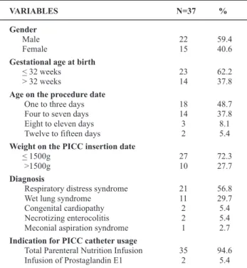
As for the characterization of the newborns submitted to the PICC catheter insertion procedure (Tables 1 and 2), it is verified that male newborns were predominant, born with up to 32 gestational weeks (average of 31.6 weeks), weigh-ing 1,500 grams or less (average weight 1,289 grams) and the catheter was inserted in the first week of the newborn’s life for the infusion of total parenteral nutrition.
Gender Male Female
Gestational age at birth < 32 weeks > 32 weeks
Age on the procedure date One to three days Four to seven days Eight to eleven days Twelve to fifteen days
Weight on the PICC insertion date < 1500g
>1500g Diagnosis
Respiratory distress syndrome Wet lung syndrome
Congenital cardiopathy Necrotizing enterocolitis Meconial aspiration syndrome Indication for PICC catheter usage
Total Parenteral Nutrition Infusion Infusion of Prostaglandin E1
22 15
23 14
18 14 3 2
27 10
21 11 2 2 1
35 2
VARIABLES N=37 %
59.4 40.6
62.2 37.8
48.7 37.8 8.1 5.4
72.3 27.7
56.8 29.7 5.4 5.4 2.7
94.6 5.4
Table 1 - Characterization of the newborns submitted to the PICC
catheter insertion procedure, HCFMUSP Nursery São Paulo -2006
Table 2 Descriptive measurements of the variables characterizing newborns submitted to PICC implantation, HCFMUSP Nursery
-São Paulo - 2006
Variable Average Median Mode Maximumvalue Minimumvalue Standarddeviation
Gestational age at birth 31.6 31.4 27.8 39.8 25.8 3.6
Weight in grams 1.289.2 1.200.0 1.040.0 2.570.0 525.0 541.5
Regarding the type of the PICC catheter material, the more rigid polyurethane catheters were used in 35 (94.6%) newborns, while the softer silicon catheters were used in only two (5.4%) of the newborns.
All catheters had a 2 French caliber, with lengths vary-ing from 48 to 50 centimeters.
Of the 37 newborns submitted to the procedure of PICC catheter installation, the catheter was successfully inserted in 72.3% (27 newborns). In 20.7% (ten newborns), the pro-cess resulted in failure related to venous puncturing, the non-progression of the catheter or other reasons.
Before the insertion of the PICC catheter, its length is measured, considering the superior vena cava as the place of insertion. However, during the catheter insertion proce-dure, the tip can progress towards an unplanned place, being misplaced into another venous branching or even in a place behind or beyond what was planned. Table 3 shows the initial positioning of the PICC catheter tip identified through radiographic images.
Table 3 – Initial position of the PICC catheter tip, HCFMUSP
nursery - São Paulo - 2006
Initial position of the
PICC catheter tip N %
Non-central
Right atrium 13 48.2
Axillary vein or innominate vein 4 14.8
Jugular vein 3 11.1
Central
Superior vena cava 7 25.9
Total 27 100.0
Data in Table 3 show that the catheter tips were placed in the superior vena cava for only seven (25.9%) newborns. Of the 27 inserted catheters, four (14.8%) had their tips in peripheral regions, i.e., placed in axillary or innominate veins; three others (11.1%) had their tips placed in jugular veins and were removed by pathway deviation. It should be noted that radiologic images showed that the catheter tips were placed in the right atrium in 13 (48.2%) newborns, which leads to the conclusion that the measurement of the catheter length from the place of insertion into the supe-rior vena cava had been overestimated, since the tip went beyond the superior vena cava and was placed into the right atrium, needing traction maneuvers to relocate it to the correct position. Table 4 presents data about the placement of catheter tips in newborns subject to traction.
Table 4 – PICC catheter tip location after traction maneuver,
HCFMUSP Nursery, São Paulo - 2006
Post-traction catheter tip location
Lower third of vena cava superior Middle third of vena cava superior Upper third of vena cava superior
Total
9 3 1
13
N %
69.2 23.1 7.7
100.0
Out of 27 catheters inserted, 24 were fixated with their tips placed centrally or peripherally, as shown in Table 5:
Central Midline Midclavicular
PICC catheter tip
location at fixation N %
Total 24 100.0
20 3 1
83.3 12.5 4.2
Table 5 - PICC catheter tip location at the moment of fixation,
HCFMUSP Nursery, São Paulo - 2006
Data in Table 5 show that the tips were centrally relo-cated and fixated in the superior vena cava in the catheters submitted to traction maneuvers.
It is observed that the prevalence of central catheters was 83.3%, while 16.7% were kept in peripheral locations.
DISCUSSION
According to the characterization data of the popula-tion, newborns submitted to PICC catheter insertion are mostly low-weight preterms who need this device to as-sure their growth and development, since the organs re-lated to suction and nutrition are not fully developed yet.
Prematurity is one of the main causes of hospitaliza-tion in neonatal units, accountable for high rates of
mor-bidity and death in the perinatal period(10).
Having central vascular access in newborns hospitalized in neonatal intensive care units, especially preterm new-borns requiring parenteral feeding for long hospitalization periods, is a fundamental measure for the survival and
re-covery of these neonates(1).
A prospective cohort study verified the incidence and location of the PICC catheter tip in newborns admitted at the Neonatal Intensive Care Unit (NICU) in a hospital in Saudi Arabia from 2002 to 2004. The average gestational age of the newborns in that population was 27.7 weeks, and av-erage weight was 1,040 grams. Avav-erage age of the neonate
at the date of catheter insertion was 12.6 days(3). The
pro-files of the newborns submitted to PICC catheter insertion are similar, with a predominance of low-weight preterm newborns.
The clinical diagnosis prevailing in newborns submitted to PICC was the Respiratory Distress Syndrome (RDS), or hyaline membrane disease (HMD), with 56.8% (Table 1). RDS affects mostly premature neonates, weighing between 501 and 1,500 grams. Prematurity, male gender and
peri-natal asphyxia are risk factors for RDS(11).
cava thrombosis compared to polyurethane and silicon; higher incidence of thrombophlebitis in the group of new-borns using silicon catheters than polyurethane catheters; lower resistance of silicon catheters when compared to the polyurethane catheters, which in turn present higher fre-quency of fractures; higher bacteremia rates in Teflon and polyethylene catheters. Through the study, advantages and
disadvantages were seen in each type of catheter(12). These
data corroborate those of the present study, which identi-fied the usage of both polyurethane and silicon catheters, with a predominance of the former, due to its lower cost than silicon catheters. However, it should be noted that a 20.7% failure rate in the insertion procedure leads to ques-tioning on whether the type of catheter material could be related to successful insertions of the PICC catheter or not.
Reasons for failure of PICC catheter insertion punctures are related to the newborn, directly and indirectly. Factors related to the neonate’s anatomy and physiology are con-sidered direct causes, and indirect causes are those related to the skill of the nurse performing the procedure.
The confirmation of the catheter tip location can be vi-sualized through injections of radiopaque contrast, thoracic
radiography and ultrasound(13).
One study found success rates for the PICC catheter implantation of 84% when 41 PICC catheter insertion pro-cedures were evaluated prospectively in newborns admit-ted at a neonatal ICU(3).
When the catheter tip is not placed correctly, it can entail serious complications. One is the cardiac arrhythmia that results when the catheter is below the right atrium or below the right ventricle(14).
The risk of thrombi formation and phlebitis increases when
the catheter tip is at the entrance of the superior vena cava(15)
Of the newborns whose catheters received traction, nine (69.2%) had their tips relocated to the lower third of
supe-rior vena cava and four (30.8%) were in the middle and upper thirds of the vena cava (Table 4).
One study about the location of the PICC catheter tip found frequencies varying from 25% to 40% in the location of the PICC catheter tip during venous puncture attempts. The tips placed in axillary, subclavian and innominate veins presented a 60%-chance of thrombosis, and 21% for the superior vena cava(16).
The length of the PICC catheter to be inserted depends on the chosen vein and limb, from 10 to 15 centimeters on the average(17).
The nurse is responsible for assisting all patients with PICC catheters. This includes checking the placement of the
catheter tip according to thoracic radiography(18).
CONCLUSION
Overall, newborns submitted to PICC catheter insertion were male, with gestational age = 32 weeks at birth, weight lower than 1,500 grams, submitted to the PICC catheter in-sertion procedure in their first week of life, indicating infu-sion of total parenteral nutrition. The prevalence of success in catheter implantation was 64.9%, and the catheter tip was placed centrally in 54.1% of the procedures, which is below the findings in literature. Incorrect initial placement of the catheter tip was related to the introduction of catheter length above the necessary, which led the nurses to perform ex-traction maneuvers to relocate the tip. This data points to the need to review the techniques used to measure catheter length. Another point worth noting is the need for new stud-ies, comparing the success rates of polyurethane catheter insertion with silicon catheters, since the success prevalence rate in the catheter insertion found in this study was lower than those in literature. Studies using silicon catheters are predominant in literature, while the use of polyurethane catheters was predominant in this study.
REFERENCES
1. Sastre JBL, Colomer BF, Cotallo GDC, Aparício AR. Estudio prospectivo sobre catéteres epicutáneos en neonatos. An Esp Pediatr. 2000;53(2):138-47.
2. Racadio JM, Doellman DA, Johnson ND, Bean JA, Jacobs BR. Pediatric peripherally inserted central catheters: complication rates related to catheter tip location. Pediatrics. 2001;107(2):E28.
3. Tawil KA, Eldemerdash A, Hathlol KA, Laimoun BA. Peripherally inserted central venous catheters in newborn infants: malpo-sitioning and spontaneous correction of catheter tips. Am J Perinatol. 2006;23(1):37-40.
4. Chlebicki MP, Teo EK. Review of peripherally inserted central catheters in the Singapore acute-care hospital. Singapore Med
5. Camara D. Minimizing risks associated with peripherally inserted cen-tral catheter in the NICU. Am J Mater Child Nurs. 2001; 26(1):17-21.
6. Toma E. Avaliação do uso do PICC – Cateter Central de Inser-ção Periférica – em recém-nascidos [tese]. São Paulo: Escola de Enfermagem, Universidade de São Paulo; 2004.
7. Fricke BL, Racadio JM, Duckworth T, Donnelly LF, Tamer RM, Johnson ND. Placement of peripherally inserted central catheters without fluoroscopy in children: initial catheter tip position. Radiology. 2005;234(3):887-92.
9. Nadroo AM, Glass RB, Lin J, Green RS, Holzman IR. Changes in upper extremity position cause migration of peripherally inserted central catheters in neonates. Pediatrics. 2002;110(1):131-6.
10. Gaíva MAM, Gomes MMF. Cuidando do neonato: uma aborda-gem de enfermaaborda-gem. Goiânia: AB; 2003. O prematuro; p. 35-41.
12. Evans M, Lentsch D. Percutaneously inserted polyurethane central catheters in the NICU: one unit’s experience. Neonatal Netw. 1999;18(6):37-46.
13. Kaye R, Sane SS, Towbin RB. Pediatric intervention: an update - part II. J Vasc Interv Radiol. 2000;11(7):807-22.
14. Geddes LAB, Nichols HA. An overview of peripherally inserted central catheters. Adv Pract Nurs J. 2005;5(3):1-9
15. Vesely TM. Central venous catheter tip position: a continuing controversy. J Vasc Interven Radiol. 2003;14(5):527-34.
16. Galloway S, Bodenham A. Long-term central venous access. Br J Anaesth. 2004;92(5):722-34.
17. Pérez EEUM, Faunes M, Avaca M, Torres V, Galleguillos J, Solis L, et al. Uso de cateter central insertado periféricamente por via percutánea em recién nacido. Pediatr Día. 2002;18(1):27-31.