• Nenhum resultado encontrado

Sao Paulo Med. J. vol.132 número1

N/A
N/A
Protected

Academic year: 2018

Share "Sao Paulo Med. J. vol.132 número1"

Copied!
8
0
0

Texto

(1)

ORIGINAL ARTICLE

DOI: 10.1590/1516-3180.2014.1321597

Follow-up of women with atypical squamous

cells cannot exclude high-grade squamous

intraepithelial lesions (ASC-H)

Acompanhamento de mulheres com células escamosas atípicas não

pode excluir lesão intraepitelial de alto grau (ASC-H)

Fanny López-Alegría

I

, Dino Soares De Lorenzi

II

, Orlando Poblete Quezada

III

Primary Healthcare Clinics, Santiago, Chile

ABSTRACT

CONTEXT AND OBJECTIVE: The concept that the presence of atypical squamous cells cannot exclude high-grade squamous intraepithelial lesions (ASC-H) was introduced in the 2001 Bethesda System of cer-vical cytology classiication. This nomenclature deines cercer-vical cancer precursor lesions. The objective of this study was to investigate the colpocytological-histological results from a three-year follow-up con-ducted on a cohort of women with reports of ASC-H who were attended during 2005-2006 at clinics of the Southern Metropolitan Healthcare Service of Santiago, Chile.

DESIGN AND SETTING: Prospective cohort study at primary healthcare clinics in Santiago, Chile. METHODS: Colpocytological-histological follow-up was conducted over a three-year period on 92 wom-en with cytological reports of ASC-H who were attwom-ended at primary healthcare clinics during 2005-2006. RESULTS: At the end of the follow-up period, high-grade lesions were evaluated and the following out-comes were observed: seven women presented invasive cancer (7.6%), 49 presented high-grade lesions (53.3%), 26 presented low-grade lesions (28.2%) and 10 presented normal results (10.9%). The “Conditional Probabilities Tree Diagram” was used to show the results from tests and the times of lesion detection. It demonstrated that, after a irst report of ASC-H, clinical management needed to be interventionist. CONCLUSION: The follow-up on our cohort of women showed that the majority of uncertain ASC-H diag-noses (82.6%) had abnormal colposcopic results and that during the follow-up using ASC-H smears, two out of every three women developed high-grade lesions.

RESUMO

CONTEXTO E OBJETIVO: O conceito de que à presença de células escamosas atípicas não se pode excluir lesão intraepitelial de alto grau (ASC-H) foi introduzido pelo Sistema de Bethesda 2001, na classiicação de citologia cervical. Esta nomenclatura deine lesões precursoras do câncer cervical. O objetivo deste estudo foi investigar os resultados colpo-cito-histológicos de três anos de acompanhamento realizado em uma coorte de mulheres com relatórios de ASC-H que receberam atendimento no período 2005-2006 em clínicas do Serviço Metropolitano de Saúde Sul de Santiago, Chile.

TIPO DE ESTUDO E LOCAL: Estudo de coorte prospectivo em unidades básicas de saúde de Santiago, Chile. MÉTODOS: Foi conduzido um acompanhamento colpo-cito-histológico por um período de três anos em 92 mulheres com laudos citológicos de ASC-H, que receberam atendimento nas unidades básicas de saúde de 2005-2006.

RESULTADOS: No inal do período de acompanhamento, as lesões de alto grau foram avaliadas e os re-sultados foram observados: sete mulheres apresentaram câncer invasivo (7,6%), 49 apresentaram lesões de alto grau (53,3%), 26 apresentaram lesões de baixo grau (28,2%) e 10 apresentaram resultados normais (10,9%). O “Diagrama de Árvore Condicional de Probabilidades” foi utilizado para mostrar os resultados dos testes e o período de detecção das lesões, demonstrando que, depois de um primeiro relatório de ASC-H, o manejo clínico deve ser intervencionista.

CONCLUSÃO: O acompanhamento de nossa coorte de mulheres mostra que a maioria dos diagnósticos incertos de ASC-H (82,6%) tiveram resultado colposcópico anormal e, durante o acompanhamento de esses esfregaços ASC-H, duas de cada três mulheres desenvolvem lesões de alto grau.

IPhD. Professor, Department of Nursing, School of

Nursing, Universidad Andres Bello, Santiago, Chile.

IIMD. Professor, Department of Obstetrics and

Gynecology, Universidade de Caxias do Sul, Rio Grande do Sul, Brazil.

IIIMedical Technologist. Cytology Laboratory,

Complejo Asistencial Barros Luco, Santiago, Chile.

KEY WORDS:

Neoplasms squamous cell. Uterine cervical neoplasms. Vaginal smears.

Biopsy. Follow-up studies.

PALAVRAS-CHAVE:

(2)

OR IG IN A L A R T IC L E | López-Alegría F, De Lorenzi DS, Quezada OP

16 Sao Paulo Med J. 2014; 132(1):15-22 INTRODUCTION

he concept that the presence of atypical squamous cells can-not exclude high-grade squamous intraepithelial lesion (ASC-H) was introduced by the 2001 Bethesda System of cervical cytology classiication. his system addresses the need to deine a cytolog-ical category between atypcytolog-ical squamous cells of undetermined signiicance (ASCUS) and high-grade squamous

intraepithe-lial lesion (HSIL).1 he deinition of this nomenclature resulted

from the ASCUS/LSIL Triage Study (ALTS) carried out between 1996 and 2000, in which interpretations of ASCUS were subcat-egorized as “equivocal LSIL” (ASCUS-L) and “equivocal HSIL” (ASCUS-H). hrough a consensus among expert pathologists, it was concluded that ASCUS-H represented a cytological category

that difered from ASCUS-L and HSIL.2 ASCUS-H has been seen

to present a risk association of 27.2% with cervical cancer pre-cursor lesions, which is higher than the risk association for the ASCUS-L category of 11.4%, but lower than the risk association for HSIL of 44.8%.2,3

Subsequent studies, such as a study by Sherman et al., detected high-grade lesions (HSIL) in 41% of the women with

initial ASC-H smears.4 Supporting previous indings, Patton

obtained a high positive predictive value for lesions of cervical intraepithelial neoplasia (CIN II/III), derived from the ASC-H

diagnosis category in a general population of women.5

Bonvicino et al. used computed data from two medical cen-ters in San Antonio, Texas, United States, in which 260 women diagnosed with ASC-H underwent cytological-histological follow-up, and found that 25.4% had a high-grade lesion at the

end of the follow-up period.6 Using the same methodology in

Brazil, Cytryn et al. found high prevalence of high-grade lesions

among a total of 57 ASC-H cases.7 hus, the abovementioned

follow-up studies demonstrated that there are connections

between ASC-H smears and high-grade lesions.4,6,7

In Chile, the 2001 Bethesda System was adopted in 2005 via Ordinance Number B232 1771 issued by the Department of Non-Communicable Diseases, within the Division of Disease Prevention and Control. his ordinance standardized the use of

the nomenclature that had started to be used in previous years.8

In 2006, the National Program for Research and Control of Cervical Cancer collected 779,068 Pap smears nationwide, from which 14,608 (1.9%) were found to be atypical using the new nomenclature.9

Although other studies such as Gaete et al.10 and Yazigi et al.11

were conducted in Chile, they were limited because they did not discriminate the type of atypia given that they were conducted using cytological reports that were produced before the 2001 Bethesda System was incorporated in Chile. he scarce scientiic evidence regarding the ASC-H category among the female popu-lation of Chile motivated the present study.

OBJECTIVE

he objective of this study was to investigate the cytological-histological results from a three-year follow-up conducted on a cohort of women with reports of ASC-H who were attended during 2005-2006 at clinics within the Southern Metropolitan Healthcare Service in Santiago, Chile.

METHODS

he present research comprises a prospective cohort study on women who were followed up over a three-year period. During 2005-2006, 88,438 cervical cytological smears were collected by midwives using Ayre spatulas and cytobrushes in primary health-care clinics, in the southern area of Santiago, Chile.

hese smear screenings were ixed and sent to Barros Luco Hospital Laboratory (central hospital). In this laboratory, the cytological smears were stained by means of the conventional Pap smear technique and were classiied using the national nomen-clature system, which is equivalent to the 2001 Bethesda system.8

hese tests were processed by a team of cytotechnologists with an average of 20 years of experience. Subsequently, these records were registered in the “Cytological Expert Diagnosis Archive System” at the Chilean Ministry of Health. his provided the cytological-histological database that we searched to obtain data for the present research study.9

he ASC-H cases were selected (n = 106) and the following selection criteria were applied: the women needed to be with-out uterine pathological conditions, withwith-out prior cervical pro-cedures and with normal Pap smear results for the last three years. hese criteria resulted in a cohort of 92 women who were followed up colpocytologically-histologically for a three-year period or until their cases were resolved.

The clinical management of these women included gyne-cological examinations, cytological tests (Pap smears), col-poscopic examinations and biopsies. HPV testing was not performed due to cost constraints. All of the follow-up tests were performed by professionals at the central hospital’s Cervical Pathology Clinic.

he variables studied included the following: the women’s age at the time of ASC-H identiication, the number and type of cytological, colposcopic and histological results and the length of follow-up (months) among the women with ASC-H cytology, which ended with diagnostic conirmation, deined as diagnosis of a more severe lesion (CIN II+) or negative conirmation.

(3)

Follow-up of women with atypical squamous cells cannot exclude high-grade squamous intraepithelial lesions (ASC-H) | ORIGINAL ARTICLE

intraepithelial lesion (LSIL); high-grade squamous intraepithelial lesion (HSIL); or squamous cell carcinoma.

To deine the histological variable (biopsy), cervical intraepi-thelial neoplasia (CIN) grades were used and divided as follows: cervical intraepithelial neoplasia of low grade (CIN I); cervi-cal intraepithelial neoplasia of moderate grade (CIN II); cervicervi-cal intraepithelial neoplasia of high grade (CIN III); carcinoma in situ

(CIS); or invasive carcinoma.

For the colposcopic variable, a standard protocol was used, which included conventional visual examination, application of 5% acetic acid and identiication of the squamocolumnar junc-tion. his variable was deined as follows: negative (-) colposcopy, when colposcopic indings did not show severe lesions or the need for a biopsy; or positive (+) colposcopy, when colposcopic indings showed lesions of severity that required biopsy.

he data collected were analyzed electronically using Microsot Excel (version 2007) and the “Conditional Probabilities Tree Diagram,” which shows the number, type, outcome and time interval between patient tests.

RESULTS

Out of the 88,438 cervical exfoliative smears collected, 752 (0.85%) were atypical Pap smears. he latter contained atypical squamous cells, which were divided into atypical squamous cells of undetermined signiicance (ASC-US) (619 cytological tests, 0.69%) and atypical squamous cells that cannot exclude high-grade squamous intraepithelial lesion (ASC-H) (106 cytological tests, 0.11%) (Table 1).

Ater applying the selection criteria to the group of women with ASC-H Pap smears, the cohort consisted of 92 women, with a mean age of 38.2 years and an age range from 19 to 71 years.

he follow-up results from this cohort were illustrated using

the “Conditional Probabilities Tree Diagram” (Figure 1), which

begins with the ASC-H Pap smear (“phase 0”) and illustrates the tests performed on the women and their outcomes over time. Squares signify a biopsy and circles indicate a Pap smear.

It needs to be borne in mind that the ASC-H cytological ind-ing is deined as a suspected case (positive Pap smear) in the cer-vical cancer program. hese 92 women who were attended at pri-mary healthcare clinics were managed in accordance with the central hospital’s “management algorithm for specialist or cervical pathology clinic treatment from the time of the irst atypical Pap”, in order to make the diagnosis and/or proceed with treatment.

During the irst contact with this clinic, the patient’s history was taken, a gynecological examination was made and colpos-copy was performed. Colposcolpos-copy is considered to be the irst procedure for diagnostic conirmation in the management algo-rithm. As shown in “Phase 1”, 76 cases (82.6%) showed abnormal colposcopic indings and required biopsy for histological anal-ysis, while in 16 cases (17.4%), the initial colposcopic indings were normal and were followed up by a Pap test.

he average length of time taken for the patients to complete these medical procedures was 3.15 months, with a minimum of two days and a maximum of 19.6 months.

Subsequently, the cytological-histological results collected in the previous stage were known as “Phase 2”. For the group that started with a histological study, the results were distributed as follows: 31 biopsies showed CIN I; 35 showed CIN II/III; three showed carcinoma; and seven were negative. For the group that started with a Pap smear, three of these tests indicated HSIL, 11 were negative for neoplastic cells and two indicated ASC-US. Given these results, and in accordance with the derivation algo-rithm, a reassessment was performed and the previous proce-dures were repeated. Pap and colposcopy were obligatory, while the histological examination was made according to the col-poscopic indings.

In “Phase 3” and as a result of the repeated examinations, 12 women were found (through biopsy) to have CIN I; 34 women were found to have CIN II/III; three women were found to have carcinoma and four women were negative. In the cases with Pap smears, 38 tests were performed and most of them (33) showed normal outcomes. hese results mostly corresponded to patients

Cytological inding Result Number %

Negative cytological indings

Satisfactory 77,411 87.53

Less than optimal 4,923 5.56

Inadequate 4,145 4.68

Atypical

Atypical squamous cells of undetermined signiicance (ASCUS) 619 0.69

Atypical squamous cells cannot exclude high-grade SIL (ASC-H) 106 0.11

Atypical glandular cells of undetermined signiicance (AGC-US) 25 0.02

Atypical glandular cells suggestive of adenocarcinoma in situ 2 0.00

Positive cytological indings

Low-grade Pap 503 0.56

High-grade Pap 638 0.72

Invasive cancer 66 0.07

Total 88,438 99.94

(4)

ORIGINAL ARTICLE | López-Alegría F, De Lorenzi DS, Quezada OP

18 Sao Paulo Med J. 2014; 132(1):15-22

who had been treated by means of surgical conization consequent to the diagnostic-treatment biopsies that were performed prior to gathering material for Pap smears. Following conization, and in accordance with the Chilean Ministry of Health’s guidelines, two Pap smears must be performed, with an interval of six months, before the patient returns to the primary healthcare clinic.

In “Phase 4”, on the one hand, the outcomes from the earlier 14 biopsies are shown: four were classiied as carcinoma, six as high-risk lesions and four as low-risk lesions. On the other hand, cytological outcomes are shown, and these mostly related to patients according to the process of epidemiological monitoring.

In “Phase 5,” there were also results with high-grade lesions from a total of 20 women. In “Phases 6 and 7”, the cases were pre-dominantly monitored by means of Pap smears and there were four patients with CIN II/III and one with CIN I who was diag-nosed via biopsy.

In this diagnostic search process, 166 oncotic cytological tests were performed via conventional Pap smears, with an average of 1.8 examinations per woman and a minimum of zero and maxi-mum of ive cytological tests per woman. With regard to histo-logical examinations, the total numbers of uterine cervix biopsies guided by colposcopy was 158, with an average of 1.7 per woman, and a minimum of zero and maximum of four. he total number of cytological-histological examinations that patients required to obtain a deinitive diagnosis was as follows:

• 1 biopsy = 27 women (29.3%); • 2 biopsies = 46 women (50%); • 3 biopsies = 8 women (8.7%); • 4 biopsies = 3 women (3.3%);

• 2 Papanicolaou smears = 8 women (8.7%);

The average length of time from the ASC-H Pap smear to the final diagnosis of the most severe lesion was 5.29 months in the biopsy group and 12.26 months in the Pap smear group.

In evaluating this three-year cytological-histological follow-up period, the 92 women were found to have the following lesion distribution:

• 7 women had invasive carcinoma (7.6%); • 49 women had high-grade lesions (53.3%); • 26 women had low-grade lesions (28.2%);

• 10 women had normal cytological outcomes (10.9%).

DISCUSSION

ASC-H is a type of atypical smear characterized by follow-up that includes a range of clinical procedures to reach a deinitive diag-nosis. Knowledge of the prevalence of women carrying ASC-H is essential for estimating the proportion of women at risk of devel-oping a high-grade lesion.

One of the irst studies aimed at investigating the preva-lence of this new cytological classiication category was carried out by the College of American Pathologists in the United States in 2002-2003: this was a nationwide survey and it revealed that ASC-H represented approximately 0.2% of cytological interpre-tations.12 hese irst studies indicated that using this category was

relatively congruent with the frequency provided by the 2001

Bethesda System.1 hus, studies including this new category were

started worldwide, thereby providing ranges of ASC-H preva-lence. his prevalence ranged from 0.22% out of a total of 27,367 Pap smears performed in a screening program in India, up 8.8% out of a total of 12,188 Pap smears in South Africa.13,14

In Latin America, and speciically in Brazil, Yamamoto et al. found ASC-H smear prevalences of 0.23% in 2007 and 0.54% in 2008, out of a total of 56,179 smears collected through the

Cervical Cancer Research and Control Program.15 Within this

line of research, our study results, which demonstrated ASC-H prevalence of 0.11%, or 106 Pap smears out of a total of 88,438 between 2005 and 2006, are congruent with the established 2001 Bethesda System (Table 2).5,13-24

Biopsy Bp Cytology Pap 5 CIN I 1 CIN I 2 CIN I 1 CIN I 1 CIN I 1 CIN I 31 CIN I 7 CIN II/III 1 CIN II/III 1 CIN II/III 2 CIN II/III 1 CIN II/III 7 CIN II/III 35 CIN II/III 23 CIN II/III 1 CIN II/III 1 CIN II/III 1 CIN II/III 1 CIN II/III 1

CIN II/III CIN II/III1 CIN II/III1 1 CIN II/III 1 CIN II/III 1 CIN II/III 1 CIN II/III 2 CIN I 1 CIN I 2 CIN I 1 CIN I 1 CIN I 2 ASCUS 1 ASCUS 2 HSIL 16 Pap 1 CIN II/III 1 CIN II/III 3 CIN I 76 Bp 1 CIN II/III 1 CIN II/III 4 Neg 1 Neg 1 Neg 1 Neg 1 Neg 1 HSIL 1 HSIL 1 Neg 7 Neg 1 Neg 1 Neg 2 Neg 11 Neg 7 Neg 1 Neg 1 Neg Neg1

1 Neg 1

Neg 1 Neg Neg1 7

Neg

19

Neg ASCUS2 CA1

1 CA 1 CA 1 CA 1 CA 1 CA 1 CA 1 CA 1 CA 1 CA 3 CA 92 ASC-H 2 CA 1 Neg H11 13 Neg 14 Neg 1 HSIL 1 HSIL 1 Neg 3 Neg 3 Neg 3 Neg 6 Neg 7 Neg Phase 2 Phase 1 Phase 0 Phase 3 Phase 4 Phase 5 Phase 6 Phase 7

Neg = negative; ASCUS = atypical squamous cells of undetermined signiicance; LSIL = low grade squamous intraepithelial lesion; HSIL = high grade squamous intrapithelial lesion; CIN I = mild dysplasia; CIN II/III = moderate and severe dysplasia; CA = carcinoma.

(5)

Follow-up of women with atypical squamous cells cannot exclude high-grade squamous intraepithelial lesions (ASC-H) | ORIGINAL ARTICLE

Concerning the percentage of ASC-H among the total volume of atypical squamous cells (ASC), the 2001 Bethesda System indicates that between 5 and 10% (approximately)

should be expected.1,3 In our study, the ASC-H prevalence (106)

represented 14% of the total atypia (752 women). he national survey conducted by the College of American Pathologists (2002-2003) found that 5% of the squamous atypias

corre-sponded to ASC-H.12 In Brazil, Yamamoto et al. obtained 2,622

atypical Pap smears in 2007-2008 and 210 (0.8%) of these were

ASC-H smears.15

For the clinical management of patients with ASC-H smears, the American Society for Colposcopy and Cervical Pathology (ASCCP) published an algorithm in relation to clinical manage-ment of these patients in order to unify the criteria.25 In Chile, and

taking this algorithm into consideration, the Chilean Ministry of Health implemented its “Clinical Guidance for Cervical Cancer” (2005 and update 2010), which includes low charts for clinical decision-making, such as the “management algorithm for spe-cialists or Cervical Pathology Clinics (CPC) from the time of the irst atypical Pap smear according to the 2001 Bethesda System”

and the “algorithm of diagnostic conirmation”. In the irst algo-rithm, it is established that women with atypical smears must be managed at a cervical pathology clinic.26,27 he follow-up for

our population study was started at a cervical pathology clinic. he population’s demographic proile comprised young women (average age: 38.2 years), which was similar to the populations of several other studies, which had averages ranging from 29 to 38.1 years. It is noteworthy that these outcomes corresponded to women in the screened population (Table 3).6,13,17,18,28

Reference Place and period Total Pap tests Number of ASC-H cases Prevalence of ASC-H %

Louro et al.18 Birmingham, USA

January 2000 to December 2001 43,840 368 0.84

Selvaggi19 Wisconsin, USA

March to September, 2002 9,214 25 0.27

Duncan and Jacob22 Tennessee, USA

October 2002 to March 2004 60,390 414 0.69

Elsheikh et al.20 Indiana, USA

July 2001 to July 2003 129,911 7,698 0.24

Barreth et al.17 Alberta, Canada

January to December 2002 241,841 727 0.30

Saad et al.16  Pittsburgh, USA

January 2003 to June 2004 152,495 800 0.52

Gupta et al.13  Noida, India

January 2001 to December 2004 27,367  60 0.22

Patton et al.5  Tennessee, USA

March 2003 to December 2006 150,702 591 0.39

Okonda et al.14 Swaziland, Southern Africa

June 2004 to May 2006 12,188 1,072 8.8

Alsharif et al.21 Minnesota, USA

July 2004 to December 2007 235,645 1,219 0.52

Yamamoto et al.15 São Paulo, Brazil

2007 30,869 72 0.23

Rekhi et al.23 Mumbai, Parel, India

January 2005 to December 2008 9,190 29 0.31

Giorgi Rossi et al.24 Italy

December 2007 to September 2008 3,410 10 0.29

Present study Santiago, Chile

2005 to 2006 88,438 106 0.11

Table 2. Prevalence of atypical squamous cells that cannot exclude high-grade squamous intraepithelial lesions (ASC-H) in population screening studies

Reference Average age

Louro et al.18 36.8

Barreth et al.17 29.0

Gupta et al.13 38.1

Bonvicino et al.6 35.6

McHale et al.28 32.8

Present study 38.2

(6)

ORIGINAL ARTICLE | López-Alegría F, De Lorenzi DS, Quezada OP

20 Sao Paulo Med J. 2014; 132(1):15-22

Reference Women with ASC-H with histological

follow-up Women with CIN II or higher

Selvaggi19 22 15 women (68%)

Louro et al.18

Pap test preparation types with ASC-H: 190 conventional smear 28 liquid-based preparation (ThinPrep)

Prevalence of CIN II+ according to Pap test preparation type: Liquid-based preparations = 9 (45%)

Conventional smears = 70 (46.1%)

There was no statistically signiicant diference in the incidence of CIN II or higher on subsequent biopsies after interpretation of ASC-H based on the preparation type

Duncan and Jacob22 99 40 women (40.4%)

Barreth et al.17

Follow-up on 517 women: Histological = 454 Cytological alone = 63

Prevalence of CIN II+ or HSIL according to examination type: Histological = 363 (79.9%)

Cytological alone = 11 (17.5%)

Saad et al.16

Cytological and histological follow-up on women:

127 perimenopausal 90 postmenopausal

Prevalence of CIN II+ or HSIL according to menopausal period: perimenopausal = 28 (22.0%)

postmenopausal = 5 (6%)

Elsheikh et al.20

Comparative: Women with ASCUS = 410 Women with ASC-H = 110

Prevalence of CIN II+ according to epithelial cell abnormalities: ASCUS = 35 women (8.5%)

ASC-H = 49 women (44.6%)

Srodon et al.3

Histological follow-up on HPV (+) women

ASCUS = 266 ASC-H = 45

Prevalence of CIN II+ in HPV(+) patients: ASCUS = 27 (10.2%) ASC-H = 18 (40.0%)

McHale et al.28 Histological follow-up on 229 women

Prevalence of CIN II/III according to time:

At the initial colposcopy, 23 of the patients (10%) had histological evidence of CIN II/III.

The cumulative risk of CIN II/III was 12.2% at 12 months

Gupta et al.13

Colposcopic-histological follow-up: ASCUS = 218 women

ASC-H = 52

Prevalence of CIN II/III or higher according to epithelial cell abnormalities: ASCUS = 7 (3.2%)

ASC-H = 16 (30.8%)

Bonvicino et al.6 Histological follow-up on 122 women

Number of follow-up cervical biopsies for a deinitive diagnosis of CIN II+ First = 35 (72%)

Second = 7 (14%) Third = 4 (8%) Fourth = 3 (6%) Total: 49 women (40%) with CIN II+

Mokhtar et al.29 Histological follow-up on 123 women

Prevalence ratio of CIN II/III according to age group > 40 years = 65.1%

< 40 years = 47.5%

Patton et al.5

Histological follow-up on 195 women Postmenopausal = 89 women

Pregnant = 44 women Postpartum= 27 women Contraceptive use = 35 women

Prevalence of CIN II+: Postmenopausal: 20 women (22.5%)

Pregnant: 35 women (79.6%) Postpartum 18 women (66.7%)

Contraceptive use: 21 (60%)

The diagnosis of ASC-H in postmenopausal Pap smears has a low predictive value in stark contrast to the pregnant, postpartum, and contraceptive-use categories

Alsharif et al.21 Histological follow-up on 691 women

Prevalence of CIN II+ according to cytologic categories: LSIL = 370 (16.1%)

LSIL-H = 112 (33.1%) HSIL = 468 (69.0%) ASC-H = 182 (26.3%)

Cytryn et al.7 Histological follow-up on 57 women

Prevalence of CIN II/III according to age group of women : < 50 years = 10 (22.2%)

50 years or older = 1 (8.3%) Total: 11 women (19.3%) biopsy with CIN II+

Present study 92 Prevalence of CIN II+:

56 women (60.9%)

Table 4. Outcomes from histology follow-up of women with smears showing atypical squamous cells that cannot exclude high-grade squamous intraepithelial lesions (ASC-H)

(7)

Follow-up of women with atypical squamous cells cannot exclude high-grade squamous intraepithelial lesions (ASC-H) | ORIGINAL ARTICLE

In the present study, women with ASC-H atypia underwent on average 1.8 cytological tests per woman (0 to 5 Pap smears) and half of them (46 women) underwent a minimum of two biopsies, in order to reach the deinite CIN II+ diagnosis. It is possible to compare these outcomes with Bonvicino’s data, in which among 260 ASC-H smears, there was an average cytologi-cal-histological follow-up of 1.35 Pap smears per woman, with a range of 1 to 4, and the biopsy follow-up consisted of an average of 0.64 biopsies per woman. In other words, the majority of the women (72%) underwent only one biopsy for a diagnosis of CIN II+ to be reached.6

he average length of follow-up in the present study was 5.29 months, to reach the most severe diagnosis (CIN II+), using only the biopsy. For conirmation of this cytological-histolog-ical diagnosis, a period of 12.26 months was needed, during which two normal cytological examinations were needed, with an interval of six months between them. hese results are con-trary to those of Bonvicino, in which the CIN II/III diagnosis

required 18.5 months.6

In our study, 60.9% (56 women) had a deinitive high-grade diagnosis (CIN II+) that was veriied by biopsy. Comparing this ratio with the literature, we found that it was interme-diate between the studies by Saad et al.16 and Barreth et al.,17

which found that 6% and 79.9% of the women had high-grade

lesions, respectively. Table 4 shows 14 studies in which

difer-ent variables were analyzed. For instance, Louro et al.18 made a

comparison between conventional collection and liquid-based smears, and found that in spite of being diferent techniques for cytological sample collection, they resulted in similar

dein-itive diagnoses of CIN II.18 his is consistent with the results

obtained from the majority of the studies observed, which dem-onstrated CIN II+ prevalences of more than 40%, according to the biopsy results.3,5,6,17-24,29

Our results conirmed that at the time of the irst ASC-H report, immediate intervention with colposcopy was necessary, followed by a biopsy. his validates the medical management of ASC-H that is required by the “management algorithm from the time of the irst atypical Pap,” which is included in the Chilean Ministry of Health’s Clinical Guidelines for Cervical Cancer.

CONCLUSIONS

he follow-up of our cohort of women showed the following:

• he majority of the uncertain ASC-H diagnoses (82.6%) also

had abnormal colposcopic results.

• During the follow-up on ASC-H smears, two out of every

three women developed high-grade lesions (CIN II+), which were detected ater an average of 3.15 months.

• Detection of high-grade lesions in a female population

through screening relects the importance of public health

programs within the National Cervical Cancer Research and Control Program in Chile.

REFERENCES

1. Solomon D, Davey D, Kurman R, et al. The 2001 Bethesda System: terminology for reporting results of cervical cytology. JAMA. 2002;287(16):2114-9.

2. Sherman ME, Solomon D, Schifman M, ASCUS LSIL Triage Study Group. Qualiication of ASCUS. A comparison of equivocal LSIL and equivocal HSIL cervical cytology in the ASCUS/LSIL Triage Study. Am J Clin Pathol. 2001;116(3):386-94.

3. Srodon M, Parry Dilworth H, Ronnett BM. Atypical squamous cells, cannot exclude high-grade squamous intraepithelial lesion: diagnostic performance, human papillomavirus testing, and follow-up results. Cancer. 2006;108(1):32-8.

4. Sherman ME, Castle PE, Solomon D. Cervical cytology of atypical squamous cells-cannot exclude high-grade squamous intraepithelial lesion (ASC-H): characteristics and histologic outcomes. Cancer. 2006;108(5):298-305.

5. Patton AL, Duncan L, Bloom L, Phaneuf G, Zafar N. Atypical squamous cells, cannot exclude a high-grade intraepithelial lesion and its clinical signiicance in postmenopausal, pregnant, postpartum, and contraceptive-use patients. Cancer. 2008;114(6):481-8.

6. Bonvicino A, Huitron S, Fadare O. Papanicolaou test interpretations of “atypical squamous cells, cannot exclude high-grade squamous intraepithelial lesion’’: an investigation of requisite duration and number of colposcopic procedures to a deinitive diagnosis of high-grade dysplasia in routine practice. Cancer. 2007;111(6):477-81. 7. Cytryn A, Russomano FB, Camargo MJ, et al. Prevalence of cervical

intraepithelial neoplasia grades II/III and cervical cancer in patients with cytological diagnosis of atypical squamous cells when high-grade intraepithelial lesions (ASC-H) cannot be ruled out. Sao Paulo Med J. 2009;127(5):283-7.

8. Gobierno de Chile. Ministerio de Salud. División Prevención Y Control de Enfermedades. Depto Enfermedades no Transmisibles. Terminología para el informe citológico de muestras cervicales (PAP). Available from: http://www.redsalud.gov.cl/archivos/cancer/Informa_ cambio_Nomenclatura_2005.pdf. Accessed in 2013 (Apr 16).

9. Base de datos Cito-Expert: Consolidado nacional citologías e histologías [Acceso en línea, restringido a servicios de salud pertenecientes a la red pública de atención del Minsal, Chile]. Chile: Laboratorio Nacional de Referencia. Ministerio de Salud Chile. 10. Gaete JL, Fuhrer K, Soto R, Rojas JL. Pap atípico escamoso: ¿un falso

negativo? [Squamous atypical vaginal smears: a false negative?] Clin Cienc. 2003;1(6):13-24.

(8)

ORIGINAL ARTICLE | López-Alegría F, De Lorenzi DS, Quezada OP

22 Sao Paulo Med J. 2014; 132(1):15-22

12. Davey DD, Neal MH, Wilbur DC, et al. Bethesda 2001 implementation and reporting rates: 2003 practices of participants in the College of American Pathologists Interlaboratory Comparison Program in Cervicovaginal Cytology. Arch Pathol Lab Med. 2004;128(11):1224-9. 13. Gupta S, Sodhani P, Chachra KL, Singh V, Sehgal A. Outcome of

‘‘Atypical squamous cells’’ in a cervical cytology screening program: implications for follow up in resource limited settings. Diagn Cytopathol. 2007;35(11):677-80.

14. Okonda S, Wright C, Michelow P. The status of cervical cytology in Swaziland, Southern Africa: a descriptive study. Cytojournal. 2009;6:14.

15. Yamamoto LSU, Pereira SMM, Etlinger D, et al. Frequência de diagnóstico de lesões do colo uterino por faixa etária em mulheres atendidas no Programa de Rastreamento Viva Mulher no período de 2004 a 2008 [Review on the occurrence of uterine cervix lesions according to age group among women enrolled in the Viva Mulher – Prevention Program during the period from 2004 to 2008]. Rev Inst Adolfo Lutz. 2009;68(1):126-32.

16. Saad RS, Dabbs DJ, Kordunsky L, et al. Clinical signiicance of cytologic diagnosis of atypical squamous cells, cannot exclude high grade, in perimenopausal and postmenopausal women. Am J Clin Pathol. 2006;126(3):381-8.

17. Barreth D, Schepansky A, Capstick V, et al. Atypical squamous cells-cannot exclude high-grade squamous intraepithelial lesion (ASC-H): a result not to be ignored. J Obstet Gynaecol Can. 2006;28(12):1095-8. 18. Louro AP, Roberson J, Eltoum I, Chhieng DC. Atypical squamous cells, cannot exclude high-grade squamous intraepithelial lesion. A follow-up study of conventional and liquid-based preparations in a high-risk population. Am J Clin Pathol. 2003;120(3):392-7.

19. Selvaggi SM. Reporting of atypical squamous cells, cannot exclude a high-grade squamous intraepithelial lesion (ASC-H) on cervical samples: is it signiicant? Diagn Cytopathol. 2003;29(1):38-41. 20. Elsheikh TM, Kirkpatrick JL, Wu HH. The signiicance of ‘‘low-grade

squamous intraepithelial lesion, cannot exclude high-grade squamous intraepithelial lesion’’ as a distinct squamous abnormality category in Papanicolaou tests. Cancer. 2006;108(5):277-81.

21. Alsharif M, Kjeldahl K, Curran C, et al. Clinical signiicance of the diagnosis of low-grade squamous intraepithelial lesion, cannot exclude high-grade squamous intraepithelial lesion. Cancer. 2009;117(2):92-100.

22. Duncan LD, Jacob SV. Atypical squamous cells, cannot exclude a high-grade squamous intraepithelial lesion: the practice experience of a hospital-based reference laboratory with this new Bethesda system diagnostic category. Diagn Cytopathol. 2005;32(4):243-6. 23. Rekhi B, Ajit D, Joseph SK, Gawas S, Deodhar KK. Evaluation of atypical

squamous cells on conventional cytology smears: An experience from a screening program practiced in limited resource setting. Cytojournal. 2010;7:15.

24. Giorgi Rossi P, Chini F, Bisanzi S, et al. Distribution of high and low risk HPV types by cytological status: a population based study from Italy. Infect Agent Cancer. 2011;(6):2.

25. Wright TC Jr, Cox JT, Massad LS, et al. 2001 Consensus Guidelines for the management of women with cervical cytological abnormalities. JAMA. 2002;287(16):2120-9.

26. Ministerio de Salud. Guía clínica cancer cervicouterino 2. 1st ed.

Santiago: Minsal; 2005. Available from: http://www.redsalud.gov. cl/archivos/guiasges/CancerCervicouterino.pdf. Accessed in 2013 (Mar 15).

27. Ministerio de Salud. Guía clínica cancer cervicouterino. Revisión y Actualización Santiago: Minsal; 2010. Available from: http://www. redsalud.gov.cl/portal/url/item/720bfefe91e9d2ede04001011f010 f2.pdf. Accessed in 2013 (Mar 15).

28. McHale MT, Souther J, Elkas JC, Monk BJ, Harrison TA. Is atypical squamous cells that cannot exclude high-grade squamous intraepithelial lesion clinically signiicant? J Low Genit Tract Dis. 2007;11(2):86-9.

29. Mokhtar GA, Delatour NL, Assiri AH, et al. Atypical squamous cells, cannot exclude high-grade squamous intraepithelial lesion: cytohistologic correlation study with diagnostic pitfalls. Acta Cytol. 2008;52(2):169-77.

Acknowledgements: Barbara Rivera Lopez was responsible for style correction of the manuscript

Sources of funding: None Conlict of interest: None

Date of irst submission: September 12, 2012 Last received: December 21, 2012

Accepted: April 24, 2013

Address for correspondence: Fanny López Alegría Capullo, 2245

Providencia — Santiago — Chile CEP 7510-196

Imagem

Table 1. Exfoliative cervical cytological examinations from primary healthcare clinics in Santiago, Chile, 2005 - 2006
Figure 1. Follow-up of women with high-grade squamous intraepithelial lesions (ASC-H) in Chile.
Table 2. Prevalence of atypical squamous cells that cannot exclude high-grade squamous intraepithelial lesions (ASC-H) in population  screening studies
Table 4. Outcomes from histology follow-up of women with smears showing atypical squamous cells that cannot exclude high-grade  squamous intraepithelial lesions (ASC-H)

Referências

Documentos relacionados

Este relatório tem como finalidade apresentar em que consistiu o Plano de Ação da EB1 do Afonsoeiro, para o ano letivo de 2015/2016 e avaliar as atividades realizadas

As part of this group, relative indications for anal cytology could be: women presenting high-grade cervical intraepithelial neoplasia, women with visible lesions in perianal region

Materials and methods: Sixty-six patients were included (34 with cervical intraepithelial neoplasia grade 1, and 32 with grade 2); an immunohistochemical study with p16

The objective was to describe the prevalence and factors associated with uterine cervical cancer (CA) and high-grade squamous intraepithelial lesions (HSIL) in adolescents..

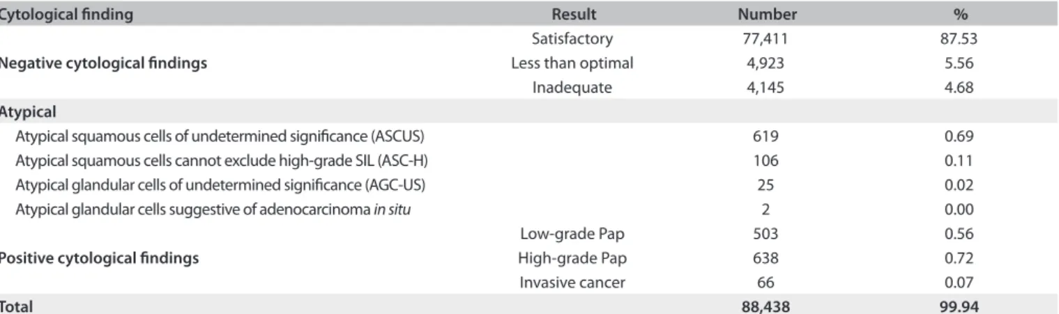
The full spectrum of carcinogenic lesions that occurs in a normal cervix includes Low-Grade Squamous Intraepithelial Lesions (LSIL), High-Grade Squamous Intraepithelial Lesions

A amostra foi constituída por 67 adolescentes portadores de fissura labiopalatal (G1), matriculados no Hospital de Reabilitação de Anomalias Craniofaciais da Universidade de

Percentage variation (R 2 ) and residual standard deviations (RSD) of weight of rib tissue coposition (g) accounted for by cold carcass weight, subjective measurements

It also has value after the cytology showing atypical squamous cells of undetermined signi fi cance (ASC-US) or low-grade squamous intraepithelial lesions (LSILs) as a triage test