Implementation of the ANA HEp-2 consensus
guidelines in Brazilian clinical laboratories
Implantação das diretrizes dos consensos de FAN HEp-2
nos laboratórios clínicos brasileiros
Glaucielen G. Silva1; Clayson M. Gomes2; Alessandra Dellavance3; Paulo Luiz C. Francescantonio2; Luís Eduardo C. Andrade3, 4; Wilson M. Cruvinel2
1. Faculdade dos Carajás, Pará, Brazil. 2. Pontifícia Universidade Católica de Goiás (PUC Goiás), Goiás, Brazil. 3. Fleury Medicina e Saúde, São Paulo, Brazil. 4. Escola Paulista de Medicina/Universidade Federal de São Paulo (EPM/UNIFESP), São Paulo, Brazil.
First submission on 03/07/17; last submission on 26/10/17; accepted for publication on 01/12/17; published on 20/12/17
ABSTRACT
Introduction: The detection of autoantibodies in HEp-2 cells represents a relevant tool for the diagnosis of autoimmune diseases, especially rheumatic autoimmune diseases. As a result of the methodological advances, the technique gradually increased the sensitivity, as well as the need for standardization. Objective: To evaluate the implementation of the Brazilian Consensus recommendations for autoantibody determination in HEp-2 cells. Methods: A structured form in a virtual platform was illed in by experts in clinical laboratories that carry out the methodology across the country. The questionnaire addressed the adoption of the Brazilian Consensus guidelines, detailing the technical aspects, quality control, the strategy for reading slides and the release of reports. Results: The study included 53 laboratories responsible for more than 300,000 antinuclear antibody (ANA) tests/month; more than half (58.5%) reported fully adopting the recommendations of the Brazilian Consensus. The majority (83.1%) used the 1:80 for screening dilution, and 75.5% of laboratories, perform education and quality control programs. Only 39.6% reported using more than one kit brand to perform the test, and 32.1% did not report observing all phases of the cell cycle during slide reading. The study also detected some heterogeneity among participants in the identiication of patterns. Conclusion: The results conirm the adoption of the Brazilian Consensus recommendations by most of participating laboratories, although with variable extent. There is need for improvement in some aspects, especially those related to the quality control.
Key words: indirect luorescent-antibody technique; autoantibodies.
INTRODUCTION
Autoimmune rheumatic diseases are relatively common in the general population. It is estimated that approximately one in 12 women and one in 20 men will develop such disease during their lifetime(1). In the last decades, due to the methodological
advances, the changes in the techniques used for the diagnosis of autoimmune diseases were substantial(2). The test for lupus
erythematosus cells, developed by Malcolm Hargraves and Robert Morton, in 1948, was considered as a criterion for the diagnosis of systemic lupus erythematosus by the American College of Rheumatology until 1997(3, 4).
In 1950, the antibody detection by indirect immunoluorescence (IIF) technique(5) was developed, which was then used to investigate
autoantibodies in mouse liver imprints, and allowed the recognition of ive classical patterns of luorescence(2), used to guide the diagnosis
of autoimmune rheumatic diseases(2, 6). Such methodology was
gradually replaced from 1980 onwards by the cell line derived from human laryngeal carcinoma, named HEp-2 cells [American Type Culture Collection (CCL-23)](2), as soon as advantages of this
substrate were evidenced, such as presentation of human speciic antigens, and generally in high concentration, expressed in the different phases of the cell cycle (interphase, prophase, metaphase, anaphase and telophase), a better nucleus/cytoplasm ratio in favor of nucleus and presence of nucleolus and cytoplasmic organelles(7).
With the change of substrate for the HEp-2 cells, it was possible to recognize more than 20 luorescence patterns(6), which led to
the need for training, standardization of the methodological
procedures and the nomenclature, which were, until then, very heterogeneous, at national and international scenarios(7, 8). Sensitive
to this demand, Brazil pioneered the process of standardization of the nomenclature for the morphological classiication of autoantibody luorescence patterns, serving as a world reference(8).
The I Brazilian Consensus was held in August 2000, in Goiânia (GO), with the main objective of promoting the nomenclature standardization, improving the diagnosis of autoimmune diseases in the country(7). It was mainly motivated by the lack
of slide reading parameters, the absence of organization of the patterns in classiication groups and the lack of standardization of nomenclature of the patterns(7). Several recommendations have
been suggested in order to seek solutions to such problems, as
demonstrated in Table.
The II Consensus contemplated further advances, such as the creation of mixed patterns group and the presentation of the main clinical relevance for each pattern(9). In the III Consensus,
adaptations have to be made in the terminology of the mixed patterns and nuclear homogeneous pattern, as well as an updating of the clinical relevance, resulting in the creation of guidelines for
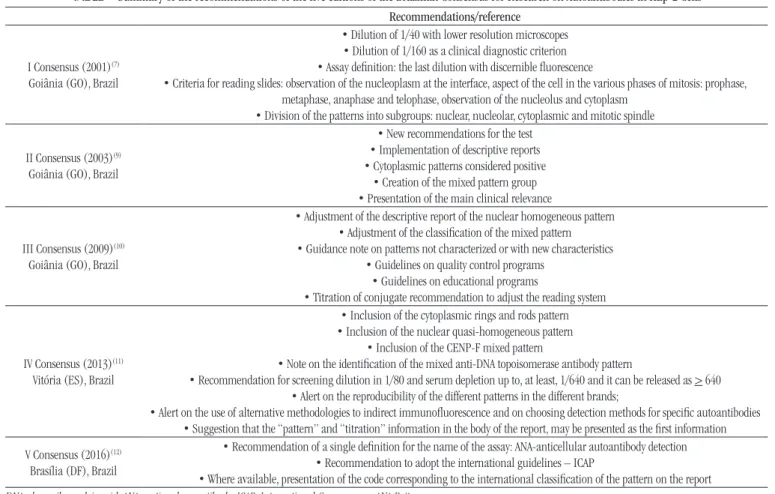
TABLE − Summary of the recommendations of the ive editions of the Brazilian Consensus for Research on Autoantibodies in HEp-2 Cells
Recommendations/reference
I Consensus (2001)(7)
Goiânia (GO), Brazil
• Dilution of 1/40 with lower resolution microscopes • Dilution of 1/160 as a clinical diagnostic criterion • Assay deinition: the last dilution with discernible luorescence
• Criteria for reading slides: observation of the nucleoplasm at the interface, aspect of the cell in the various phases of mitosis: prophase, metaphase, anaphase and telophase, observation of the nucleolus and cytoplasm
• Division of the patterns into subgroups: nuclear, nucleolar, cytoplasmic and mitotic spindle
II Consensus (2003)(9)
Goiânia (GO), Brazil
• New recommendations for the test • Implementation of descriptive reports • Cytoplasmic patterns considered positive
• Creation of the mixed pattern group • Presentation of the main clinical relevance
III Consensus (2009)(10)
Goiânia (GO), Brazil
• Adjustment of the descriptive report of the nuclear homogeneous pattern • Adjustment of the classiication of the mixed pattern • Guidance note on patterns not characterized or with new characteristics
• Guidelines on quality control programs • Guidelines on educational programs
• Titration of conjugate recommendation to adjust the reading system
IV Consensus (2013)(11)
Vitória (ES), Brazil
• Inclusion of the cytoplasmic rings and rods pattern • Inclusion of the nuclear quasi-homogeneous pattern
• Inclusion of the CENP-F mixed pattern
• Note on the identiication of the mixed anti-DNA topoisomerase antibody pattern
• Recommendation for screening dilution in 1/80 and serum depletion up to, at least, 1/640 and it can be released as > 640 • Alert on the reproducibility of the different patterns in the different brands;
• Alert on the use of alternative methodologies to indirect immunoluorescence and on choosing detection methods for speciic autoantibodies • Suggestion that the “pattern” and “titration” information in the body of the report, may be presented as the irst information
V Consensus (2016)(12)
Brasília (DF), Brazil
• Recommendation of a single deinition for the name of the assay: ANA-anticellular autoantibody detection • Recommendation to adopt the international guidelines – ICAP
• Where available, presentation of the code corresponding to the international classiication of the pattern on the report DNA: deoxyribonucleic acid; ANA: antinuclear antibody; ICAP: International Consensus on ANA Patterns.
the control of the quality of the assay. Proposals were also made to carry out studies to incorporate new patterns(10).
In September 2012, the IV Consensus provided to the scientiic community 10 new recommendations related to characterization of the patterns, technical procedure, quality control, use of alternative methods and organization of the reports(11). The V
Consensus was carried out in 2016, under the inluence of the International Consensus on ANA Patterns (ICAP), with the purpose of harmonizing the guidelines of the Brazilian Consensus with the international ICAP recommendations(8, 12). The Table summarizes
the main recommendations of the Brazilian Consensus for Autoantibodies Screening in HEp-2 Cells(7, 9-12).
Thus far, almost two decades after the establishment of the Brazilian guidelines for the examination, a detailed evaluation of the adherence of Brazilian clinical laboratories to the the antinuclear antibody (ANA) HEp-2 consensus recommendations has not been performed. In 2014, the international standardization process started(8), adding more complexity in the standardization of
of the process of standardisation of reports, quality control and interpretation of the test are stimulated in the Brazilian laboratories.
OBJECTIVES
After 16 years of the national standardization process for autoantibody research in HEp-2 cells, the present study aimed to evaluate the implementation and adherence of the Brazilian consensus guidelines in clinical laboratories across the country, focusing on the adoption of technical and quality control recommendations of the test under the scope of technical procedure, slides reading and the classiication of luorescence patterns.
METHODS
The work consisted of an electronic form study directed to the clinical laboratories that perform the evaluation of autoantibodies in HEp-2 cells. A virtual platform was developed, hosted on the oficial site of the Brazilian Consensus (www.hep-2.com.br), with online access to the questionnaire. The laboratories were invited to participate voluntarily in the research by e-mail, and only one expert per laboratory was allowed to ill out the form.
The research system was developed using the HTML5, CSS3, JQuery and PHP 5 languages, MySQL database, hosted Apache 2.2 web server on Linux Operating System plataform. The questionnaire was composed of 25 questions targeted at performing the assay, quality control of the test, slides reading process and writing of reports. Those laboratories that afirmed to carry out the test by outsourcing were excluded.
A descriptive analysis of the results was carried out, as well as the construction of tables and graphs, using the Microsoft® Excel
2016 and the GraphPad Software© 2016 programs.
RESULTS
A total of 53 Brazilian laboratories from 12 federal states and the Federal District participated in the study. Two were from Bahia, 13 from São Paulo, three from Rio de Janeiro, six from Paraná, nine from Minas Gerais, seven from Rio Grande do Sul, three from the Federal District, two from Pará, two from Goiás, one from Espírito Santo, one from Ceará, one from Pernambuco,
one from Paraíba and one from Santa Catarina. One laboratory did not report the state to which it belongs.
Participants were divided into six groups according to the monthly average of ANA/HEp-2 tests declared on the form: 1) G0 – laboratories that did not inform the average number of exams performed per month (n = 2; 3.8%); 2) G1 – laboratories performing up to 99 tests/month (n = 5; 9.4%); 3) G2 – laboratories that perform between 100 and 499 tests/month (n =
23; 43.4%); 4) G3 – laboratories that perform between 500 and 1,000 tests/month (n = 3; 5.7%); 5) G4 – laboratories that reported an average between 1,000 and 9,999 tests/month (n = 14; 26.4%); 6) G5 – laboratories that afirmed performing an average routine greater than 10,000 test/month 10.000 (n = 6; 11.3%). Based on the information presented by the 53 participating laboratories, it was possible to estimate that they are responsible for more than 300,000 ANA/HEp-2 tests per month (Figure 1).
FIGURE 1 − Brazilian laboratories participating in the study distributed into groups, according to the average ranges of examinations performed per month
50
40
30
20
10
0
% G0: not informed
G1: up to 99 tests/month G2: 100-499 tests/month G3: 500-999 tests/month G4: 1,000-9,999 tests/month G5: > 10,000 tests/month G0 G1 G2 G3 G4 G5
3.8 9.4
43.4
5.7 26.4
11.3
Participating laboratories informed about the medical specialties that most order ANA/HEp-2 screening, besides rheumatology. This demand was attributed to different medical specialties such as clinical medicine (20.1%), dermatology (13.4%), gynecology (9.4%), endocrinology (7.4%), gastroenterology (6.0%), infectology (5.4%), hematology (5.4%), orthopedics (5.4%), cardiology (4.7%), neurology (4%), pediatrics(4%), nephrology (3.4%), ophthalmology (3.4%), oncology (2.0%), hepatology (1.3%), obstetrics (1.3%), pneumology (1.3%), geriatrics (0.7%), immunology (0.7%) and otolaryngology (0.7%).
Regarding the academic qualiications of the professional responsible for the slides reading, the participants reported different training. Biochemical/pharmaceutical professionals were the most mentioned, corresponding to 38.4%, followed by biomedical and biologists, 22.2% and 17.2%, respectively. Other professionals were mentioned, including pathologist (8.6%), rheumatologist (6.2%) and undergraduate professionals (7%).
Regarding the terminology used to name the test in the six options presented by the III Consensus, 26.4% of the laboratories reported adopting the ANA-HEp2 option and 22.6% reported using screening for autoantibodies against cellular antigens – ANA. Another 20.8% use ANA – autoantibody screening; 7.6% reported adopting the terminology ANA – screening for antibodies against components of nucleus, nucleolus, cytoplasm and mitotic spindle and 20.8%, ANA – antinuclear antibody. One participant (1.8%) did not inform about the nomenclature used.
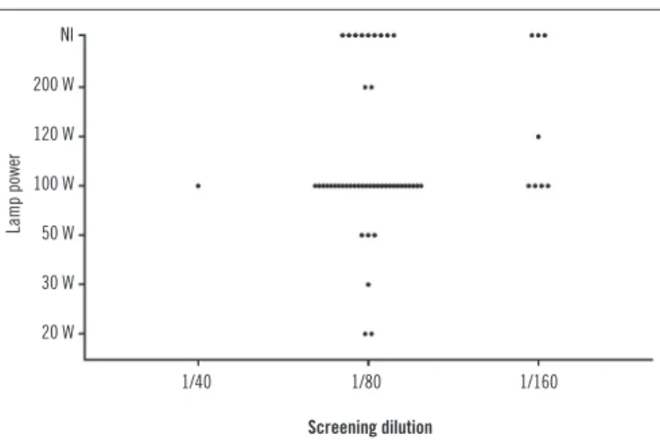
When questioned about the screening dilution, 83.1% of the laboratories stated that they performed 1:80 dilution, seven (13.3%) reported using 1:160 dilution and one laboratory (1.8%) reported using 1:40 dilution. When asked about the power of the lamp used, 33 participating laboratories (62.3%) reported using a 100 Watts lamp. The remaining participants reported using wattage of 20 Watts (3.8%), 30 Watts (1.8%), 50 Watts (5.7%), 120 Watts (1.8%) and 200 Watts (3.8%). Another 11 laboratories (20.8%) did not inform.
An analysis comparing the screening dilution used and the power for the luorescence lamp demonstrated that 27 laboratories (51.0%) use 100 Watts lamp with 1:80 screening dilution. All laboratories using 1/160 screening dilution use lamps at 100 Watts or higher. and, as shown in Figure 2, a signiicant part of the laboratories included in the study use different parameters of screening dilution versus lamp power.
In relation to the dilution for titration of the positive samples, 25 laboratories (47.3%) stated that they diluted the positive samples up to 1:640; in such case, the test is released as titre ≥ 640. For 18.8% of the laboratories, the maximum dilution performed was 1:1280 and 17% reported performing the maximum dilution at 1:2560, and the result is released as ≥ 1280 and ≥ 2560, respectively. Eight participants (15.1%) stated that serum is diluted to the last titre with noticeable luorescence, and one participant (1.8%) did not respond on the screening and titration dilutions used.
Participants responded questions related to the routine technical procedures, such as conjugate titration, availability in the laboratory of more than one kit brand, keeping a serum bank to evaluate the kit’s ability in discriminating different dilutions and patterns, and on the use of serum control with minimal luorescence. The results of this stage of the research are shown
in Figure 3A.
Questioned about the slides reading with a 40×, 100× objective or both, 41 laboratories (77.4%) reported that they read the slides using only the 40× objective, while three (5.7%) used only 100× objective and eight (15.1%) use both. One participant (1.8%) did not inform the objective used for slides reading.
As shown in Figure 3B, considering the evaluation of the
criteria for slide reading, 50 participating laboratories (94.3%) reported to observe luorescence in different cell compartments (nucleus, nucleolus, cytoplasm and mitotic spindle). A total of 36 laboratories (67.9%) reported observing cells at all stages of development (interphase, prophase, metaphase, anaphase and
FIGURE 2 − Relationship between screening dilution and lamp power of the microscope used to read the indirect immunofluorescence slides in 53 Brazilian laboratories
NI: not informed; W: Watts. NI
200 W
120 W
100 W
50 W
30 W
20 W
Lamp power
1/40 1/80 1/160
Screening dilution
64.2 35.8
45.3 54.7
54.7 45.3
60.4 39.6
77.4 22.6
A)
B)
No Yes
No Yes Defines cutoff point based on minimum
fluorescence control
Use of a serum bank for support in the identification of patterns
Use of a serum bank with serum in different dilutions
Uses more than one brand of kits
Label the conjugate for each new kit lot
0 20 40 60 80 %
0 20 40 60 80 100 %
FIGURE 3 − Evaluation of the Brazilian laboratories regarding the technical procedures of quality control and strategy for slides reading
A) laboratory responses in relation to the technical quality assurance procedures of the assay; B) laboratory responses regarding the technical procedures for slides reading.
Classifies the chromosome metaphase plate in positive or negative
Observe cells in all phases of mitosis
Evaluates fluorescence of core, nucleolus, cytoplasm to mitotic spindle
7.5
92.5
32.1
67.9
5.7
telophase) and 49 (92.5%) reported categorizing the chromosome at metaphase plate as positive or negative. In the elaboration of the reports, 10 laboratories (18.9%) reported providing information on the possible autoantibodies associated with the pattern presented and possible clinical conditions consistent with the indings.
Regarding the procedure for the preparation of the reports, 43 laboratories (81.1%) stated to issue them according to the consensuses recommendation, where the presentation of the pattern name is followed by the titration and the classiication of the positivity of the cellular compartments, while seven participants (13.2%) stated to report only the pattern and titration name. Three other laboratories (5.7%) did not inform.
It was also identiied that 67.9% of the participating laboratories considered cytoplasmic pattern as a positive test, and in the case of mixed patterns, 75.5% of the laboratories reported releasing the maximum titre present for each cell compartments, distinguishing the titre from each luorescent cell compartment.
When questioned about the use of educational programs and quality control, 40 laboratories (75.5%) reported using them, while 13 (24.5%) reported not using.
As shown in Figure 4, the last stage of the evaluation was the
request of the analysis, by the participating laboratories, of ive igures representative of patterns of the four main groups, nuclear homogeneous (AC-1), cytoplasmic rings and rods (AC-23), mitotic spindle centriole pattern 24), nucleolar homogeneous (AC-8) and nuclear dense ine speckled pattern (AC-2). Images were classiied correctly by most participating laboratories. However, there were a small percentage of classiications that differed from those expected in the ive images, 1 (3.6%), 23 (7.5%), AC-24 (7.2%), AC-8 (5.7%) and AC-2 (22.7%). And still according to Figure 4, in all ive images represented, there was a percentage of laboratories that did not characterize the image nor informed the corresponding pattern, in the cases of images AC-1, AC-23 and AC-8 (3.9%), AC-24 (5.7%) and AC-2 (22.7%).
DISCUSSION
Fifty-three laboratories from 12 Brazilian states and the Federal District participated in the study, comprehending an average of 300,000 tests/month. It was demonstrated that all the Brazilian laboratories participating in the study adopt the recommendations of the consensuses integrally or partially. The bias in adherence to the recommendations, observed in some laboratories, refers to the clinical pathology services that,
7.5
FIGURE 4 − Pattern images presented to study participants for classification, as recommended by the Brazilian Consensus
AC-1: nuclear homogeneous pattern; AC-4: nuclear fine dot pattern; NQH: nuclear quasi-homogeneous pattern; NI: not informed; AC-23: cytoplasmic rings and rods pattern; AC-22: polar cytoplasmic pattern; NEG: negative results; AC-24: mitotic spindle centriole pattern; AC-25/26: mitotic spindle pattern NuMA-type; MS: mitotic spindle pattern; MM: mixed mitotic spindle pattern; CP: cytoplasmic dot pattern; Nu: Nucleolar pattern; AC-8: nucleolar homogeneous pattern; NP: nuclear dot pattern; AC-2: nuclear dense fine dot pattern.
A)
B)
C)
D)
E) 100
80
60
40
20
0
%
%
%
%
%
AC-1 AC-4 NQH NI 3.9 92.5
1.8 1.8
100
80
60
40
20
0
100
80
60
40
20
0
80
60
40
20
0
80
60
40
20
0
AC-23 AC-22 NEG NI 3.9 88.6
1.8 5.7
AC-24 AC-25/26 MS MM + CP NEG NI
5.7 87.1
1.8 1.8 1.8 1.8
Nu AC-8 AC-8 + NP AC-4 + Nu NI
AC-2 NQH AC-4 AC-1 AC-1 + AC-4 NI
32 58.4
1.8 3.9 3.9
69.8
13.1
3.9 3.9 1.8
as reported, did not adhere, for example, to the recommended screening dilution of 1/80, the adoption of measures to control the quality of the test, the recommendation for slides reading, among other aspects discussed ahead.
and resulted in a higher rate of false positives(6, 10, 13). Our results
show a high diversity of medical specialties that order the examination, which causes low pre-test probability and favors the generation of false positive results, in opposition to the recommendation that the test should be performed only in the presence of sound clinical evidence(10, 11).
Such an alert is relevant, considering studies with healthy individuals (597 workers in Japan and 500 blood donors in São Paulo), which show a frequency of autoantibodies of 20% and 22.6%, respectively(14, 15) in this population, reinforcing the
need for awareness in ordering the examination by the medical community.
The fact that approximately 60% of the laboratories participating in the study fully adopt the recommendations of the Brazilian Consensus and the others adopt it partially, demonstrates that the Consensus has fulilled its educational role, which has occurred along ive editions by the elaboration of vast teaching material in the form of physical and digital atlases and the oficial website(7, 9-12).
Considering the technical aspects of the examination, it was evidenced that different professional categories perform the test, which reinforces the need to discuss these contents within the scope of the different undergraduate courses that technically act in the area of clinical pathology.
The present study also shows that there is no consensus regarding the deinition adopted to nominate the assay, which motivated the adoption by the V Consensus of a single terminology to designate the test in the services that perform the exam, being deined as nomenclature: ANA – antinuclear antibody screening tests(12).
Another aspect addressed were the screening dilutions that can directly inluence the rate of positive and
false-negative results(10, 16, 17), knowing that a low dilution may result
in increased false positives, as well as a high dilution, in false
negative(17). The IV Consensus recommended dilution of 1:80 for
the screening dilution(11). In our study, 83.1% of the laboratories
afirmed following this recommendation and 13.3% carried out the screening using 1:160 dilution. The screening dilution is very valuable in the speciicity of the test, since one in three healthy subjects will have ANA positive if they were screened at 1:40 dilution, but the positivity will be one in 20, if performing a 1:160 dilution(18). Likewise, although the 1:80 dilution is
used, the ANA positivity may be 12.9% in healthy patients(19).
Brito et al. (2013)(16) stated that the ideal screening dilution is
1:160, as it produced a 53% reduction in the number of false
positive results. Healthy patients tend to have low titres and patients with autoimmunity have moderate to high titres(18). It is
known that at 1:80 dilution the results considered to be important are those with titre greater than 160(11, 18, 19). It is suggested that
patients who have luorescence patterns, but in smaller titres, may be monitored by clinicians for the onset of autoimmune
disease(20), and the ability of the test to precede the emergence
of systemic lupus erythematosus (SLE) symptoms has already been described, and may still be presented indeinitely negative in some cases(6, 11, 19).
The power of the lamp plays a relevant role in the reading of the luorescence slides(10). In the present study, 62.3% of
the participants reported using 100 Watts lamp. The higher the lamp power, the lower the concentration of the conjugate needed to visualize the reaction(10). Therefore, low dilution associated
with high potency may cause a high number of false positive results. On the other hand, high dilution associated with low lamp power will produce false negative results. The same applies to the screening dilution of the serum sample. It was reported among the study participants the use of 100 Watts lamp with screening dilution at 1:40, which increases the chance of false positives, or even the use of screening dilution at 1:80 with 20 Watts lamp, which enhances the occurrence of false negatives. Such situations should be evaluated by the services based on the recommendations of the Consensus(10), and the professionals
who technically perform the exam must be aware of this detail.
In the III and IV Consensus, participants were recommended to perform test quality assurance strategies(10, 11). One of the key
points to ensure the quality control of a service is the evaluation of the optical system, considering the lamp power, the quality of the kit and the titration of the conjugate as a tool to balance the system(10, 11). The conjugate titration is recommended for
each new kit of different batch(10). In the results presented,
only 22.6% of the laboratories reported performing conjugate titration for each new kit, and the other participants reported using conjugate ready for use. However, the Consensus states that in cases of kits from the same batch, the maintenance of the titration should occur by the use of low intensity controls(10). In
response to the recommendation provided in the III Consensus, we veriied that 35.8% of the participants had a serum bank with minimal reactivity sample for control(10). The aspect
Regarding the titre deinition, the Consensus recommends that the positive sample should be diluted to a minimum of 1:640, and the test may be released as titre > 640, or diluted at higher dilutions for differentiation of mixed patterns(11). The
results show that almost half of the participants adopt this recommendation and the others apply even greater dilutions, taking full account of this parameter.
It is recommended, since the irst Consensus, that a stratiied reading of all cell compartments should be performed, due to the existence of nuclear, nucleolar, cytoplasmic and mitotic spindle patterns(7, 10, 11, 16). We veriied in our results that only 5.7% of the
participants did not follow this guideline, and another 7.5% did not evaluate the reactivity in the chromosome metaphase plate. A total of 32.1% did not conirm performing the reading of all phases of cell division, although this is of great importance for the recognition of mitotic spindle patterns, besides helping to classify and differentiate some nuclear patterns(10, 11). Although
in a small part, this aspect points to the need to offer training and improvement programs for professionals who technically respond for the test performing.
Following the guidelines of report design, 81.1% of the laboratories afirmed to issue reports presenting in the upper part the name of the pattern, the titre of the autoantibody and just below a presentation of reactivity of the different cellular compartments, as recommended by the Consensus(11). When
invited to issue the descriptive report of ive igures representative of patterns, based on the recommendations of the Brazilian and the International Consensus(7-12), most laboratories were able to
recognize and classify the luorescence patterns.
The irst image was compatible with the presence of native anti-deoxyribonucleic acid (DNA), anti-histone and anti-chromatin antibodies, SLE markers(10, 21, 22), classiied as
AC-1 – nuclear homogeneous by the International Consensus(8).
With very low divergence, most laboratories choosed this classiication. This divergence in the reports, although small, can be explained by the dificulty and need of training in the differentiation of nuclear patterns with a positive metaphase plate. In the second image, the AC-23 pattern was recognized by 88.6% of the laboratories. This pattern is related to the treatment against hepatitis C virus (HCV) with interferon alpha
and ribavirin(11, 23, 24). It was integrated into the classiication
tree of the cytoplasmic patterns in the IV Consensus, remarking the fact that the proteins responsible for the presentation of this pattern are not present in all commercial substrates(11). One of
the study participants reported negativity of the pattern because the kit used in their laboratory would not detect the pattern.
It is recommended to state in the report that AC-23 pattern is substrate-dependent(11).
The third image caused more confusion regarding the classiication of the pattern. A representative igure of the AC-24 pattern was presented, recognized by the majority of participants. This pattern is expressed in the presence of the anti-alpha-phenolase antibody(10) and is of clinical relevance only in high
titres(10). This pattern can only be visualized if the luorescent
point isolated in the cytoplasm is observed at one of the poles in the resting cell (interphase) that is divided into two and migrates to the opposite pole of the nucleus as the cell begins its division process.
In the classiication tree, the Consensus stratiies the characterization of some patterns in mandatory or optional. Regarding the nucleolar group, the differentiation in AC-8, AC-9 or AC-10 is optional(10-12). In this study, about 90.4% of the participants
mentioned the nucleolar pattern. These results demonstrate good technical quality of the participating laboratories in the recognition of this group of patterns, considering the mandatory requirements suggested by the Brazilian Consensus.
The ifth image corresponds to the nuclear dense ine speckled pattern(11). The analysis of the answers evidenced a
divergence in the classiication. The laboratories considered the image as nuclear quasi-homogeneous (NQH), AC-2, AC-4 and AC-1. The nucleolar mixed and nuclear ine speckled pattern were also mentioned. The results show more dificulty to the laboratories in differenting this pattern, which is expected, since the pattern is found in the optional Brazilian Consensus classiication guide(11). Again, dificulty in differentiating
nuclear patterns with a positive metaphase plate was observed, and the error in the classiication of the aforementioned patterns may limit the potential of the test, therefore this group of patterns deserves special attention.
Regarding the participation of 53 Brazilian laboratories, it should be taken into account that the study was intended to services that perform the technique, and those who said to outsource the test were excluded from the analysis. It should be noted that the average of monthly ANA/HEp-2 tests, informed by each participant, were added together, the study consolidated information on more than 300 thousand tests/month.
This study is the irst evaluation of the inluence of the Brazilian Consensuses for the detection of autoantibodies in HEp-2 cells in the laboratory practice. The results conirm signiicant advances and deine as priorities the organization of educational courses and improvement programs, the intensiication of measures of test
REFERENCES
1. Crowson CS, Matteson EL, Myasoedova E, et al. The lifetime risk of adult-onset rheumatoid arthritis and other inlammatory autoimmune rheumatic diseases. Arthritis Rheum. 2011; 63(3): 633-9. PubMed PMID: 21360492.
2. Dellavance A, Andrade LEC. Das células LE às células HEp-2: perspectiva histórica e avaliação crítica do teste de imunoluorescência indireta para pesquisa de anticorpos antinúcleo. Rev Bras Med [Internet]. 2008; 1(1): 7-21. Available at: http://www.moreirajr.com.br/revistas.asp?id_ materia=3500&fase=imprime.
3. Hasehick JH, Bortz DW. Normal bone marrow inclusion phenomena induced by lupus erytrematosus plasma. J Invest Dermatol. 1949; 13: 47-9. PubMed PMID: 18134649.
quality control and other relevant measures that must be achieved for the purpose of continuous qualiication.
ACKNOWLEDGEMENTS
To the Brazilian Society of Clinical Pathology and Laboratory Medicine [Sociedade Brasileira de Patologia Clínica e Medicina
Laboratorial (SBPC/ML)], the 50th Brazilian Congress of
Clinical Pathology and Laboratory Medicine, the REDELAB, the Diagnóstico Clínico, S.A., Controllab, and the Brazilian Regional and Federal Biomedicine Councils.
RESUMO
Introdução: A pesquisa de autoanticorpos em células HEp-2 representa uma relevante ferramenta no auxílio diagnóstico de
doenças autoimunes, especialmente as reumáticas. Em virtude dos avanços metodológicos, a técnica aumentou gradativamente
a sensibilidade, bem como a necessidade de padronização. Objetivo: Avaliar a implantação das recomendações dos consensos
brasileiros de pesquisa de autoanticorpos em células HEp-2. Métodos: Preenchimento de formulário em plataforma virtual
direcionada aos laboratórios clínicos que realizam a metodologia. Os participantes responderam a um questionário sobre a adoção das diretrizes dos consensos brasileiros, detalhando os aspectos técnicos, o controle de qualidade, a leitura de lâminas e a emissão
de laudos. Resultados: Participaram do estudo 53 laboratórios responsáveis por mais de 300 mil testes de fator antinuclear (FAN)/
mês; mais da metade (58,5%) informou adotar integralmente as recomendações dos consensos. A maioria (83,1%) utiliza a diluição 1:80 para triagem, e 75,5% dos laboratórios, programas de educação e controle de qualidade. Apenas 39,6% utilizam mais de uma marca de kit para a realização do teste, e 32,1% não relataram observar todas as fases do ciclo celular na leitura
da lâmina. O estudo detectou ainda discreta heterogeneidade entre participantes na identificação de padrões. Conclusão: Os
resultados evidenciam a adoção das recomendações dos consensos de forma absoluta pela maioria dos laboratórios participantes, bem como a necessidade de aperfeiçoamento em alguns aspectos relevantes para a qualidade do ensaio.
Unitermos: técnica indireta de fluorescência para anticorpo; autoanticorpos.
4. Altit G, Brochu P, Jacob SV, Buithieu M. LE cells in pleural luid. J Clin Rheumatol. 2012; 18(5): 273-4. PubMed PMID: 22832304.
5. Coons AH, Kaplan MH. Localization of antigenin tissue cells II. Improvements in a method for the detection of antigen by means of luorescent antibody. J Exp Med. 1950; 91: 1-13. PubMed PMID: 15395569. 6. Dellavance A, Andrade LEC. Como interpretar e valorizar adequadamente o teste de anticorpos antinúcleo. J Bras Patol Med Lab. 2007; 43(3): 157-68.
7. Dellavance A, Gabriel Júnior A, Cintra AFU, et al. I consenso nacional para padronização dos laudos de FAN HEp-2. J Bras Patol e Med Lab. 2001; 38(3): 207-16.
Jan; 6: 412. Available at: http://www.pubmedcentral.nih.gov/articlerender. fcgi?artid=4542633&tool=pmcentrez&render ype=abstract.
9. Dellavance A, Gabriel Júnior A, Cintra AFU, et al. II Consenso Brasileiro de Fator Antinuclear em Células HEp-2. Deinições para a padronização da pesquisa de autoanticorpos contra constituintes do núcleo (FAN HEp-2), nucléolo, citoplasma e aparelho mitótico e suas associações clínicas. Rev Bras Reumatol [Internet]. 2003; 43(3): 129-40. Available at: http://www.scielo.br/scielo.php?script=sci_arttext&pid =S0482-50042003000300002.
10. Francescantonio PLC, Andrade LEC, Cruvinel WM, et al. III Consenso Brasileiro para Pesquisa de Autoanticorpos em Células HEp-2: perspectiva histórica, controle de qualidade e associações clínicas. J Bras Patol e Med Lab. 2009; 45(3): 185-99.
11. Francescantonio PLC, Cruvinel WM, Dellavance A, et al. IV consenso brasileiro para pesquisa de autoanticorpos em células HEp-2. Rev Bras Reum. 2013; 54(1): 44-50.
12. Consenso Brasileiro de FAN HEp-2 [Internet]. 2016. Available at: www. hep-2.com.br.
13. Dellavance A, Leser PG, Andrade LEC. Análise crítica do teste de anticorpos antinúcleo (FAN) na prática clínica. Rev Bras Reumatol [Internet]. 2007 Aug; 47(4): 265-75. Available at: http://www.scielo.br/ scielo.php?script=sci_arttext&pid=S0482-50042007000400005&lng=pt &nrm=iso&tlng=pt.
14. Watanabe A, Kodera M, Sugiura K, et al. Anti-DFS70 antibodies in 597 healthy hospital workers. Arthritis Rheum [Internet]. 2004 Mar; 50(3): 892-900. Available at: http://doi.wiley.com/10.1002/art.20096.
15. Fernandez SAV, Lobo AZC, Oliveira ZNP, Fukumori LMI, Prigo AM, Rivitti EA. Prevalence of antinuclear autoantibodies in the serum of normal blood dornors. Rev Hosp Clin Fac Med Sao Paulo [Internet]. 2003; 58(6): 315-9. Available at: http://www.ncbi.nlm.nih.gov/ pubmed/14762490
16. Brito FA, Santos SME, Ferreira GA, et al. Detecção de anticorpos antinucleares por imunoluorescência indireta em células HEp-2:
deinindo a diluição de triagem adequada para o diagnóstico das doenças reumáticas autoimunes. Rev Bras Reumatol [Internet]. 2013; 54(21): 13-20. Available at: www.reumatologia.com.br.
17. Egner W. The use of laboratory tests in the diagnosis of SLE. J Clin Pathol. 2000 Jun; 53(6): 424-32. PubMed Central: PMC1731203. 18. Tozzoli R, Bizzaro N, Tonutti E, et al. Guidelines for the laboratory use of autoantibody tests in the diagnosis and monitoring of autoimmune rheumatic diseases. Am J Clin Pathol [Internet]. 2002; 117: 316-24. Available at: http://ajcp.oxfordjournals.org.sci-hub.cc/ content/117/2/316.long.
19. Bohan A. Seronegative systemic lupus erythematosus. J Rheumatol [Internet]. 1979; 6(5): 534-40. Available at: http://www.ncbi.nlm.nih. gov/pubmed/93147.
20. Amoura Z, Koutouzov S, Chabre H, et al. Presence of antinucleosome autoantibodies in a restricted set of connective tissue diseases: antinucleosome antibodies of the IgG3 subclass are markers of renal pathogenicity in systemic lupus erythematosus. Arthritis Rheum. 2000; 43(1): 76-84. PubMed PMID: 10643702.
21. Wagner A, Souza S, Keusseyan SP, et al. Anticorpos antinucleossomo e síndrome antifosfolipídica: estudo observacional. Rev Bras Reum. 2012; 52(3): 357-65.
22. Franca NR, Dellavance A, Rodrigues SH, Perazzio SF, Silva NP, Andrade LEC. Quasi-homogeneous ANA-HEp-2 pattern relects an autoantibody proile intermediate to the homogeneous and dense ine speckled nuclear patterns. Arthritis & Rheumatism [Internet]. 2011; 63 Suppl(10):2307.
23. Felisberto M, Jorge AS, Menolli RA, Gnutzmann LV, Nesi V. Indução do padrão citoplasmático em forma de “bastões e anéis” através do tratamento da hepatite C: relato de caso. Rev Bras Reumatol. 2015; 55(4): 181-4. 24. Carcamo WC, Satoh M, Kasahara H, et al. Induction of cytoplasmic rods and rings structures by inhibition of the CTP and GTP synthetic pathway in mammalian cells. PLoS One [Internet]. 2011; 6(12): e29690. Available at: http://www.ncbi.nlm.nih.gov/pubmed/22220215.
CORRESPONDING AUTHOR
Wilson de Melo Cruvinel