einstein. 2012;10(3):371-3 CASE REPORT
Femoral nerve neuropathy after the psoas hitch procedure
Neuropatia do nervo femoral após
psoas hitch
Antonio Cardoso Pinto1, José Rafael Macea1, Marcello Toscano Pecoraro1
1 Faculdade de Ciências Médicas da Santa Casa de São Paulo – FCMSCSP, São Paulo (SP), Brazil.
Corresponding author: Antonio Cardoso Pinto – Rua Cerro Corá, 1917B – Alto de Pinheiros – São Paulo (SP), Brazil – Zip code: 05061350 – Phone: (55 11) 3022-2044 – E-mail: antonio.cardoso@sbu.org.br Received on: Feb 19, 2012 – Accepted on: Jun 2, 2012
ABSTRACT
Femoral nerve neuropathy as a complication from abdominopelvic surgery was firstly described in 1896, by Gumpertz, in a case report of femoral nerve injury following hysterectomy. The authors report two cases of femoral nerve neuropathy following psoas hitch vesicopexy in ureteral reimplantation, and to discuss the etiology and clinical manifestations of this complication. Femoral nerve neuropathy secondary to psoas hitch is a rare complication, although it should be considered during the surgical procedure, as well as in postoperative care.
Keywords: Peripheral nervous system diseases/etiology; Psoas Muscle; Ureter/surgery; Postoperative complications; Suture techniques; Case reports
RESUMO
A neuropatia do nervo femoral como complicação de cirurgia abdominopélvica foi descrita, pela primeira vez, em 1896, por Gumpertz, em um relato de caso de lesão do nervo femoral após histerectomia. Os autores relatam dois casos de neuropatia do nervo femoral após reimplantação ureteral, com técnica de psoas hitch em vesicopexia, e discutem a etiologia e as manifestações clínicas dessa complicação. A neuropatia do nervo femoral secundária à técnica de psoas hitch é uma complicação rara, embora deva ser levada em consideração durante o procedimento cirúrgico, bem como no cuidado pós-operatório.
Descritores: Doenças do sistema nervoso periférico/etiologia; Músculo Psoas; Ureter/cirurgia; Complicações pós-operatórias; Técnicas de sutura; Relatos de casos
INTRODUCTION
Femoral nerve (FN) neuropathy may occur as a complication from abdominopelvic surgery. Gumpertz was the first to report a case of FN injury after hysterectomy, in 1896(1).Ever since, the morbidity of this condition has been considerably high following
hysterectomy, aortic surgery, appendicectomy and herniorrhaphy. In the scientific literature concerning the urology field, it has been following kidney transplantation, radical perineal prostatectomy, pelvic lymphadenectomy, radical cystectomy and percutaneous nephrolithotomy.
Some procedures, such as bladder fixation to the psoas major muscle (PMM or psoas hitch) and/or the Boari’s technique to treat short distal ureters, involve considerable risks to present this complication, since the FN is formed amidst the psoas major fibres, emerges in the iliac region and continues deeply through the iliac fascia between the PMM and iliac muscles(1,2) (Figure 1). This article reports two cases of FN neuropathy following psoas hitch vesicopexy in ureteral reimplantation, and discusses the etiology and clinical manifestations of this condition.
CASE 1
einstein. 2012;10(3):371-3
372 Pinto AC, Macea JR, Pecoraro MT
decreased tactile sensation of the anteromedial region of the thigh. Although such findings are compatible with FN neuropathy, they were initially attributed to anesthesia side-effects. However, the diagnosis was confirmed by an electromyography investigation, which finally revealed the FN injury. The pathological examination showed ureteral endometriosis. The patient was treated conservatively and the symptoms resolved completely after 1 year.
CASE 2
A 28-year-old male patient, treated in 2009 for severe nephritic colic resilient to treatment. A computed tomography (CT) of the abdomen and pelvis showed a 9mm calculus in the distal portion of the left ureter, with significant ureteric dilation involving the downstream portion. The patient was submitted to ureterolithotripsy but the ureteral dilatation did not subside. Considering the presumptive diagnosis of congenital megaureter, the patient underwent Politano-Leadbetter ureteric reimplantation with fixation of the bladder to the PMM (psoas hitch), also performed with 2-0 polyglycolic acid sutures. The anatomical-pathological findings confirmed the presumptive diagnosis. The day after this surgery the patient reported significant left leg weakness, impossibility to climb stairs, as well as decreased tactile sensation of the anterior-medial region of the thigh and medial region of the leg. Considering the experience with the previous case, physical therapy was recommended and a significant improvement of the motor and sensory symptoms was obtained; the patient left the
hospital on the 7th postoperative day, being able to
walk without help, still with some difficulty to climb stairs and to sustain the left leg in elevated position. Electromyography showed severe FN lesion. The conservative treatment has continued after hospital discharge. The case evolved with severe atrophy of the quadratus femoralis muscle (QFM), gradually reverted along the rehabilitation process; 1 year after the procedure, there as a complete recovery of tactile sensation and adequate deambulation could be achieved; climbing stairs, however, was still a difficult task for this patient.
DISCUSSION
The psoas hitch technique is performed as part of the ureteroneocytostomy procedure in an attempt to obtain an anastomosis without tension and to prevent kinking of the lower ureter. However, the FN, which
emerges inferiorly to the site that usually holds the fixation, must be avoided. Injuries to this nerve following psoas hitch are a potential complication and few case reports are to be found in the literature, of which we obtained five cases.
The FN is formed by the anterior divisions of the primary ventral branches from L2 to L4, and is the thickest and longest nerve of the lumbar plexus. It is responsible for the motor innervation of the iliopsoas, pectineus, sartorius and QFM, as well as for the sensory innervation of the skin of the anterior region of the thigh, medial region of the leg and dorsal portion of the foot.
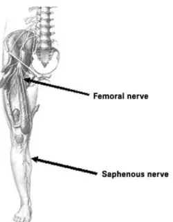
This nerve lies deep in the fascia of the PMM, and is positioned inside a groove between the PMM and illiacus muscles, following toward the gap between these muscles and branching to both of these muscles (Figure 1). Approximately 8cm below the inguinal ligament, the FN divides into numerous muscular and cutaneous branches, and sends a long terminal sensory branch, the saphenous nerve, which continues to supply the foot region. Initially this nerve enters the adductor canal along with the femoral vessels, and then goes to the medial knee with the sartorius. After sending an infrapatellar branch to the medial knee, it follows the great saphenous vein toward the medial surface of the leg and foot(3,4) (Figure 2).
Neuromuscular examination of the lower limbs reveals possible dysfunctions related to injury of this nerve. Patient may present with muscular weakness during knee extension and hip flexion resulting in difficulty to perform specific tasks, such as climbing or descending stairs, or kicking a ball. Alteration of
Source: Netter FH. Atlas de anatomia humana. 8a ed. Porto Alegre: Artes Médicas; 1996.
373
Femoral nerve neuropathy after the psoas hitch
einstein. 2012;10(3):371-3
sensitivity occurs in the lateral and anterior thigh and medial leg. The patellar reflex may be abnormal; there may be atrophy of the QFM.
If nerve injury is suspected, an electromyogram can confirm the diagnosis and to establish the injury extent.
Different forms of FN injury may occur during surgery, including direct or indirect compression, ischemia, and transection. The most common cause of FN neuropathy during urologic procedures is often attributed to the use of retractors, which are poorly placed in the pelvic cavity, thus compressing the FN nearby its emergence from within the PMM, causing edema of the epineurium and nerve compression. Usually, in such cases, the patient presents with paresis of the QFM in the immediate postoperative period, which regresses in a few weeks, with little long term damage.
Techniques for distal ureter reconstruction, in which the bladder is fixed to the PMM, should avoid the FN. The site where the fixation is usually performed is superior to the emergency of the FN, therefore, along its path within the PMM. If the suture is too deep, there is a considerable risk of nerve injury. Kowalczyk et al.(1) stated that such sutures should be superficial and preferably not exceed a depth of 3mm(1).
Once a neuropathy is diagnosed, treatment options should be evaluated. Conservative treatment such
as physical therapy and early mobilization should be used, but the main decision relies on the expectation of spontaneous recovery or an exploratory surgery to assess the suture and to remove it if necessary.
In the cases reported herein, although severe lesions were confirmed by electromyography, the regression of symptoms was mainly related to incomplete ligation or nerve transection, and also to the usage of absorbable sutures. Kowalczyk et al.(1) described three similar cases: in those in which an absorbable suture was used there was total regression of the neuropathy. In contrast, in the case that a nonabsorbable suture was used the injury has remained, thus requiring surgical exploration for removal.
Crook et al.(2) described two cases of FN neuropathy following psoas hitch in which surgical re-exploration was performed. The sutures possibly affecting the FN were removed and both patients showed an improvement of the symptoms, which were almost absent in the 6 months of follow-up. The authors noted that the removal of those sutures did not compromise the ureteral implant integrity. Therefore, the use of absorbable sutures is justified by the fact that surgical re-exploration would not be necessary should the patient present FN neuropathy after the psoas hitch.
FN neuropathy following psoas hitch has few case descriptions in literature. However, due to its potential morbidity regarding lower limb motor function and sensitivity, it should be considered by urologists performing such procedure, and promptly diagnosed and assessed, if present.
CONCLUSION
FN neuropathy secondary to the psoas hitch is an acknowledged complication and, although rare, should be considered during the procedure and in postoperative follow-up.
REFERENCES
1. Kowalczyk JJ, Keating MA, Ehrlich M. Femoral Nerve neuropathy after the psoas hitch procedure. Urology. 1996;47(4):563-6.
2. Crook TJ, Steinbrecher HA, Tekgul S, Malone PSJ. Femoral nerve neuropathy following the psoas hitch procedure. J Ped Urol. 2007;3(2):145-7.
3. Schünke M, Schulte E, Schumacher U, editores. Prometheus - Atlas de Anatomia. Rio de Janeiro: Guanabara Kooga; 2007.
4. Lockhart RD, Hamilton GF, Fyfe FW. Anatomia do corpo humano. 2.ed. São Paulo: Guanabara Koogan; 1983.
Source: Netter FH. Atlas de anatomia humana. 8ª ed. Porto Alegre: Artes Médicas; 1996.

