Abstract
Submitted: May 9, 2017 Modiication: July 13, 2017 Accepted: August 1st, 2017
Debris extrusion and foraminal
deformation produced by
reciprocating instruments made of
thermally treated NiTi wires
Objective: To evaluate the amount of apically extruded debris, percentage of foraminal enlargement and apical foramen (AF) deformation that occurred during root canal preparation with different reciprocation systems: Reciproc, WaveOne (M-Wire), and ProDesign R (Shape Memory Technology Wire) at two different working lengths (WLs): 0.0 and 1.0 mm beyond the AF. Material and methods: The AF of 120 root canals in 60 mesial roots of mandibular molars were photographed with stereomicroscope and randomly assigned into four groups: manual, Reciproc (REC), WaveOne (WO), and ProDesign R (PDR); subsequently, they were further subdivided according to the WL (n=15). Teeth were instrumented, coupled to a dual collecting chamber, and then another photograph of each AF was captured. Extrusion was analysed by determining the weight of extruded debris. Each AF diameter was measured in pre- and post-instrumentation images to determine deformation, which was analysed, and afterwards the inal format of AFs was classiied (circular/oval/deformed). Results: We found no signiicant differences when analysing each system at different WLs. When considering each WL, REC and WO showed highest extrusion values (P<.05); for AF enlargement, differences were observed only for WO, when it was used beyond the AF; differences were observed among M-Wire groups beyond the AF (P<.05). AF deformation was observed in all groups; PDR showed the lowest AF deformation values at both WLs; M-Wire groups showed 50% strain beyond the AF. Conclusion: Authors concluded that beyond the apical limit, the alloy and taper are important aspects when considering extrusion and deformation.
Keywords: Endodontics. Nickel. Titanium. Root canal preparation.
Myrna Maria Arcanjo FROTA1 Ricardo Affonso BERNARDES2 Rodrigo Ricci VIVAN3 Nilton VIVACQUA-GOMES4 Marco Antonio Hungaro DUARTE3 Bruno Carvalho de VASCONCELOS1
1Universidade Federal do Ceará, Programa de Pós-Graduação em Odontologia, Fortaleza, Ceará,
Brasil.
2Associação Brasileira de Odontologia, Taguatinga, Distrito Federal, Brasil.
3Universidade de São Paulo, Faculdade de Odontologia de Bauru, Departamento de Dentística, Endodontia e Materiais Odontológicos, Bauru, São Paulo, Brasil.
4Faculdade de Odontologia São Leopoldo Mandic, Fortaleza, Ceará, Brasil.
Corresponding address: Bruno Carvalho de Vasconcelos Programa de Pós-Graduação em Odontologia -
Introduction
Since their introduction by Walia, Brantley, Gerstein24 (1988), nickel-titanium (NiTi) alloy
endodontic instruments have undergone several
changes to produce further improvement in their
properties4,13. Because NiTi is very sensitive to thermal
and mechanical treatments, different manufacturing
strategies are capable of producing alloys with
differentiated superelastic, resistance and memory
characteristics12. These treatments have provided
instruments made of R-phase NiTi (SybronEndo;
Orange, CA, USA), M-Wire NiTi (Dentsply-Tulsa Dental;
Tulsa, OK, USA and VDW GmbH; Munich, Germany)
and CM-Wire NiTi (Coltène-Whaledent; Cuyahoga Falls, OH, USA), which have improved characteristics
of resistance to torsional fracture and cyclic fatigue
when compared with instruments manufactured with
conventional NiTi alloy3,8,12,22. These characteristics
supposedly promote the efficiency of
chemical-mechanical preparation and reduce the risk of fracture
and iatrogenic errors.
Parallel developments of NiTi alloys and different kinematics have been proposed as a way of providing
safer, simpler and faster preparation. The reciprocating
motion proposed by Yared26 is one of the most
successful examples in this regard. The kinematics associated with the use of M-Wire instruments, such
as Reciproc (VDW GmbH) and WaveOne
(Dentsply-Maillefer; Ballaigues, Switzerland), have allowed safe,
quick and eficient root canal preparations with a single
instrument12,13,21,26.
The Reciproc and WaveOne instruments have
similar D0 and taper: #25 and 0.08, respectively.
However, the cross-sections are different, which favour their singular characteristics of resistance to torsional
and lexural fracture. As they have a larger metallic
mass, the WaveOne (triple-helix cross-section and
hollow triangle) is more resistant to torsion; and, in turn, the Reciproc (“S” shaped cross-section) is
more lexible8. This information reinforces the need
for thorough knowledge of the instruments to enable
the best indication of the systems in different clinical conditions.
Another clinically relevant aspect of these
instruments/kinematics is their greater tendency to
extrude debris through the apical foramen (AF) during the mechanical preparation of the root canal system,
which can lead to postoperative pain5,23. This inding is
not unanimous in the literature; however, it seems to
be related to the design of the instruments (larger or smaller area for debris accumulation between the coils)
and kinematics (release of the scrapings collected
when the direction of movement is reversed)1,3,17,21.
Recently, a new reciprocating instrument was developed, the ProDesign R (Easy Dental Equipment;
Belo Horizonte, MG, Brazil), manufactured with a
NiTi-based shape-memory alloy (SMT-wire), quite similar
to CM-wire. This instrument has D0 #25 and constant taper (.06) over its active part9. Despite the constant
external taper, the instrument has a reduced volume
along the active part of its core, which, supposedly,
increases the capacity for collecting dentin scrapings
produced during preparation, as well as lexibility in
the intermediate portions14. Its cross-section is “S”
shaped; however, it is sharper than that of Reciproc,
which would give it greater power cutting ability. Unlike other reciprocating systems that use lower angles, the
ProDesign R operates in a counterclockwise rotation of
330° followed by relief rotation of 30° in the opposite
direction14. Because it is made of SMT-Wire NiTi, it has
a controlled memory, which allegedly favours a more
centralized root canal preparation10. To date, only one
study has been available in the literature, presenting
promising results related to its bending and cyclic fatigue resistence16.
Regarding the deinition of the apical limit of
instrumentation, Endodontics has been dedicated to
investigating possible variations, and understanding the need to disinfect the entire root canal system, not
only to a historically predetermined limit (i.e., 1.0 mm
short of the AF), but throughout its entire extension,
which means right up to the AF. Thus, apical limits considering the root canal length (RCL) of the tooth,
or even beyond this, as being an ideal apical stop
have appeared in the literature17,19,25. Although not
consolidated, this practice could allow irrigation in the
apical region and promote a more eficient mechanical
debridement of the apical portion, including the AF,
optimizing the disinfection of the root canal and
favouring its repair14,19.
However, a major concern about extending the
apical limits (i.e., beyond AF) is the possibility that
larger quantities of debris, bacteria and irrigators
could be extruded through the AF compared with those that could occur during conventional
in periapical repair6. Furthermore, this method could
cause possible changes to the format of the AFs due
to the limitation of the lexibility of the instruments
available at present6. Thus, this study evaluated
the apical extrusion produced by NiTi instruments
manufactured with M-Wire (Reciproc and WaveOne) and SMT-Wire (ProDesign R) alloy, correlating it to
the percentage of foraminal enlargement and AF
deformation during root canal preparations at two
different apical limits (WL1=0.0 mm; WL2=1.0 mm beyond the AF). Null hypotheses tested were (1)
there would be no differences between instruments
regarding apical extrusion of debris, and AF expansion
and deformation; and (2) that the WL would have no
inluence on the apical extrusion of debris, and AF
expansion and deformation.
Material and methods
The sample calculation was performed with the
G*Power v. 3.1 for Mac program (Heinrich Heine; Universität Düsseldorf, Düsseldorf, Germany) by
selecting the Wilcoxon-Mann-Whitney t-test. We
considered data of a previous study, which used
unirradicular teeth17, thus establishing effect size
in this study (1.03). The alpha type error of .05, a
beta power of .80 and a ratio N2/N1 of 1 were also
stipulated. Thirteen samples per group were indicated
as the ideal size required for noting significant differences. Because of the risk of tooth loss during
the chemical-mechanical preparation, we stipulated a
sample of ifteen canals per group.
After approval by the Local Ethics Committee (Protocol #1.935.069), a total of 120 root canals in
type IV Vertucci mesial roots – only those with slight
curvatures (10 to 25°) – of 60 mandibular molars
were included in this study. We replaced root canals in which foraminal patency was not possible or with
AF diameter greater than 200 µm.
After standard coronal access (#1012 and #3081,
KG Sorensen; São Paulo, SP, Brazil), K-type iles
(Dentsply-Maillefer) were inserted into the root canals
until their tips were viewed throughout the AF with the
aid of a clinical microscope (Alliance; São Paulo, SP,
Brazil) at 16× magniication. When foraminal patency
was not achieved, another trial was performed with
C-Pilot iles #10 (VDW GmbH). In the patent canals, with iles placed in the AFs, the RCLs were recorded and
digital periapical radiographs (FIT – Digital Radiograph
Sensor, Micro Imagem; Indaiatuba, SP, Brazil) were taken to determine the curvatures according to the
Schneider15 methodology. The occlusal portions of the
teeth were also adjusted (sanded) by dental wear to
standardize the WLs (20.0±1.0 mm).
Teeth were then placed in a silicone mould (3D,
Angelus; Londrina, PR, Brazil) and taken to the
stereomicroscope (Stemi 2000C, Carl Zeiss; Jena,
Germany) to capture the initial digital photographs of their AFs with AxioVision 4.8 software (Carl Zeiss)
using 40× magniication. These images were analysed
with Image J software (National Institutes of Health;
Bethesda, MD, USA) in which the initial area of AFs (A0) was determined. To enable analysis of apical
debris extrusion, instrumentation of the root canals
was performed with teeth connected to a container
model with a dual chamber3, so that each canal had its own collection vial. Prior to itting the model, the
collector vial was carefully weighed (W0) on a precision
scale (0.0001 g) (AUW 320, Shimadzu Corp.; Tokyo,
Japan) to determine the amount of extruded material; weights were measured in triplicate.
Irrespective of the group or subgroup, distilled
water was used as the irrigating solution inserted into
the canals through a 5.0 mL plastic syringe (Ultradent Products; South Jordan, UT, USA) with a 30-gauge
needle (NaviTip, Ultradent Products). A penetration
depth of 5.0 mm short of the WL established and a
dynamic irrigation procedure were performed with particular attention to avoid needle locking onto the
root canal walls. After each complete removal of
the instrument or drill, irrigation associated with a
K-ile was performed to recap the foraminal patency.
Considering variation in the number of pecking
movements of instruments/instrumentations, a total
volume of 10 mL irrigation solution was proportionally
distributed during the preparation procedures. Two subgroups were established for each group,
with the sole purpose of varying the WL: WL1=0.0 mm
from RCL (i.e., at the AF) and WL2=RCL+1.0 mm. To
eliminate possible heterogeneity in the samples of the four groups and their two subgroups (n=15), teeth
were randomly divided between the groups. A single
experienced operator performed the preparations
according to the following sequence:
G1.1 and G1.2 – Manual groups (used as controls) The preparation of the cervical and middle thirds
#2 used in descending order until the lowest reached
two-thirds of WL or reached the bend of the canal. Instrumentation of the apical third was carried out
with hand K-Flexoile (#50 – #25, Dentsply-Maillefer), using instrument #25 as apical ile; instruments were
used with balanced force motion.
G2.1 and G2.2 – Reciproc groups
Reciproc R25 instruments driven by motor VDW
Silver (VDW GmbH) in “Reciproc All” function were
used with gentle in-and-out movements (pecking); the range of motion was limited to 3.0 mm. After
each sequence of three pecks, the instrument was
completely removed from the root canal and cleaned
with gauze.
G3.1 and G3.2 – WaveOne groups
In this group of root canals, preparation was
performed similarly to that described for the Reciproc
groups; however, we used the electric motor function “WaveOne All”.
G4.1 and G4.2 – ProDesign R groups
This group of root canals was prepared in a manner
similar to that described for the other reciprocating
iles; however, we used the electric Endo Easy SI motor
(Easy Dental Equipment) in the “ProDesign R” function.
Irrespective of the group, canals received inal
irrigation with 2.0 mL of irrigating solution. Then the
tooth set/cover was removed, and the apical portion
of the mesial root was gently irrigated with 5.0 mL
of distilled water to remove any debris sticking to the
external surface. Vials were collected and taken to a
dry-heat oven where they were kept at 140°C for 5 h to evaporate their liquid contents. They were again
weighed in triplicate (W1), which determined the
weight of the material extruded during the root canal
system preparation (Ex=W1−W0).
Simultaneously to determining the weight of
the extruded contents, teeth were once again
assembled in silicone moulds and photographed
with a stereomicroscope to verify the inal area of
AFs (A1). The percentage of foraminal enlargement
(PFE) produced by instrumentation was determined
by the difference between initial and final areas
(PFE=A1−A0). This last image captured was also analysed to determine the inal shapes of foramens;
for this purpose, the largest and smallest diameters
of the AF were assessed. Because of the difference
between these diameters, AFs were classiied as
circular (<.02 mm), oval (>.02 mm and <.06 mm)
or deformed (>.06 mm), as adapted from Marroquim
and Al-Sayed11 (2007).
Data obtained for each of the parameters evaluated were submitted to a normality test that attested the
non-normal distribution of values (Shapiro-Wilks).
The Mann-Whitney test was used to compare each
technique in the two WLs; and Kruskal-Wallis and Dunn tests were used for comparisons between the
techniques in each WL; for the foraminal deformation
analysis, the Chi-square test was applied. For all tests
the level of signiicance was set at P<.05.
Results
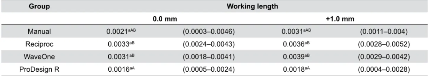
Table 1 shows the median values and range
(minimum and maximum) of apical extrusion values
for the different preparation techniques found with
the two apical limits tested. No statistically signiicant
differences were found when comparing the values
attained by each technique for the two apical limits
(0.0 mm and +1.0 mm) (P>.05). However, when
techniques were compared at the same limit, ProDesign
R produced signiicantly lower apical extrusion values
in comparison with the other reciprocating techniques
(P<.05), and with values similar to those of the control
(P>.05).
Group Working length
0.0 mm +1.0 mm
Manual 0.0021aAB (0.0003–0.0046) 0.0031aAB (0.0011–0.004) Reciproc 0.0033aB (0.0024–0.0043) 0.0036aB (0.0028–0.0052) WaveOne 0.0031aB (0.0018–0.0041) 0.0039aB (0.0029–0.0042) ProDesign R 0.0016aA (0.0005–0.0024) 0.0018aA (0.0004–0.0028)
a,bDifferent superscript lowercase letters indicate statistically signiicant differences according to the Mann-Whitney test (P<.05), considering
each preparation technique.
A,BDifferent superscript uppercase letters indicate statistically signiicant differences according to the Kruskal-Wallis and Dunn tests
(P<.05), considering each working length level.
The median values and range (minimum and
maximum) of the percentage of foraminal enlargement produced by the instrumentation techniques are
presented in Table 2. With the exception of the groups
that used the WaveOne Primary (G3), in which the
expansion beyond the AF reached 358.8%, no other
technique showed signiicant differences between
the two subgroups, i.e., the same system in the two
Group Working length
0.0 mm + 1.0 mm
Manual 30.93aA (14.75–98.46) 73.02aA (56.12–98.39)
Reciproc 145.2aA (56.53–187.66) 201.11aB (120.03–250.72) WaveOne 121.29aA (33.79–169.21) 358.80bB (298.08–450.34) ProDesign R 47.48aA (27.99–93.42) 96.14aAB (58.72–187.21)
a,bDifferent superscript lowercase letters indicate statistically signiicant differences according to the Mann-Whitney test (P<.05), considering
each preparation technique.
A,BDifferent superscript uppercase letters indicate statistically signiicant differences according to the Kruskal-Wallis and Dunn tests
(P<.05), considering each working length level.
Table 2 - Median (range) percentage increase of apical foramen diameter produced by the root canal preparation techniques at different working lengths
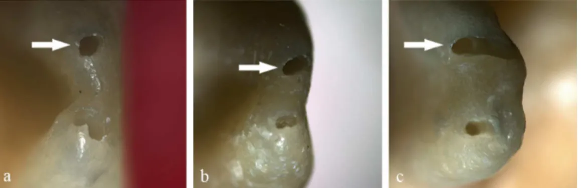
Figure 1- Apical foramen coniguration examples after mechanical preparation using different techniques and working lengths (a- circular, b- oval, c- deformed)
WLs (P>.05). When we analysed the percentage of
enlargement produced with the apical limit in the AF, we found no differences between techniques; the
control group (manual) had the lowest percentages
of enlargement. However, considering the preparation
beyond the AF, signiicant differences were observed
between control and G2 and G3 (P<.05); there were no
differences between G4 and the other groups studied.
Figure 1 presents post-preparation foraminal
aspects produced and illustrates the classiication
performed. While the statistical analysis did not show
signiicant differences, indings suggest the inluence
of the WL used when considering the instruments
made of M-Wire (Reciproc and WaveOne) in which the incidence of deformed foramens when the canals
were prepared beyond the AF (RCL+1.0 mm) reached
50%. The same behaviour was not observed in teeth
prepared with the instrument made of SMT-Wire, in which the percentages of deformed AFs were
equivalent in the two WLs (10%); this group had even
lower values than those of the manual preparation
(20%). Even when used at the level of the AF (0.0 mm), M-Wire NiTi alloy instruments had only 50%
(G2) and 30% (G3) of circular AFs; the SMT-Wire
instrument showed 70% of circular AFs. Figure 2 shows
the compilation of the data obtained in the evaluation of foraminal deformation.
Discussion
Null hypotheses tested were both partially
rejected, since we observed signiicant differences in
apical debris extrusion and foraminal enlargements. However, for foraminal deformation, these differences
were not statistically signiicant.
To understand the behaviour of apical extrusion
produced by the instrument, we determined the
percentage of foraminal enlargement and classiication
of these AFs regarding their formats. In addition, this
study evaluated the possible inluence on the patterns of these indings, in relation to two apical instrumentation
limits, AF and beyond it (WL2=RCL+1.0 mm). To
date, no study has evaluated the extrusion produced
by any reciprocating instrument made of SMT-Wire
NiTi-based alloy. Similarly, there were no references
in the literature to inal foraminal design associated
with apical extrusion or in preparations made up to
the AF or beyond it.
Mesial root canals of mandibular molars with
slight curvatures and AF patents with diameters
lower than 200 µm were used. This coniguration was
intended to approximate the conditions of the study
to clinical reality, without risking the homogeneity
of the sample. Studies with more constricted root
canals or larger curvatures may ind results differing
from those observed here. The dual chamber model
used here is enshrined in literature and has been
used by several studies that evaluated the extrusion produced by various preparation techniques, either by
extrusion of debris1,3,17,18,22 or bacteria20,21. Similarly,
observation of the shape of AFs by recording their
format in photographic documentation has also been used in the literature6,11; however, to this day, only two classiications have been used (circular and oval). The
authors of this study decided to include the deformed
proile according to the indings that suggested that
this form was an occurrence commonly observed in
some groups.
Results suggest a greater inluence of the
NiTi-based alloy type regarding the extrusion of debris and foraminal format produced by reciprocating systems.
Concerning the enlargement of AF, the apical limit
seemed to have had a more intense inluence, possibly
due to the increased metal mass of the instrument, depending on the taper, to surpass the AF. However,
the overall analysis of the observations made here has
helped to gain better understanding of the indings.
The Reciproc and WaveOne instruments caused the highest apical extrusion values, with no difference
between them. This similarity has previously been
pointed out by De Deus, et al.3 (2015) and Topçuoğlu,
et al.22 (2016). Silva, et al.17 (2016) evaluated the
apical extrusion promoted by Reciproc instruments
in two working lengths, at the AF and 1.0 mm
shorter; however, there was no information about
their behaviour in lengths beyond the AF threshold at which this similarity was also observed. Likewise,
to this day, no comparisons of these instruments
with those made of SMT-Wire were found in the
literature. Nevertheless, they were also systems that produced the highest values of foramen enlargement
and percentage of deformed foramen, irrespective
of the WL considered, which could possibly explain
why they extruded more debris. Due to having less
metal mass and thus more lexibility, it could be
considered that Reciproc instruments should provide
been offset by their greater cutting power, thereby
producing foraminal distortions equivalent to those of the WaveOne instrument. It must be considered
that the research method used here obliged the use
of distilled water as irrigant, therefore, the clinical
results of the use of sodium hypochlorite, or even a less irritant solution as irrigant, could be strongly
considered, mainly in over-extended WLs.
Another factor was the difference in the taper of
the ProDesign R in comparison with WaveOne and Reciproc instruments. The ProDesign R has a .06 taper,
consequently less metallic mass than the WaveOne
and Reciproc systems that have a .08 taper. Results
observed for the ProDesign R instrument have no parallel in the literature; however, considering the
comparison with other reciprocating instruments, the
authors could state that since it produced the best
results, it was a feasible option for use during root canal system preparation. Not only did this instrument
offer the best results of foraminal extrusion of debris,
it also differed from other mechanized instruments in
both WLs, offering similar results to those presented by
the manual instrumentation (control). This inding was
consistent with the analysis of expansion and foraminal
deformation, which showed that this instrument had
the lowest percentage of foraminal enlargement – 47.48% (RCL) and 96.14% (RCL+1.0 mm) –, in spite
of showing no statistically signiicant differences in
comparison with other mechanized systems. This
system also produced less AF deformation – 20% oval and 10% deformed at both WLs. Possibly, these
indings could be the remarkable result attributable
to the NiTi-based alloy of which the instrument is
made; other factors could be related, such as the cross-section, rotation angles and smaller taper of
the instrument. Higher foraminal enlargement and
deformation values may favour higher values of root
illing material extrusion into the apical tissues, which
could harm the apical repair process7.
Undeniably, it would have been better if this
evaluation had been carried out with instruments of
similar design (size/taper); however, manufacturers
consider that those used in this study are better
suited for use in root canals similar to those used in
this research. Moreover, all of the iles had a #25 tip,
the Reciproc and WaveOne had a .08 taper at their tips, and the Reciproc and ProDesign R presented “S”
shaped cross-sections, indications and similarities
that could justify this evaluation. Furthermore, it
should be understood that the NiTi-based alloy, of
which the instruments are manufactured, should not be considered the only factor to justify the results,
because it would be dificult to separate the alloy from the design of the iles.
According to results, the authors observed that performing chemical-mechanical preparation with the
WL established in the AF and beyond it could produce
deleterious changes to original anatomy and may
compromise the success of endodontic treatment, as mentioned by Çapar, et al.2 (2015). However,
the introduction of instruments made with
shape-memory technology may be an option for preparations
performed at the foraminal level. There is no literature discussing the ideal percentage of foraminal
enlargement to achieve maximum decontamination of
the apical region; however, this study shows that in
cases in which the Endodontist, in spite of knowing the risks involved, chose to overextend the preparation in
an attempt to increase the AF debridement – or this
occurred accidentally during instrumentation – highly
lexible instruments, such as those made of SMT-Wire
NiTi-based alloy, promoted less foraminal deformation
and extrusion of debris.
Conclusions
Under the conditions of this study, the authors were
able to conclude that all the instrumentation systems produced apical debris extrusion and foraminal
deformation; however, rather than the apical limit
used, the NiTi-based alloy and the taper were the
factors that inluenced the results of the reciprocating
instruments. The ProDesign R system, made with
shape-memory technology, and the .06 taper, showed
the best results.
Conlict of interest statement
The authors deny any conlict of interests related
to this study.
Acknowledgement
The authors wish to thank the State of São Paulo
Research Foundation for their partial support of this
study (#2016/00245-1).
The authors wish to thank Mrs. Lais de Souza
References
1- Bürklein S, Schäfer E. Apically extruded debris with reciprocating
single-ile and full-sequence rotary instrumentation systems. J Endod.
2012;38(6):850-2.
2- Çapar İD, Uysal B, Ok E, Arslan H. Effect of the size of the apical enlargement with rotary instruments, single-cone illing, post space preparation with drills, iber post removal, and root canal illing removal
on apical crack initiation and propagation. J Endod. 2015;41(2):253-6. 3- De-Deus G, Neves A, Silva EJ, Mendonça TA, Lourenço C. Calixto
C, et al. Apically extruded dentin debris by reciprocating single-ile and multi-ile rotary system. Clin Oral Investig. 2015;19(2):357-61.
4- Duque JA, Vivan RR, Cavenago BC, Amoroso-Silva PA, Bernardes
RA, Vasconcelos BC, et al. Inluence of NiTi alloy on the root canal
shaping capabilities of the ProTaper Universal and ProTaper Gold rotary instrument systems. J Appl Oral Sci. 2017;25(1):27-33.
5- Gambarini G, Testarelli L, De Luca M, Milana V, Plotino G, Grande
MN, et al. The inluence of three different instrumentation techniques
on the incidence of postoperative pain after endodontic treatment. Ann Stomatol (Roma). 2013;4(1):152-5.
6- González-Sánchez JA, Duran-Sindreu F, de Noé S, Mercandé M, Roig M. Centring ability and apical transportation after overinstrumentation
with ProTaper Universal and ProFile Vortex instruments. Int Endod J. 2012;45(6):542-51.
7- Holland R, Mazuqueli L, Sousa V, Murata SS, Dezan Júnior E, Suzuki
P. Inluence of the type of vehicle and limit of obturation on apical and periapical tissue response in dogs' teeth after root canal illing with
mineral trioxide aggregate. J Endod. 2007;33(6):693-7.
8- Kim HC, Kwak SW, Cheung GS, Ko DH, Chung SM, Lee W. Cyclic fatigue and torsional resistance of two new nickel-titanium instruments
used in reciprocation motion: Reciproc versus WaveOne. J Endod. 2012;38(4):541-4.
9- Limas Easy ProDesign R [internet]. 2017. [cited 2016 May 08].
Available from: http://www.easy.odo.br/limas/limas-easy-prodesign-r/.
10- Marceliano-Alves MF, Sousa-Neto MD, Fidel SR, Steier L, Robinson
JP, Pécora JD, et al. Shaping ability of single-ile reciprocating and heat-treated multiile rotary systems: a micro-CT study. Int Endod J.
2015;48(12):1129-36.
11- Marroquín BB, El-Sayed MA, Willershausen-Zönnchen B. Morphology of the physiological foramen: I. Maxillary and mandibular
molars. J Endod. 2004;30(5):321-8.
12- Pereira ES, Viana AC, Buono VT, Peters OA, Bahia MG. Behavior
of nickel-titanium instruments manufactured with different thermal treatments. J Endod. 2015;41(1):67-71.
13- Plotino G, Grande NM, Cotti E, Testarelli L, Gambarini G. Blue treatment enhances cyclic fatigue resistance of vortex nickel-titanium
rotary iles. J Endod. 2014;40(9):1451-3.
14- Rodrigues RC, Zandi H, Kristoffersen AK, Enersen M, Mdala I,
Ørstavik D, et al. Inluence of the apical preparation size and the
irrigant type on bacterial reduction in root canal-treated teeth with
apical periodontitis. J Endod. 2017;43(7):1058-63.
15- Schneider, SW. A comparison of canal preparations in straight and
curved root canals. Oral Surg Oral Med Oral Pathol. 1971;32(2):271-5. 16- Silva EJ, Rodrigues C, Vieira VT, Belladonna FG, De-Deus G,
Lopes HP. Bending resistance and cyclic fatigue of a new heat-treated reciprocating instrument. Scanning. 2016;38(6):837-41.
17- Silva EJ, Teixeira JM, Kudsi N, Sassone LM, Krebs RL, Coutinho-Filho
TS. Inluence of apical preparation size and working length in debris
extrusion. Braz Dent J. 2016;27(1):28-31.
18- Silva PB, Krolow AM, Pilownic AJ, Cassarin RP, Lima RK, Leonardo
RT, et al. Apical extrusion of debris and irrigants using different irrigation needles. Braz Dent J. 2016;27(2):192-5.
19- Souza RA. The importance of apical patency and cleaning of the apical foramen on root canal preparation. Braz Dent J. 2006;17(1):6-9.
20- Teixeira JM, Cunha FM, Jesus RO, Silva EJ, Fidel SR, Sassone
LM. Inluence of working length and apical preparation size on apical
bacterial extrusion during reciprocating instrumentation. Int Endod J. 2015;48:648-653.
21- Tinoco JM, De-Deus G, Tinoco EM, Saavedra F, Fidel RA, Sassone
LM. Apical extrusion of bacteria when using reciprocating single-ile and rotary multiile instrumentation systems. Int Endod J.
2014;47(6):560-6.
22- Topçuoğlu HS, Zan R, Akpek F, Topçuoğlu G, Ulusan Ö, Akti A, et
al. Apically extruded debris during root canal preparation using Vortex
Blue, K3XF, ProTaper Next, and Reciproc instruments. Int Endod J. 2016;49(12):1183-7.
23- Uezu MK, Britto ML, Nabeshima CK, Pallotta RC. Comparison of debris extruded apically and working time used by ProTaper Universal
rotary and ProTaper retreatment system during gutta-percha removal. J Appl Oral Sci. 2010;18(6):542-5.
24- Walia HM, Brantley WA, Gerstein H. An initial investigation of the
bending and torsional properties of Nitinol root canal iles. J Endod.
1988;14(7):346-51.
25- Wu MK, Wesselink PR, Walton RE. Apical terminus location of root
canal treatment procedures. Oral Surg Oral Med Oral Pathol Oral Radiol Endod. 2000;89(1):99-103.