Abstract
Submitted: June 3rd, 2016
0RGL¿FDWLRQ-DQXDU\
Accepted: January 29, 2017
I n vit ro
and
in vivo
evaluations of
glass-ionomer cement containing
chlorhexidine for Atraumatic
Restorative Treatment
Objectives: Addition of chlorhexidine has enhanced the antimicrobial effect of glass ionomer cement (GIC) indicated to Atraumatic Restorative Treatment (ART); however, the impact of this mixture on the properties of these materials and on the longevity of restorations must be investigated. The aim of this study was to evaluate the effects of incorporating chlorhexidine (CHX) in the in vit ro biological and chemical-mechanical properties of GIC and in vivo clinical/ microbiological follow-up of the ART with GIC containing or not CHX. Material and Methods: For in vit ro studies, groups were divided into GIC, GIC with 1.25% CHX, and GIC with 2.5% CHX. Antimicrobial activity of GIC was analyzed
A randomized controlled trial was conducted on 36 children that received ART m ut ans st rept ococci (MS) counts and the survival rate of restorations was checked after 7 days, 3 months and one year after ART. ANOVA/Tukey or Kruskal-Wallis/ Mann-Whitney tests were performed for in vit ro tests and in vivo microbiological analysis. The Kaplan-Meier method and Log rank tests were applied to estimate survival percentages of restorations (p<0.05). Results: Incorporation of 1.25%
affecting F release and mechanical characteristics, but 2.5% CHX was cytotoxic. Survival rate of restorations using GIC with 1.25% CHX was similar to GIC. A
1.25% CHX increased the in vit ro antimicrobial activity, without changing chemical-mechanical properties of GIC and odontoblast-like cell viability. This combination improved the in vivo short-term microbiological effect without affecting clinical performance of ART restorations.
Ke yw or ds: Dental atraumatic restorative treatment. Chlorhexidine. Glass ionomer cements.
Cristiane DUQUE1
Kelly Limi AIDA1
Jesse Augusto PEREIRA1
Gláucia Schuindt TEIXEIRA2
Angela Scarparo CALDO-TEIXEIRA2
Luciana Rodrigues PERRONE2
Karina Sampaio CAIAFFA1
Thais de Cássia NEGRINI3
Aline Rogéria Freire de CASTILHO4
Carlos Alberto de Souza COSTA5
http://dx.doi.org/10.1590/1678-7757-2016-0195
1Univ. Estadual Paulista (UNESP), Faculdade de Odontologia de Araçatuba, Departamento de
Odontologia Infantil e Social, Araçatuba, SP, Brasil.
2Universidade Federal Fluminense (UFF), Instituto de Saúde de Nova Friburgo, Departamento de
Odontologia, Nova Friburgo, RJ, Brasil.
3Universidade Federal do Rio Grande do Sul (UFRGS), Faculdade de Odontologia, Departamento de
Odontologia Conservadora, Porto Alegre, RS, Brasil.
4Universidade Estadual de Campinas (UNICAMP), Faculdade de Odontologia de Piracicaba,
Departamento de Odontologia Infantil, Piracicaba, SP, Brasil.
58QLY(VWDGXDO3DXOLVWD81(63)DFXOGDGHGH2GRQWRORJLDGH$UDUDTXDUD'HSDUWDPHQWRGH
)LVLRORJLDH3DWRORJLD$UDUDTXDUD63%UDVLO
Corresponding address: &ULVWLDQH'XTXH Departmento de Odontologia Infantil e Social. Faculdade de Odontologia de Araçatuba. Universidade Estadual Paulista (UNESP).
Introduction
Early childhood caries (ECC), mainly in developing
countries, is the most prevalent chronic disease in childhood and, consequently, a pending public health
problem6. Depending on the severity of ECC and the
number of dental sources of infection, this disease
causes functional, aesthetic and psychosocial disorders that reduce the quality of life of children and their
families6. The decay process of ECC generally tends
repair the longer it remains untreated. An alternative for the treatment of ECC is the Atraumatic Restorative
Treatment (ART). ART is a definitive restorative
treatment which consists of removing demineralized tooth tissues using minimal intervention to preserve
the tooth structure and restoring the dental cavity with
glass ionomer cement (GIC)9. The correct execution
of ART procedures may change the balance of the oral microbiota, reducing cariogenic microorganisms7.
This factor is relevant, because children affected by
ECC have high counts of cariogenic bacteria in saliva,
such as mutans streptococci and lactobacilli, and other species such as Candida albicans3. Additionally,
residual microorganisms can be found in dentin after
partial caries removal procedures with ART. Some
researchers have suggested the incorporation of antimicrobial agents into glass ionomer cements4,5,27.
Chlorhexidine (CHX) presents a wide spectrum of
activity against Gram positive bacteria, especially
mutans streptococci, Gram negative, aerobic and facultative anaerobic bacteria, and fungi8. Studies
have suggested that the incorporation of chlorhexidine
salts into glass ionomer cements (GIC) increases
their antimicrobial activity without compromising their physical-chemical properties11,25,26. On the other
hand, other studies have shown that the inclusion of
chlorhexidine into glass ionomer cements promoted
induced negative effects on the biocompatibility and
mechanical properties of the restorative material13.
One clinical study evaluated the long-term outcome of ART using glass ionomer cement containing CHX15.
Therefore, the objectives of this study were 1) to
evaluate the in vit ro
concentrations of CHX on biological and physical-chemical properties of a GIC and 2) to investigate in
vivo clinical/microbiological follow-up of the ART with
GIC containing CHX.
Material and Methods
Dental materials
GIC used was Ketac Molar Easy Mix® (KM, 3M
ESPE, Seefeld, Bavaria, Germany). This material was
Westphalia, Germany) without altering liquid/powder
26 (2008).
I n vit ro
study
Antimicrobial activity
Microorganism s and grow t h condit ions
St rept ococcus m ut ans (ATCC 25175), Lact obacillus
acidophilus (ATCC#IAL-523) and Candida albicans
(ATCC 40176) were obtained from Oswaldo Cruz
Foundation (FIOCRUZ, Rio de Janeiro, RJ, Brazil). S. m u t an s and L. aci d o p h i l u s were cultured on
Mitis Salivarius Agar (Difco Laboratories, Detroit,
MI, USA) with 0.2 UI bacitracin and Rogosa Agar
(Difco Laboratories, Detroit, MI, USA) for 24-48 h at 37°C in 5% CO2. Candida albicans were grown in
Sabouraud Dextrose Agar (Difco Laboratories, Detroit,
MI, USA) for 24-48 h at 37°C in aerobic conditions. Subsequently, colonies were transferred to Brain-Heart
Infusion broth (BHI; Difco Laboratories, Detroit, MI,
USA) for 18-24 h at the same conditions. Cultures
were adjusted to 1-5x10⁸ cells/mL in order to obtain an inoculum for subsequent tests.
Agar diffusion t est
This test was conducted according to Castilho, et
al.5 (2012). Twelve 5-mm-diameter wells were made
with 1.25% CHX, and KM with 2.5% CHX. All materials were handled under aseptic conditions according to the
manufacturer’s instructions, inserted into wells using
a syringe (Centrix Inc., Shelton, CT, USA) and light
Brazil) for 30 s. Light output was periodically checked
(approximately 500 W/cm2). Positive control used was
0.2% CHX. After 2 h of material diffusion, the plates were incubated for 24 h in each microorganism’s
conditions. Then, inhibition zones around the materials
were measured using a digital caliper.
%LR¿OPDVVD\V
previous described by Hu, et al.12 (2013). Five
cylindrical of each KM group containing or not CHX
4 mm diameter) and individually suspended in 24-well
plates (Corning Inc., New York, NY, USA) containing 2
mL of BHI broth supplemented with 1% sucrose and 2
μl of inoculum. The plates were incubated in 5% CO2
at 37°C for 24 h. After this period, GIC samples were
washed, immerged in 500μl of 0.9% NaCl solution
and sonicated in an ultrasonic cell disruptor at 7 W .
This solution was diluted and plated on BHI agar and
incubated for 48 h at 37°C. Then, bacterial colonies were counted and expressed in colonies forming units/
mL (CFU/mL). Three independent assays (n=15) were
performed for the analysis.
Cyt ot oxicit y assays
These assays were conducted in accordance with
Castilho, et al.5 MDPC-23
odontoblast-like cells were used. The cells were seeded (30,000 cells/cm2/well) in sterile 24-well plates and maintained 2 and 95%
air at 37°C
PA, USA). Ten round-shaped samples of each group (2x4 mm) were prepared in stainless-steel molds,
light-cured for 30 s and maintained for 1 h at 37°C
in relative humidity. The specimens were then
inserted into sterile 24-well plates containing DMEM
h. After that, 800 μL of the extract from each well
was applied to previously cultured MDPC-23 cells for 24 h. Cell metabolism was analyzed using methyl
tetrazolium (MTT) assays. The means were calculated
for the groups and transformed into percentages, and
metabolism.
Measurement of mechanical properties
Com pressive t ensile st rengt h and m icrohardness t est s5
Ten specimens from each group were prepared in cylindrical molds for compressive strength (4x2
mm) and surface microhardness tests (3x6 mm).
Compressive tensile strength tests were performed in
an Instron universal test machine (4411, Instron Co., Canton, MA, USA) in a vertical position using a load at
a crosshead speed of 1.0 mm/min until failure occurred
and the values were calculated by dividing the load (F) by the cross-sectional area and converted to MPa.
Microhardness was measured using a microhardness
tester (Shimadzu HMV-2000 Micro Hardness Tester;
Shimadzu Corporation, Kyoto, Keihanshin, Japan), under a static load (Knoop) of 50 gf for 5 s. Five
indentations were randomly performed, 500 μm apart, on the top surface of the material and hardness means
were obtained for each sample.
Measurement of chemical properties
Fluoride release23
Six specimens of each group were made with 5 mm and 2 mm diameter, with a surface area of
0.71 cm2. Each specimen was placed in 4 ml of
deionized water under agitation at room temperature
for 24 h. An equal volume of TISAB II (acetate buffer 1.0 M, pH 5.0, containing NaCl 1.0 M and
1,2-cyclohexanediaminetetraacetic 0.4%) was added
to the tubes. The specimens were daily washed with
deionized water, dried with absorbent paper and transferred to new tubes containing 4 ml of deionized
water. The solutions from 24 h and 7 days were
Orion Research, Inc., Beverly, MA, USA) connected to
a digital ion-analyzer (Orion 720A, Orion 9609-BN,
Orion Research, Inc., Beverly, MA, USA), previously
calibrated with standard solutions of 0.0625 to 1 or 1 to 16 mg F-/ml in TISAB II, and expressed in mg F-/cm2.
I n vivo
study
St udy design
The present study was designed as a randomized
controlled clinical trial with parallel groups. One
hundred and tirty six three to six-year-old children from
four public primary schools of Nova Friburgo (Rio de Janeiro, Brazil) whose parents signed a written consent
were examined for dental caries status using the
criteria developed by the WHO. Inclusion criteria were
(1) good general health; (2) cooperative behavior; (3) at least one cavitated dentin carious lesion (occlusal
or occluso-proximal cavities) in primary molars that
had an opening wide enough for the smallest ART
were children with mixed dentition, teeth with pulpal
history of sensitivity and/or spontaneously pain. The
study was approved by Research Ethics Committee of the Federal Fluminense University (reference
number 056/2010) and registered at the Clinical Trials
NCT02459730). Parents and/or caretakers were informed in writing about the investigation and
treatments. Children whose parents or caretakers
failure rates reported for conventional approximal
ART restorations using high-viscosity glass ionomer
cements (HVGIC) in primary posterior teeth (29%)
after one year1. For ART restorations with HVGIC
containing CHX, there were no reliable failure rate
data. It was considered a positive outcome if the
results were similar to those with HVGIC, showing clinical equivalence. A hypothetical minimal difference
of 20% among groups were considered with a
probability of type I error of 5% and a power of 80%. A minimum of 41 restorations were calculated per
sample was increased in 20% resulting in at least 49
restorations per group.
ART procedures
An independent dentist randomly distributed
children in two groups. ART restorations and clinical
evaluation were performed by a trained and previously calibrated pediatric dentist (CD), aided by two trained
graduate students (LRP and KSC), using a portable
bed and an operating light. The mean kappa value
for the intra-examiner reproducibility was 0.78. Restorations were performed according to the ART
approach described by Frencken, Taifour and van´t
Hof9
removing infected dentin with hand instruments. No local anesthesia was used. Relative moisture isolation
was performed with cotton wool rolls. Then, the
cavities were conditioned with liquid from the material,
washed and dried with cotton pellets. They were
with one of the randomly selected materials: (1) Ketac
Molar Easy Mix® containing 1.25% CHX (KM+CHX;
n=17 children; 49 restorations), or (2) Ketac Molar
Easy Mix® as a control group (KM; n=19 children;
68 restorations). Each tooth was considered as the
sampling unit. However, all carious teeth indicated to
ART in each child were treated exclusively with one of the materials tested. After the removal of material
excess and adjustment of the occlusion using the
carver instrument, the restoration was coated with a layer of petroleum jelly. Multiple-surface cavities were
The dentist gave instructions to caregivers for children
not to eat solid food for one hour.
Follow up
An independent dentist, previously trained and
calibrated, evaluated the restorations after 7 days,
3 months and 1 year of treatment. Following the
ART criteria adopted for approximal restorations, as
proposed by Roeleveld, et al.24 (2006), restorations
were considered as a success (codes 00 and 10),
failure (codes 11-40) or unavailable (codes 50-91).
All carious teeth were treated with the same GIC
used in molars for each patient, but only molars were considered for statistical analysis. New restorations
were carried out to replace failed restorations but they
were not considered in subsequent analysis. Children were encouraged and instructed on dental hygiene,
and received all other necessary oral care.
Microbiological assays
Unstimulated whole saliva and pooled supragingival
surfaces, except from the interior of the cavities, were
collected from each subject. A sterile plastic disposable
(Greiner, Frickenhausen, Germany) was used to collect
22
into 1 mL microtubes containing Tris-EDTA buffer (10
performed at least 1 h after feeding and the tubes
were transported on ice and processed within 2 h. The samples were homogenized and the suspensions were
serially diluted in 0.9% NaCl solution. Each dilution
was cultivated in triplicate on the surface of Mitis
Salivarius Agar (Difco Laboratories, Detroit, MI, USA) with sucrose and 0.2 U/ml bacitracin for isolation of
mutans streptococci (MS). All plates were incubated
at 37°C for 48 h in 5% CO2 atmosphere. After 48 h
of incubation, the number of CFU was counted using a stereoscopic microscope and the results were
expressed as CFU/mL.
Statistical analysis
Data were submitted to normality and homogeneity of variance tests, using the SPSS (version 17)
were analyzed by Kruskal-Wallis and Mann-Whitney
tests. ANOVA and Tukey tests were used to evaluate data from agar diffusion tests, cytotoxicity, mechanical
Kruskal/Wallis and Mann-Whitney tests were used to
compare differences among material groups in the same period of time (7 days, 3 months or 1 year of
evaluation) for microbiological analysis. The Wilcoxon
test was used to compare microbiological differences
tests were applied to estimate survival percentages
of restorations2. All statistical tests were considered
Results
I n vit ro
study
Antimicrobial activity
Mean values of the results for the agar diffusion
test are shown in Table 1. KM was not effective against all microorganisms tested. When CHX was
incorporated into KM, it presented an inhibitory
activity on all microorganisms. However, an increased
antimicrobial effect. Regarding the S. m ut ans
anti-action of KM+2.5% CHX was statistically better (p=0.007) than the observed for KM+1.25% CHX
(Figure 1).
Toxicit y on odont oblast - like cells
Figure 2 shows that KM and KM+1.25% CHX did not present a cytotoxic effect. However, when KM was
cell viability was observed.
0HFKDQLFDODQGÀXRULGHUHOHDVHSURSHUWLHV
The results of compressive strength and
microhardness tests are shown in Table 2 and the
in both concentrations, did not affect these properties when compared to control group.
I n vivo
microbiological and clinical assessments
A CONSORT flowchart of the patients and
restorations made along this study is described in
(55.6%) of them were females. The population’s mean
KM KM+1.25% CHX
KM+2.5% CHX
Streptococcus mutans
0a b b
Lactobacillus acidophilus
12:00 AM b b
Candida albicans
12:00 AM b b
Table 1-0HDQVVWDQGDUGGHYLDWLRQVRILQKLELWLRQ]RQHVPPIRU
the glass ionomer cements against the tested microorganisms, using agar diffusion tests
aDifferent lower letters indicate a statistical difference among the
groups of materials, according to the ANOVA and Tukey tests
S
Figure 1 Box-whisker plots of the S. mutansDQWLELR¿OPDFWLYLW\
of the glass ionomer cements. Bars indicate minimum and maximum values. Black and white boxes indicate lower and
XSSHUTXDUWLOHVUHVSHFWLYHO\7KHOLQHLQWKHPLGGOHRIWKHER[HV
is the median
aDifferent lower letters indicate a statistical difference among the
JURXSV RI PDWHULDOV DFFRUGLQJ WR WKH .UXVNDO:DOOLV S
DQG0DQQ:KLWQH\WHVWVS aDifferent lower letters indicate a statistical difference among the
groups of materials, according to the ANOVA and Tukey tests
S
Figure 2- Means (standard deviations) of the percentage of odontoblast-like cell viability after exposure to extracts obtained from glass ionomer cements (MTT assays)
KM KM+1.25% CHX KM+2.5% CHX p value
Compressive strength (MPa)
a a a 0.992
Knoop microhardness (KHN)
a a a 0.908
Table 2-0HFKDQLFDOSURSHUWLHVPHDQVVWDQGDUGGHYLDWLRQVRIJODVVLRQRPHUFHPHQWVFRQWDLQLQJRUQRWFKORUKH[LGLQH
age was 46.09±7.9 months. There was no statistical
difference among groups of materials in relation to age
53%), mean ± standard deviation of molar surfaces
treated (KM: 3.47±3.76; KM+CHX: 2.41±2.42) and
number of teeth with single surface restorations (KM:
(p>0.05, ANOVA and Chi-square tests). Dmfs (decay,
KM and KM+CHX groups, respectively. In relation to molar restoration retention at different follow-up
times, there were 21 failures in KM after 3 months
and 11 after one year. For the KM+CHX group, failures were observed only in the third month (n=14) and one
year after ART (n=10). However, survival percentage
of restorations were similar among groups (Table 3).
The main reason for restoration failures was partial or total fracture of restorations. Only two teeth treated
with KM had secondary caries and one tooth treated
year of ART. Microbiological analysis at follow-up times is presented in Table 4. The best antimicrobial
aThe same lower letters indicate no statistical difference among
the groups of materials, considering each time separately,
DFFRUGLQJWRRQHZD\$129$KS GS
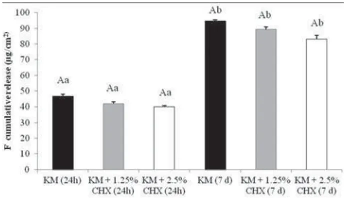
Figure 3- 0HDQV VWDQGDUG GHYLDWLRQV RI FXPXODWLYH ÀXRULGH
release (ugF/cm2) from glass ionomer cements containing or not
containing chlorhexidine after 24 h and 7 days in deionized water
performance was observed in the experimental group
(KM+CHX) at the 7th day follow-up for both saliva and
was also observed after 1 year of ART.
Discussion
Several attempts have been made to introduce antimicrobial properties to restorative materials,
including the incorporation of CHX salts into GICs,
focusing on a new perspective for arresting residual
caries after ART. Some authors demonstrated that the addition of CHX to glass ionomer cements improved the
inhibitory effect against oral microorganisms, including
St r e p t o co ccu s and La ct o b a ci l l u s species7,19,20,25.
However, there are several differences in the methodologies used in these studies, mainly in the
glass ionomer cement and chlorhexidine salt chosen
for the experiments. The present study evaluated the
inhibitory effect of adding chlorhexidine digluconate to Ketac Molar Easy Mix (KM) against S. m ut ans, L.
acidophilus and C. albicans
digluconate and diacetate forms of chlorhexidine presented antimicrobial activity7,20,25-27. However,
differences among them were found considering
the inhibition zones against S. m u t a n s and L.
acidophilus, indicating that the type of salt may affect the antimicrobial action of CHX when associated
with GICs25-27. The results of the current study
are in agreement with the microbiological results obtained by Marti, et al.20 (2014) on S. m ut ans and L.
acidophilus. Regarding Candida albicans, the current
al.26 (2008). This may be related to the glass ionomer cement chosen for the study and the agar diffusion
methodology.
A d d i t i o n a l l y, t h i s s t u d y e va l u a t e d t h e
St rept ococcus m ut ans has
been implicated as the main etiological agent of dental
formation17. This study demonstrated that the activity of GIC containing CHX against S. m ut ans
Intervals of time N child * N restorations N dropout N failed Survival % Means (SE)
KM
0 – 7 d 19 68 1 0 100 (0)
7 d – 3 m 18 67 1 21 68.19 (15.29)
3 m – 1 y 12 45 7 11 48.45 (8.36)**
KM+CHX
0 – 7 d 17 49 0 0 100 (0)
7 d – 3 m 17 49 0 14 71.43 (14.57)
3 m – 1 y 11 35 6 10 48.57 (11.43)
*Nchild – number of children at start of interval, N restorations number of restorations at start of interval, N dropout number of restorations dropout at the end of interval, N failed number of restorations that failed at end of interval
/RJUDQN&KLVTXDUH GI S
Table 3- Cumulative survival (means - %) and standard error of the means (SE) of ART restorations in primary molars treated with glass ionomer cements containing or not chlorhexidine
Saliva %LR¿OP
KM KM+CHX p value KM KM+CHX p value
Baseline 5.61 (5.50) 0.19Aa 5.13 (5.27) 0.21Aa 0.288 5.47 (5.20) 0.25Aa 5.44 (5.24) 0.29Aa 0.908
7 days 5.53 (5.47) 0.27Aa 4.52 (4.48) 0.13Bb 0.012 5.67 (5.55) 0.23Aa 4.59 (4.49) 0.22Bb 0.015
3 months 5.38 (4.98) 0.28Aa 5.34 (5.38) 0.20Aa 0.631 4.52 (4.79) 0.31Aa 4.72 (4.83) 0.17Aa 0.748
1 year 5.51 (5.54) 0.30Aa 4.44 (4.68) 0.29Aa 0.109 4.66 (5.11) 0.55Aa 4.25 (4.59) 0.29Ab 0.361
A'LIIHUHQWFDSLWDOOHWWHUVLQGLFDWHDVWDWLVWLFDOGLIIHUHQFHDPRQJWKHPDWHULDOV.0[.0&+;FRQVLGHULQJVDOLYDDQGELR¿OPVHSDUDWHO\
DFFRUGLQJWR.UXVNDO:DOOLVDQG0DQQ:KLWQH\WHVWVS
a'LIIHUHQWORZHUOHWWHUVLQGLFDWHDVWDWLVWLFDOGLIIHUHQFHDPRQJWKHJURXSVLQHDFKSHULRGRIHYDOXDWLRQDFFRUGLQJWR:LOFR[RQWHVWVS
)RU.0GD\V[PRQWKVS )RU.0&+;VDOLYD%DVHOLQH[GD\VS GD\V[PRQWKV .0&+;ELR¿OP %DVHOLQH[GD\VS %DVHOLQH[\HDUS
group. Almost all studies in the literature demonstrated
the antibacterial effectiveness of incorporating CHX
to a conventional GIC by using the agar diffusion test
and not by activity25-27. In the present
study, considering the limitations of the in vit ro
anti-reduced S. m ut ans counts adhered to the GIC surface,
CHX concentration. Although this study used CHX
digluconate and a different glass ionomer cement, the results are in accordance with those obtained by Hu, et
al.12 (2013) and Du, et al.10 (2012). It was speculated
that CHX released from the material could persist in
the environment, due to its substantivity, creating a bacteriostatic effect and interfering on bacterial
17.
Studies have demonstrated that the addition
of antibacterial agents can change the mechanical properties of glass ionomer cements20,25-27. In the
present study, the mechanical properties of GIC were
not negatively affected by the addition of CHX (1.25
or 2.5%) when compared with the control group.
by Takahashi, et al.25 (2006) and Hu, et al.12 (2013).
According to Jedrychowski, Caputo and Kerper13
(1983), glass ionomer cement deteriorates after the addition of CHX at concentrations above 5%.
Fluoride is widely used as a highly effective
anti-caries agent. Fluoride has also an antimicrobial
activity, affecting bacterial metabolism, directly as an enzyme inhibitor or by reducing the acid tolerance of
the bacteria19. by glass ionomer
cements is one of its most important properties, and is
intrinsically associated with the anti-caries effect of the cement.
by the incorporation of both concentrations of CHX,
27 (2011). Fluoride release
of GICs after the addition of 10% CHX decreased over
time, but remained measurable after 60 days11. It
was speculated that it is an interaction between the
the precipitation of salts with lower solubility, leaving
11.
Biocompatibility is a property required for GICs,
since these materials are usually applied in deep dentin and could release toxic components, which might
indirectly affect the dental pulp5,18. High concentrations
of CHX have cytotoxic effects on odontoblastic cells18.
However, those results are related to in vit ro direct
contact of CHX with cells. In this study, toxicity
against odontoblastic cells was observed only for the
combination of GIC with the highest concentration of CHX (2.5%). The current results are in agreement with
those obtained by Castilho, et al.5 (2012).
Regarding the present clinical trial, the survival
rate after one year was approximately 48% for both materials. The majority of multiple-surface
restorations for both groups (KM: 80.89% and
KM+CHX: 77.56%) could explain partially the lower survival rate values. This result was slightly higher
than 44.8% obtained by Kemoli, et al.15 (2009) and
lower than 65% obtained by Yu, et al.30 (1998),
using the same GIC in class II restorations, over a comparable period of time. Higher percentages were
obtained in some studies presented in the systematic
review of Amorim, et al.1 (2012). The authors found
a weighted mean score of 71% for survival rate of
multiple-surface restorations in primary teeth after
one year. More recently, a cumulative survival rate of
80.9% was obtained for multiple-surface restorations
using high viscosity glass ionomer cement within the same period of time10. The literature presents survival
rates of ART restorations with high viscosity GIC in
posterior teeth ranging from 74 to 100% and 31 to
100% for single or multiple surface, respectively,
1. This wide range of
survival percentages observed in the studies, mainly
for approximal surfaces, is attribute to a combination
of factors, such as cavity selection and preparation, salivary contamination, restorative material, and the
operator knowledge and clinical skills2,28. Particularly
the cervical area of cavities increases the risk of
microleakage, secondary caries formation and
restoration failure28. Besides, large cavities did not show good survival results, probably because of bulk
failures or pulpal effect16.
between GIC containing and not containing CHX, even after 1 year of treatment, confirming that
the addition of chlorhexidine digluconate did not
affect the mechanical properties of the restorative
material. A recent study showed that the addition of 0.5% CHX to GIC improved antibacterial properties
compared to conventional GIC, without affecting the
clinical performance of class I restorations in young
However, in contrast to our results, after 9 months the
restoration success with GIC containing CHX (60%)
was lower than the control group (85%)14. Differences of age, type of dentition and restorative material used
could explain the disparities found in the studies.
Although the oral hygiene index was not applied to
this study participants, it is expected that a poor oral hygiene, since high scores of dfms were observed in
a low age population (46.09±7.9 months), may have
an overall impact on the survival of restoration29.
count on both saliva and biofilm from children
at the 7th day follow-up of ART procedure with
GIC containing 1.25% CHX was observed in the present study, showing the antimicrobial action
of CHX on buccal environment. The reduction in
cariogenic microorganisms could be attributed to
both cavity sealing and the antimicrobial properties of chlorhexidine digluconate. This antimicrobial agent
has a wide spectrum of activity against Gram-positive
bacteria, especially mutans streptococci8. However,
the antibacterial effect of CHX associated with GIC seems to be limited, since after 3 months and one
year of restoration, the experimental group did not
to the control group. In a clinical trial study with a
chlorhexidine digluconate, it was found that the
antibacterial action of the material on residual dentin
lasts up to 90 days after the restorative procedure5. I n v iv o addition of 1% chlorhexidine diacetate to
GIC showed comparable results to conventional GIC
with regard to microleakage21. Differences in the
selection of materials, sampling procedures and local of CHX action could explain the controversial results
of GIC containing CHX. In this study, we used a conventional high viscosity GIC that may easily release
may keep the same product for long time in the matrix,
delaying its release. Furthermore, in this study, GIC was exposed to oral environment and it was subject
to tooth abrasion that probably accelerated the
chlorhexidine release.
The results of this study should be analyzed considering possible methodological limitations. One
of them is the dropout rate, approximately 36%, that
was higher than expected (20%), at one-year
follow-up of the intervention. The main reasons for dropout
were school transfer or traveling abroad with their
parents. This fact could interfere in the reliability of
results. Unfortunately, when the study was conducted, the schools have not been registered in the national
the schools in the national system. Another limitation
is the combination of single and multiple-surface
the comparison with other studies. The participation of
younger children whose tooth restoration is considered
of restorative treatment could also explain the low
success rate of ART restorations in the present study.
This low success rate raises the question about the longevity of approximal-ART restorations. Then,
besides the antimicrobial effect, new restorative
materials with enhanced mechanical properties could
minimize cumulative effect of failures.
Conclusions
The inclusion of CHX in GIC improves in v it r o
antimicrobial/antibiofilm action, without causing
detrimental effects on cytotoxicity, mechanical and
follow-up demonstrated that ART restoration with
GIC+CHX had a similar survival rate and better
antimicrobial performance at the 7th day when
compared to conventional GIC. GIC containing chlorhexidine could be an alternative to traditional
GIC indicated to ART, for it provides an additional
antimicrobial effect that is interesting for children
with high mutans streptococci counts during the initial adaptive phase of treatment.
Acknowledgments
This study was supported by the Rio de Janeiro
E-26/100.487/2010 and E-26/110.205/2011).
References
1- Amorim RG, Leal SC, Frencken JE. Survival of atraumatic restorative treatment (ART) sealants and restorations: a meta-analysis. Clin Oral Investig. 2012;16(2):429-41.
van Amerongen WE. Survival rate of approximal-ART restorations using a two-layer technique for glass ionomer insertion. Clin Oral Investig. 2013;17(7):1745-50.
3- Carvalho FG, Silva DS, Hebling J, Spolidorio LC, Spolidorio DM. Presence of mutans streptococci and Candida spp. in dental plaque/ dentine of carious teeth and early childhood caries. Arch Oral Biol. 2006;51:1024-8.
4- Castilho AR, Duque C, Negrini TC, Sacono NT, Paula AB, Sacramento
glass-ionomer cement containing doxycycline hyclate. Arch Oral Biol. 2012;57:131-8.
5- Castilho AR, Duque C, Negrini TC, Sacono NT, Paula AB, Souza Costa CA, et al. I n vit ro and in vivo investigation of the biological
containing chlorhexidine. J Dent. 2013;41:155-63.
6- Chaffee BW, Cheng A. Global research trends on early-life feeding practices and early childhood caries: a systematic review. J Oral Dis. 2014;2014:675658.
7- Du X, Huang X, Huang C, Frencken JE, Yang T. Inhibition of early in vivo: a pilot study. Aust Dent J. 2012;57:58-64.
8- Emilson CG. Susceptibility of various microorganisms to chlorhexidine. Scand J Dent Res. 1977;85:255-65.
9- Frencken JE, Taifour D, van´t Hof MA. Survival of ART and amalgam restorations in permanent teeth of children after 6.3 years. J Dent Res. 2006;85:622-6.
10- Hilgert LA, Amorim RG, Leal SC, Mulder J, Creugers NH, Frencken JE. Is high-viscosity glass-ionomer-cement a successor to amalgam for treating primary molars? Dent Mater. 2014;30(10):1172-8. 11- Hoszek A, Ericson D. I n vit ro
effect of glass ionomers containing chlorhexidine gluconate. Oper Dent. 2008;33:696-701.
12- Hu J, Du X, Huang C, Fu D, Ouyang X, Wang Y. Antibacterial and physical properties of EGCG-containing glass ionomer cements. J Dent. 2013;41:927-34.
13- Jedrychowski JR, Caputo AA, Kerper S. Antibacterial and mechanical properties of restorative materials combined with chlorhexidines. J Oral Rehabil. 1983;10:373-81.
14- Kabil NS, Badran AS, Wassel MO. Effect of the addition of chlorhexidine and miswak extract on the clinical performance and antibacterial properties of conventional glass ionomer: an in vivo study. Int J Paediatr Dent. 2016. doi: 10.1111/ipd.12273. Epub ahead of print.
the survival rate of proximal ART restorations in primary molars. Int J Paediatr Dent. 2009;19(6):423-30.
16- Konde S, Raj S, Jaiswal D. Clinical evaluation of a new art material:
Community Dent. 2012;2:42-7.
The virulence of St rept ococcus m ut ans
Eur J Clin Microbiol Infect Dis. 2014;33:499-515.
18- Lessa FC, Nogueira I, Vargas FS, Spolidorio DM, Hebling J, García-Godoy F, et al. Direct and transdentinal antibacterial activity of chlorhexidine. Am J Dent. 2010;23:255-9.
J Microbiol. 1995;41:955-64.
20- Marti LM, Mata M, Ferraz-Santos B, Azevedo ER, Giro EM, Zuanon AC. Addition of chlorhexidine gluconate to a glass ionomer cement: a study on mechanical, physical and antibacterial properties. Braz Dent J. 2014;25:33-7.
21- Mathew SM, Thomas AM, Koshy G, Dua K. Evaluation of the
in vivo study. Int J Clin Pediatr Dent. 2013;6:7-11.
22- Parisotto TM, Steiner-Oliveira C, Duque C, Peres RC, Rodrigues LK, Nobre-dos-Santos M. Relationship among microbiological composition and presence of dental plaque, sugar exposure, social factors and different stages of early childhood caries. Arch Oral Biol. 2010;5:365-73.
23- Pedrini D, Delbem AC, França JG, Machado TM. Fluoride release by
gel. Pesqui Odontol Bras. 2003;17(2):137-41.
caries and cervical gaps on the survival rate of Class II glass ionomer restorations. Eur Arch Paediatr Dent. 2006;7:85-91.
25- Takahashi Y, Imazato S, Kaneshiro AV, Ebisu S, Frencken JE, Tay FR. Antibacterial effects and physical properties of glass-ionomer cements containing chlorhexidine for the ART approach. Dent Mater. 2006;22:647-52.
antibacterial effects and physical properties of a chlorhexidine-containing glass ionomer cement. J Esthet Restor Dent. 2008;20:29-44.
Antibacterial activity and physical properties of conventional glass-ionomer cements containing chlorhexidine diacetate/cetrimide mixtures. J Esthet Restor Dent. 2011;23:46-55.
28- Tyas MJ. Clinical evaluation of glass-ionomer cement restorations. J Appl Oral Sci. 2006;14:10-13.