This study aimed to evaluate in vitro the antimicrobial effect of a bioadhesive chitosan-based oral membrane with chlorhexidine for local treatment of infections in the oral tissues. Five oral membranes of different compositions were tested: 5% chitosan (G1); 5% chitosan ± 0.2% chlorhexidine (G2), 5% chitosan ± 0.6% chlorhexidine (G3), 5% chitosan ± 1.0% chlorhexidine (G4), and 5% chitosan ± 2.0% chlorhexidine (G5). Also, five gel types were tested according to the following compositions: 5% chitosan gel (G6), 0.2% chlorhexidine gel (G7), 2.0% chlorhexidine gel (G8), 5% chitosan gel ± 0.2% chlorhexidine gel (G9), and 5% chitosan gel ± 2.0% chlorhexidine gel (G10). The antimicrobial action of the samples was tested against Candida albicans and Streptococcus mutans through antibiogram by measuring the inhibition halos. Data were statistically analyzed by Kruskal-Wallis and one-way ANOVA followed by Tukey test (p<0.05). The 2.0% chlorhexidine membrane (G5) and the disks containing 2.0% chlorhexidine gel (G8) showed the greatest inhibition halos for both microorganisms, with statistically significant difference when compared to others tested groups (p=0.008) only for Candida albicans inhibitions results.
All the other formulations of membranes and gels showed inhibition halos, but without statistically significant difference. The bioadhesive chitosan-based oral membrane with 2% chlorhexidine and 2% chlorhexidine gel were the most effective in inhibiting the tested microorganisms.
I n V i t r o A n t i m i c r o b i a l E f f e c t
of Bioadhesive Oral Membrane
w i t h C h l o r h e x i d i n e G e l
Annelyze Podolan Kloster1, Natalino Lourenço Neto1, Silgia Aparecida da Costa2, Thais Marchini Oliveira1, Rodrigo Cardoso de Oliveira3, Maria Aparecida Andrade Moreira Machado1
1Department of Pediatric Dentistry,
Orthodontics and Community Dentistry, Bauru Dental School, USP - Universidade de São Paulo, Bauru, SP, Brazil 2Course on Textiles and Fashion,
School of Arts, Sciences and Humanities, USP - Universidade de São Paulo, São Paulo, SP, Brazil 3Department of Biological Sciences,
Discipline of Biochemistry, Bauru Dental School, USP - Universidade de São Paulo, Bauru, SP, Brazil
Correspondence: Rodrigo Cardoso de Oliveira, Alameda Dr. Octávio Pinheiro Brisolla, 9-75, 17012-901 Bauru, SP, Brasil. Tel: +55-14-3235-8295. e-mail: [email protected]
Key Words: Layer-by-layer coatings, chitosan, chlorhexidine, anti-infective agents.
Introduction
Dentistry requires therapeutic alternatives to be applied on surgical wounds and wounds caused by physical and/or chemical trauma, periodontal and dental pulp infections, and other types of ulcerated oral lesions (1-5). The development of an alternative vehicle to allow the releasing and the contact of the drug with the affected tissue, for a longer time, will greatly contribute to repair (6-8).
Biomaterials allow interaction and repair of a biological system (1,9-10). Key features such as biodegradability and biocompatibility are fundamental for new biomaterials to be developed in medical and dental area (5-6,11-12). Chitosan is a biopolymer obtained from chitin and has interesting properties for the production and development of new “drug-delivery” biomedical products, allowing to be used as bioadhesive membranes, gels, films, and capsules (6-7,13-14). Recently, the chitosan has gained popularity for its use as a matrix molecule for drug delivery with upcoming utility and often used in medicine and dentistry (15-16). Wieckiewicz et al. (16) affirmed it can be applied in all fields of dentistry including preventive dentistry, conservative dentistry, endodontics, surgery, periodontology, prosthodontics and orthodontics.
Chlorhexidine is routinely used in dentistry to control infections, thus helping in wound repair. This drug is often
used in gel formulation in concentrations between 0.12% to 5% presenting a bactericidal and bacteriostatic properties, and is considered the “gold standard” of oral antiseptics, with the disadvantage of not having the intrinsic ability to bind to the oral tissues. In this context, the association of chlorhexidine with chitosan-based drug-delivery membrane seems to be a promising alternative for local treatment of infections in the oral tissues (2-3,5,17-20).
Therefore, the creation of oral membranes associated with chlorhexidine as a new “drug-delivery” system, malleable as the oral tissues from the mouth, with adhesion and continuous liberation of the medicament seems to be a viable solution to a constant problem observed in dentistry, which has currently available only the medicaments in gel or solution that has less contact time with the tissues and therefore end up with a low effectiveness in the control of oral mucosal infections.
In this sense, this study aimed to evaluate in vitro the antimicrobial effect of a bioadhesive chitosan-based oral membrane with chlorhexidine for local treatment of infections in the oral tissues.
Material and Methods
Antimicrobial bioadhesive oral membrane as the production of oral membranes, followed the same
procedures and development described by SILVA et al. (5). Preparation of Chitosan Gels: the chitosan gel was prepared by dissolving 5% of chitosan (m/v) (Sigma-Aldrich, St. Louis, MN, USA) in 5% of an acetic acid solution with a range between 1% to 5% (v/v). The solution was kept under stirring for a time ranging from 8 to 30 hr. Thereafter, a percentage range between 0.5 to of 3% of glycerol were added to the gels (Fig. 1).
For preparation of oral membranes, multilayer membranes were prepared with individual layers of 5% chitosan gel and the drug layer among them. Each layer was placed individually and dried inside an oven at 30 °C for 15 min. After that, the three-layer hybrid membrane was placed in an oven for 24 h for final drying (5).
Five oral membranes of different compositions were tested: 5% chitosan (G1); 5% chitosan ± 0.2% chlorhexidine (G2), 5% chitosan ± 0.6% chlorhexidine (G3), 5% chitosan ± 1.0% chlorhexidine (G4), and 5% chitosan ± 2.0% chlorhexidine (G5). Also, five gel types were tested according to the following compositions: 5% chitosan gel (G6), 0.2%
chlorhexidine gel (G7), 2.0% chlorhexidine gel (G8), 5% chitosan gel ± 0.2% chlorhexidine gel (G9), and 5% chitosan gel ± 2.0% chlorhexidine gel (G10).
Evaluations of antimicrobial action: for evaluations of antimicrobial action, samples of Candida albicans and Streptococcus mutans were used. Cultures of these microorganisms were obtained by inoculation in 5 mL of culture medium brain heart infusion (BHI) with incubation at 37°C for 24 h. After the incubation period, the microorganisms were then suspended and read for confirmation of strains by spectrophotometry at 660 nm (OD 800) turbidity of 1.5x10⁸ufc mL-¹ equivalent to 0.5 McFarland standard. Three discs with 6 mm diameter weighing 1.5gr of the membranes and of paper soaked in sterile gels were prepared for microbial testing.
Petri dishes (90x15 mm), containing either Sabouraud agar (DifcoTM, Detroit, MI, USA) or 20% Sucrose-Bacitracin (SB20-DifcoTM) were used for testing C. albicans and S. mutans, respectively. The Petri dishes were inoculated with 0.1 mL of each microorganism culture using sterile swabs on the plated medium, disks of the membranes, and gels
A.P
. Kloster et al.
to be tested. The plates were taken for incubation and growth of microorganisms in bacteriological oven at 37° C for a period of 48 h (Fig. 2).
Zones of microorganism growth inhibition around the membrane and gel disk samples were measured after the incubation period. The inhibition zone was measured by two blinded and previously calibrated evaluators, considering the circumference radius, that is, the straight line between the inhibition halo margins passing through the center of the disc/membrane. The measurement was performed with the aid of millimetric sterile rules (0.01 mm) and repeated 3 times. Data were statistically analyzed by Kruskal-Wallis and one-way ANOVA followed by Tukey test (p<0.05).
Results
Five groups of bioadhesive chitosan-based oral membrane (G1 to G5) were tested to evaluate the antimicrobial potential against C. albicans and S. mutans. The inhibition halo formed for each oral membrane was measured and the mean and standard deviation values obtained (Table 1). To compare, the antimicrobial effect of five gel types against the same microorganisms was also tested (G6 to G10) (Table 2).
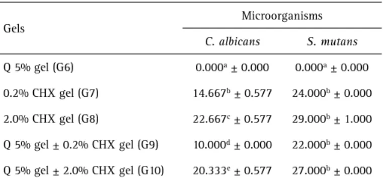
The 2.0% chlorhexidine membrane (G5) and the disks containing 2.0% chlorhexidine gel (G8) showed the greatest inhibition halos for both microorganisms, with statistically significant difference when compared to others tested groups (p=0.008) only for C. albicans inhibitions results. All the other formulations of membranes and gels showed inhibition halos, but without statistically significant difference.
Discussion
From a pharmaceutical and dental point of view, chitosan-based biomaterial production is a reality, mainly in tissue engineering and as “drug-delivery systems”
Figure 2. Incubation and growth of microorganisms in bacteriological oven at 37° C for a period of 48 h.
Table 1. Means and standard deviation of the growth inhibition halos (mm) of the different membranes
Membranes
Microorganisms
C. albicans S. mutans
Q 5% (G1) 0.000a ± 0.000 0.000a ± 0.000
Q 5% ± CHX 0.2% (G2) 10.667b ± 0.577 20.667b ± 0.577
Q 5% ± CHX 00.6% (G3) 13.000c ± 0.000 22.667c ± 1.155
Q 5% ± CHX 1.0% (G4) 14.333c ± 0.577 24.000c,d ± 0.000
Q 5% ± CHX 2.0% (G5) 16.667d ± 1.155 25.000d ± 1.000
Different letters indicate statistically significant difference among groups (vertical line).
Table 2. Means and standard deviation of the growth inhibition halos (mm) of the different gels
Gels
Microorganisms
C. albicans S. mutans
Q 5% gel (G6) 0.000a ± 0.000 0.000a ± 0.000
0.2% CHX gel (G7) 14.667b ± 0.577 24.000b ± 0.000
2.0% CHX gel (G8) 22.667c ± 0.577 29.000b ± 1.000
Q 5% gel ± 0.2% CHX gel (G9) 10.000d ± 0.000 22.000b ± 0.000
Q 5% gel ± 2.0% CHX gel (G10) 20.333e ± 0.577 27.000b ± 0.000
Antimicrobial bioadhesive oral membrane (7,14-15,18). The biocompatibility and other favorable
properties of chitosan in the health field are described by several studies (1,11-12,21), demonstrating the diverse applications of this polymer in both tissue regeneration and as a facilitating agent in drug delivering (2,5,8,20).
In dentistry, in the presence of oral mucosa lesions, the treatment options are limited to antiseptic gels and solutions, which have poor adhesion to moist mucosas, decreasing the treatment effectiveness (3,19,21). The membranes developed and tested in this study was the same tested by SILVA et al., 2017 (5) and has the main property the bioadhesiveness and have been comprehensively tested during their development, demonstrating good physical and chemical stability of the components as well as pH near to 7.0 (5).
Because these membranes would have an intimate contact with the tissues and would gradually release the antimicrobial agent, promoting the healing of the injured region, they were tested regarding biocompatibility showing good interaction with the mucosa cells, in addition to the effective capacity of drug release demonstrated by a semi-quantitative evaluation of the release of chlorhexidine digluconate in phosphate buffer solution (PBS) and by mass loss tests previously performed (2-3,5,10).
Chitosan has antimicrobial action, which was increased by the association with chlorhexidine, showing a high antimicrobial potential (22-23). In this study, we opted to use chlorhexidine as antimicrobial agent in association with chitosan due to wide application of this drug in dentistry in the control of infections of oral tissues (3,8,17,21). The rationale behind this fact was to obtain membranes with increased contact time between the drug and the oral tissues, which is greatly advantageous over oral solutions and gels. Because of the valuable properties showed by chitosan and the need to develop an unprecedented method to promote the oral mucosa repair and decontamination, this study aimed to develop the antimicrobial oral membrane by analyzing against C. albicans and S. mutans. Chitosan had the function of structural and antimicrobial component of the oral membrane. Decker et al. (24) and Ballal et al. (21) revealed that chitosan associated with chlorhexidine has antimicrobial effect. Moreover, chitosan has hemostatic action and stimulates healing. Burkatovskaya et al. (9) showed a hemostatic chitosan-based oral membrane to control bleeding and prevent microbial infection when in contact with surgical wounds. This oral membrane was bactericide compared with curative alginate and silver sulfadiazine after contact against surgical wounds contaminated with staphylococcus and pseudomonas in rats.
Chlorhexidine was chosen as antimicrobial agent because it is largely used in dentistry to control S. mutans and C. albicans (3,5,9,21,24). Chlorhexidine is an effective
mouthrinse, but compared with solutions, the gels may significantly increase the contact time with the oral mucosa, and consequently improve the antimicrobial effect. Senel et al. (7), evaluating antimicrobial gels and films to treat periodontal diseases, both compositions were prepared with chitosan (carrier) to release the antimicrobial agent to the oral mucosa. Chitosan released the antimicrobial agent for a longer time, increasing the clinical effect due to its bioadhesiveness and high viscosity.
The choice for the microorganisms to be tested was based in the main goal of the membrane application, that is, the control of oral microbiota: S. mutans (present in the dental biofilm and main microorganism of caries lesion) (25-26) and C. albicans (main microorganism of oral mucosa infections in adult wearers of prostheses) (27).
To standardize the tests proposed by this study, we used the antibiogram method (Kirby-Bauer) by which the drug diffuse through sterile paper discs on the culture medium. In dental researches, the methodology of diffusion in culture medium is rather employed to analyze the antimicrobial activity of dental materials (28-30).
The results of this present study revealed that only the association of chlorhexidine and chitosan in membranes, especially those with higher concentrations of 2% chlorhexidine and chitosan in membranes was statistically effective which agrees with other studies on the association of chitosan with chlorhexidine delivered by gels and films (2,5,23,31).
The antimicrobial capacity of pure chitosan against C. albicans and S. mutans was not proven by the results obtained in this study, with is in accordance with other studies using the same methodology and active principles (21,23,32). But others studies affirmed that pure chitosan has some antimicrobial capacity, Akncbay et al. (3) showed the antimicrobial effect of chitosan gels against periodontitis bacteria in a clinical trial, and Je and Kim. (22) also demonstrated the antimicrobial action of pure chitosan and chitosan films associated with others components in molecular tests and in direct contact with some gram-positive and gram-negative bacterias.
Based on the results the bioadhesive chitosan-based oral membrane with 2% chlorhexidine and 2% chlorhexidine gel were the most effective in inhibiting the tested microorganisms. Further studies should be conducted to prove the antimicrobial action of chitosan-based oral membranes with chlorhexidine against other oral microorganisms as well as to assess the action and release dynamics of the drug when in direct contact with the oral mucosa.
Acknowledgements
A.P
. Kloster et al.
Research Foundation (FAPESP grant #2013/07836-7).
Resumo
O objetivo deste estudo foi avaliar in vitro o efeito antimicrobiano de uma bandagem oral bioadesiva de quitosana com clorexidina para o tratamento de infecções dos tecidos orais. Cinco bandagens de diferentes composições foram testadas: Quitosana 5% (G1); Quitosana 5% ± clorexidina a 0,2% (G2), Quitosana 5% ± clorexidina a 0,6% (G3), Quitosana 5% ± clorexidina a 1,0% (G4) e Quitosana 5% ± clorexidina a 2,0% (G5). Foram testados também 5 tipos de géis nas seguintes composições: Gel de Quitosana 5% (G6), Gel de clorexidina a 0,2% (G7), Gel de clorexidina a 2,0% (G8), Gel de Quitosana 5% ± clorexidina a 0,2% (G9) e Gel de Quitosana 5% ± clorexidina a 2,0% (G10). A ação antimicrobiana das amostras foi testada contra Candida albicans e Streptococcus mutans por meio do antibiograma, medindo o halo de inibição. Os dados foram analisados pelo teste de Kruskal-Wallis e ANOVA a um critério seguido pelo teste de Tukey (p<0,05). A membrana com 2,0% de clorexidina (G5) e os discos contendo gel com 2,0% de clorexidina (G8) apresentaram os maiores halos de inibição para os dois microrganismos, com diferença estatisticamente significativa em relação aos demais grupos testados (p=0,008) apenas nos resultados de inibição de C. albicans. Todas as outras formulações de membranas e géis apresentaram halo de inibição, mas sem diferença estatisticamente significativa. A bandagem oral bioadesiva de quitosana com gel de 2% de clorexidina foi a mais efetiva em inibir os microrganismos testados.
References
1. Spin-Neto R, Pavone C, Freitas RM, Marcantonio RAC, Marcantonio-Jr E. Chitosan based biomaterials with medical and dental application: literature review. Rev Odontol UNESP 2008;37:155-161.
2. Petelin M, Pavlica Z, Bizimoska S, Sentjurc M. In vivo study of different ointments for drug delivery into oral mucosa by EPR oximetry. Int J Pharm 2004;270:83-91.
3. Akncbay H, Senel S, Ay ZY. Application of chitosan gel in the treatment of chronic periodontitis. J Biomed Mater Res B Appl Biomater 2007;80:290-296.
4. Del Carpio-Perochena A, Bramante CM, Duarte MA, de Moura MR, Aouada FA, Kishen A. Chelating and antibacterial properties of chitosan nanoparticles on dentin. Restor Dent Endod 2015;40:195-201. 5. Silva MD, Neto NL, da Costa SA, da Costa SM, Oliveira TM, Oliveira RC,
et al. Biophysical and biological characterization of intraoral multilayer membranes as potentialcarriers: A new drug delivery system for dentistry. Mater Sci Eng C Mater Biol Appl 2017;71:498-503. 6. Shahidi F, Abuzaytoun R. Chitin, chitosan, and co-products: chemistry,
production, applications, and health effects. Adv Food Nutr Res 2005;49:93–135.
7. Senel S, McClure SJ. Potential applications of chitosan in veterinary medicine. Adv Drug Deliv Rev 2004;56:1467-1480.
8. Juliano C, Cossu M, Pigozzi P, Rassu G, Giunchedi P. Preparation, in vitro characterization and preliminary in vivo evaluation of buccal polymeric films containing chlorhexidine. AAPS PharmSciTech 2008;9:1153-1158. 9. Burkatovskaya M, Castano AP, Demidova-Rice TN, Tegos GP, Hamblin MR. Effect of chitosan acetate bandage on wound healing in infected and noninfected wounds in mice. Wound Repair Regen 2008;16:425-431.
10. Spin-Neto R, Freitas RM, Pavone C, Cardoso MB, Capana-Filho SP, Marcantonio RAC. Histological evaluation of chitosan-based biomaterials used for the correction of critical size defects in rat’s calvaria. J Biomed Mater Res A 2010;93:107-114.
11. Kim IY, Seo SJ, Moon HS, Yoo MK, Park IY, Kim BC, et al. Chitosan and its derivatives for tissue engineering applications. Biotechnol Adv 2008;26:1-21.
12. Jayakumar R, Prabaharan M, Sudheesh Kumar PT, Nair SV, Tamura H. Biomaterials based on chitin and chitosan in wound dressing applications. Biotechnol Adv 2011;29:322-337.
13. Croisier F, Jerome C. Chitosan-based biomaterials for tissue engineering. Eur Polym J 2013;49:780-792.
14. Giovino C, Ayensu I, Tetteh J, Boateng JS. An integrated buccal delivery system combining chitosan films impregnated with peptide loaded PEG-b-PLA nanoparticles. Colloids Surf B Biointerfaces 2013;112:9-15. 15. Ahsan SM, Thomas M, Reddy KK, Sooraparaju SG, Asthana A, Bhatnagar
I. Chitosan as biomaterial in drug delivery and tissue engineering. Int J Biol Macromol 2018;110:97-109.
16. Wieckiewicz M, Boening KW, Grychowska N, Paradowska-Stolarz A. Clinical Application of Chitosan in Dental Specialities. Mini Rev Med Chem 2017;17:401-409.
17. Ordikhani F, Dehghani M, Simchi A. Antibiotic-loaded chitosan-Laponite films for local drug delivery by titanium implants: cell proliferation and drug release studies. J Mater Sci Mater Med 2015;26:269.
18. Needleman IG, Martin GP, Smales FC. Characterisation of bioadhesives for periodontal and oral mucosal drug delivery. J Clin Periodontol 1998;25:74-82.
19. Singla AK, Chawla M. Chitosan: some pharmaceutical and biological aspects - an update. J Pharm Pharmacol 2001;53:1047-1067. 20. Liu DZ, Chen WP, Lee CP, Wu SL, Wang YC, Chung TW. Effects of alginate
coated on PLGA microspheres for delivery tetracycline hydrochloride to periodontal pockets. J Microencapsul 2004;21:643-652.
21. Ballal NV, Kundabala M, Bhat KS, Acharya S, Ballal M, Kumar R, et al. Susceptibility of Candida albicans and Enterococcus faecalis to Chitosan, Chlorhexidine gluconate and their combination in vitro. Aust Endod J 2009;35:29-33.
22. Je JY, Kim SK. Antimicrobial action of novel chitin derivative. Biochim Biophys Acta 2006;1760:104-109.
23. Liakos I, Rizzello l, Scurr DJ, Pompa PP, Bayer IS, Athanassiou A. All-natural composite wound dressing films of essential oils encapsulated in sodium alginate with antimicrobial properties. Int J Pharmaceut 2014;463:137-138.
24. Decker EM, von Ohle C, Weiger R, Wiech I, Brecx M. A synergistic chlorhexidine/chitosan combination for improved antiplaque strategies. J Periodontal Res 2005;40:373-377.
25. Kassebaum NJ, Bernabé E, Dahiya M, Bhandari B, Murray CJ, Marcenes W. Global burden of untreated caries: a systematic review and metaregression. J Dent Res 2015;94:650-658.
26. Palmer SR, Miller JH, Abranches J, Zeng L, Lefebure T, Richards VP, et al. Phenotypic heterogeneity of genomically-diverse isolates of
Streptococcus mutans. PLoS One 2013;8:613-658.
27. Perić M, Radunović M, Pekmezović M, Marinković J, Živković R, Arsić Arsenijević V. J. Laboratory-based investigation of denture sonication method in patients with candida-associated denture stomatitis. Prosthodont 2017 [Epub ahead of print. doi: 10.1111/jopr.12610] 28. Blanscet ML, Tordik PA, Goodell GG. An agar diffusion comparison
of the antimicrobial effect of calcium hydroxide at five different concentrations with three different vehicles. J Endod 2008;34:1246-1248.
29. De Rezende GP, da Costa LR, Pimenta FC, Baroni DA. In vitro antimicrobial activity of endodontic pastes with propolis extracts and calcium hydroxide: a preliminary study. Braz Dent J 2008;19:301-305. 30. Athanassiadis B, Abbott PV, George N, Walsh LJ. An in vitro study of
the antimicrobial activity of some endodontic medicaments and their bases using an agar well diffusion assay. Aust Dent J 2009;54:141-146. 31. Aksungur P, Sungur A, Unal S, İskit AB, Squier SD, Şenel S. Chitosan
delivery systems for the treatment of oral mucositis: In vitro and in vivo studies. J Control Release 2004;98:269–279.
32. Bae K, Jun EJ, Lee SM, Paik DI, Kim JB. Effect of water-soluble reduced chitosan on Streptococcus mutans, plaque regrowth and biofilm vitality. Clin Oral Investig 2006;10:102-107.