Fasting/Refeeding Cycles Prevent Myocardial Dysfunction and
Morphology Damage in the Spontaneously Hypertensive Rats
Matheus Fécchio Pinotti,
1Amanda Martins Matias,
2Mário Mateus Sugizaki,
3André Ferreira do Nascimento,
3Maeli Dal Pai,
1,4Ana Paula Lima Leopoldo,
2Antônio Carlos Cicogna,
1André Soares Leopoldo
2Departamento de Clínica Médica, Faculdade de Medicina, Universidade Estadual Paulista (UNESP),1 Botucatu, SP - Brazil
Departamento de Desportos, Centro de Educação Física e Desportos, Universidade Federal do Espírito Santo (UFES),2 Vitória, ES - Brazil Universidade Federal de Mato Grosso (UFMT),3 Cuiabá, MT - Brazil
Departamento de Morfologia, Instituto de Biosciências da Universidade Estadual Paulista UNESP,4 Botucatu, SP - Brazil
Mailing Address: André Soares Leopoldo •
Avenida Fernando Ferrari, 514. CEP 29075-410, Goiabeiras, Vitória, ES – Brazil
E-mail: [email protected], [email protected] Manuscript receveid November 10, 2017, revised manuscript May 09, 2018, accepted May 09, 2018
DOI: 10.5935/abc.20180152
Abstract
Background: Caloric restriction is known to impair the cardiac function and morphology in hypertrophied hearts of spontaneously hypertensive rats (SHR); however, the influence of fasting/refeeding (RF) is unknown.
Objective: To investigate the fasting/refeeding approach on myocardial remodeling and function. In addition, the current
study was designed to bring information regarding the mechanisms underlying the participation of Ca2+ handling and
β-adrenergic system.
Methods: Sixty-day-old male SHR rats were submitted to food ad libitum (C), 50% food restriction (R50) or RF cycles for 90 days. Cardiac remodeling was assessed by ultrastructure analysis and isolated papillary muscle function. The level of
significance considered was 5% (α = 0.05).
Results: The RF rats presented lower cardiac atrophy than R50 in relation to C rats. The C rats increased weight gain,
R50 maintained their initial body weight and RF rats increased and decreased weight during RF. The RF did not cause
functional impairment because the isotonic and isometric parameters showed similar behavior to those of C. The isotonic
and isometric cardiac parameters were significantly elevated in RF rats compared to R50 rats. In addition, the R50 rats had
cardiac damage in relation to C for isotonic and isometric variables. While the R50 rats showed focal changes in many
muscle fibers, the RF rats displayed mild alterations, such as loss or disorganization of myofibrils.
Conclusion: Fasting/refeeding promotes cardiac beneficial effects and attenuates myocardial injury caused by caloric restriction in SHR rats, contributing to reduce the cardiovascular risk profile and morphological injuries. Furthermore, RF
promotes mild improvement in Ca2+ handling and β-adrenergic system. (Arq Bras Cardiol. 2018; 111(3):400-409)
Keywords: Rats; Hypertension; Myocardial/dysfunction; Chronic Disease; Fasting, Reffeding; Caloric Restriction.
Introduction
The major causes of chronic non-communicable diseases (NCD)-attributable mortality are cardiovascular disease, cancers, chronic respiratory disease and diabetes.1
These conditions share a small number of behavioral risk factors, which aggravate the NCD and include unhealthy diet, which is closely related to hypertension.
Caloric restriction (CR) has been recognized throughout history for promoting several beneficial effects.2,3 Nevertheless, although
CR may prevent cardiac damage in hypertrophied hearts of spontaneously hypertensive rats (SHR),4 it is common to note
body weight fluctuations typically referred to as the "yo-yo
syndrome" while on a regimented diet, and these fluctuations
have shown deleterious cardiovascular effects.5,6 Researches from
our laboratory and others have shown that, when dietary restriction is severe, it can promote morphological injuries and impairment of cardiac function in normal or SHR rats.7-12
Recently, intermittent fasting or fasting/refeeding has also shown to extend lifespan and have beneficial health effects as compared to ad libitum food consumption,3,13,14 as it
enhances cardiovascular function and improves several risk factors for cardiovascular diseases.15,16 This dietary approach
also implies a protective effect against oxidative stress, lower rates of kidney disease,17 prolongation of reproductive
function,18 and leads to the normalization of resting energy
expenditure and protein synthesis recuperation, but can cause many metabolic disturbances.19,20
In normotensive rats submitted to food restriction, chronic refeeding decreased the incidence of cardiac arrhythmia and reversed the depletion of heart proteins.21,22 Food restriction
caused cardiac function disturbances that were almost completely reversed back to normal after chronic refeeding in the isolated rat heart.23 In our laboratory, we observed that
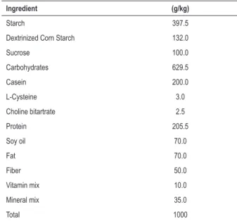
Table 1 – Ingredient composition of the Labina rat experimental diet
Ingredient (g/kg)
Starch 397.5
Dextrinized Corn Starch 132.0
Sucrose 100.0
Carbohydrates 629.5
Casein 200.0
L-Cysteine 3.0
Choline bitartrate 2.5
Protein 205.5
Soy oil 70.0
Fat 70.0
Fiber 50.0
Vitamin mix 10.0
Mineral mix 35.0
Total 1000
and attenuated the structural injuries in papillary muscles caused by CR in normotensive rats.24 Nevertheless, it is not
yet clear whether fasting/refeeding cycles are able to promote similar effects and/or reverse the cardiac damage induced by food restriction in SHR rats.7-12 Thus, the objective was
to investigate the fasting/refeeding approach on myocardial remodeling and function. In addition, the current study was designed to bring information regarding the mechanisms underlying the participation of Ca2+ handling and β-adrenergic
system. Our hypothesis is that fasting/refeeding condition would attenuate the myocardial injury caused by food restriction and would contribute to normal cardiac remodeling in SHR rats without alterations in the Ca2+ handling and
β-adrenergic system.
Methods
Animal model and experimental protocol
Sixty-day-old male SHR were distributed into three groups: control (C, n = 7); food-restriction (R50, n = 7); and fasting/
refeeding cycles (RF, n = 7). The C group was fed Labina rat chow containing 7.0% fat, 20.55% protein, 62.95% carbohydrate, 5.0% fiber and 4.5% moisture (Agribands, Brazil), and water was provided ad libitum. Table 1 shows the ingredient composition of Labina ratchow. The R50 group
received 50% of the amount of food consumed by the C group. The RF group was submitted to cycles of 50% food restriction and refeeding ad libitum weekly. All rats were maintained on this dietary regimen for 90 days and were then euthanized.
All animals were housed in individual cages in a room maintained at 23ºC with a 12-hour light/dark cycle and were weighed once a week. Initial and final body weights (IBW and FBW, respectively), the ratios between left and right ventricular weights to final body weight (LVW/FBW and RVW/FBW, respectively) and papillary muscle cross-sectional area (CSA) were also measured. All experiments and procedures were
performed in accordance with the Guide for the Care and Use
of Laboratory Animals published by the United States National
Institutes of Health and were approved by the ethics committee of Botucatu School of Medicine, UNESP, São Paulo, Brazil.
Systolic blood pressure
Systolic blood pressure evaluation was assessed by the non-invasive tail-cuff method with a Narco BioSystems Electro-Sphygmomanometer (International Biomedical, Austin, TX, USA) at the beginning and after the end of the experimental protocol. The average of two pressure readings was recorded for each animal.
Isolated muscle performance
Cardiac intrinsic contractile performance was evaluated by studying isolated left ventricular (LV) papillary muscle as described previously.9,10,12 Isometric contraction parameters,
including peak of developed tension (DT, g/mm2, defined as
peak isometric tension minus resting tension), resting tension (RT, g/mm2), time to peak tension (TPT, ms), peak isometric
tension development rate (+dT/dt, g/mm2/s) and maximum
tension decline rate (-dT/dt, g/mm2/s), time from peak tension
to 50% relaxation (RT50, ms) were determined. The isotonic parameters were percentage of shortening (PS, %), time to peak shortening (TPS, ms), maximum shortening velocity (-dL/dt, ML/s) and maximum relaxation velocity (+dL/dt, ML/s).
The mechanical behavior of the papillary muscle was evaluated under baseline conditions at 1.25 mM [Ca2+]
and after the following inotropic maneuvers: increase in extracellular Ca2+ concentration from 0.625 to 1.25, 2.5 and
5.2 mM, and β-adrenergic stimulation with 0.01, 0.1 and 1.0 µM isoproterenol. The parameters used to characterize papillary muscle were as follows: length (mm), weight (mg) and CSA (mm2). Muscle length (ML) at peak DT was defined
as Lmax in vitro and measured with a Gartner cathetometer
(Chicago, IL, USA). To compare the mechanical function between different muscle lengths, isometric and isotonic parameters were normalized to CSA and Lmax.
Morphological study
For the ultrastructural study (three animals per group), small pieces of the LV papillary muscle were fixed in Karnovsky’s fixative in 0.12 M phosphate, pH 7.2, for 1-2 hours and were postfixed in 1% osmium tetroxide in 0.1 M phosphate buffer for 2 hours.25 After dehydration in a graded ethanol series, the
samples were embedded in epoxy resin. Ultrathin sections were cut from selected areas with a diamond knife, double-stained with uranyl acetate and lead citrate, and examined using a Philips EM 301 electron microscope. The LV myocyte CSA was measured using a compound microscope attached to a computerized imaging analysis system (Image-Pro Plus 3.0, Media Cybernetics, Silver Springs, MD, USA).
Statistical analysis
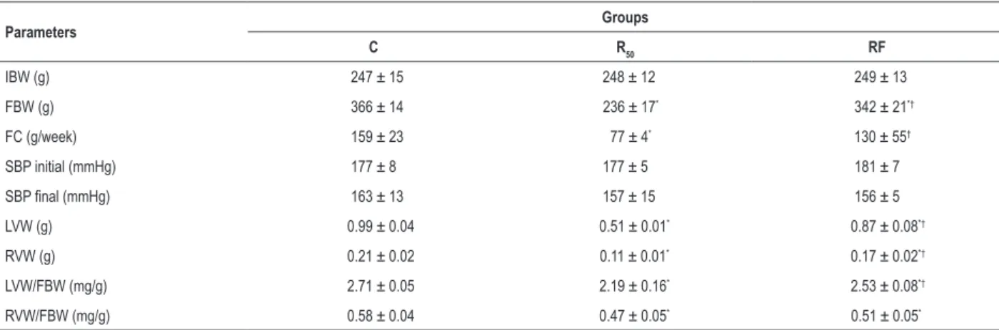
Table 2 – General characteristics of rats
Parameters Groups
C R50 RF
IBW (g) 247 ± 15 248 ± 12 249 ± 13
FBW (g) 366 ± 14 236 ± 17* 342 ± 21*†
FC (g/week) 159 ± 23 77 ± 4* 130 ± 55†
SBP initial (mmHg) 177 ± 8 177 ± 5 181 ± 7
SBP final (mmHg) 163 ± 13 157 ± 15 156 ± 5
LVW (g) 0.99 ± 0.04 0.51 ± 0.01* 0.87 ± 0.08*†
RVW (g) 0.21 ± 0.02 0.11 ± 0.01* 0.17 ± 0.02*†
LVW/FBW (mg/g) 2.71 ± 0.05 2.19 ± 0.16* 2.53 ± 0.08*†
RVW/FBW (mg/g) 0.58 ± 0.04 0.47 ± 0.05* 0.51 ± 0.05*
C: control group; R50: animals with food restriction of 50%; RF: animals with alternation between food restriction of 50% and refeeding; IBW: initial body weight;
FBW: final body weight; FC: food consumption; SBP: systolic blood pressure; LVW: left ventricle weight; RVW: right ventricle weight. Values are means ± SD (n = 7). * significant at p < 0.05 vs. C; † p < 0.05 vs. R
50. One-way ANOVA and post hoc Tukey’s test.
myocardial function at baseline condition were reported as means ± standard deviation (SD). Comparisons between groups were performed using one-way analysis of variance (ANOVA) for independent samples. A repeated-measures two-way ANOVA was utilized to evaluate the body weight evolution and the positive and negative inotropic effects on myocardial function. When significant differences were found (p < 0.05),
post hoc Tukey’s or Bonferroni’s test for multiple comparisons
was carried out. The level of significance considered was 5%. The sample size (n) was performed using the equation: n = 1 + [2C * (s/d)2], where C (z score α + z score β)2 is
dependent on the values chosen for statistical power of the test (90%; type II error) and level of significance (0.05; type I error); the standard deviation value (s) adopted was 0.25, and the minimal difference between groups (d) was 0.5. The sample size needed to detect a significant difference between groups is 6.25 rats per group; however, we decided to use 7 animals per group for most of the analyses.
Results
General and morphological characteristics of rats
Significantly higher values of FBW, LVW, RVW, LVW/FBW and RVW/FBW were found in C compared to R50 and RF rats
(Table 2). After 12 weeks, fasting/refeeding cycles promoted a substantial elevation of FBW and food consumption that were significantly greater than those in the R50 group. In relation to cardiac parameters, the RF and R50 groups presented different
behavior. Specifically, the LVW (RF: 12.12% and R50: 48.5%; p < 0.05), RVW (RF: 19.04% and R50: 47.62%; p < 0.05), LVW/FBW (RF: 6.64% and R50: 19.2%; p < 0.05) and RVW/
FBW (RF: 12.06% and R50: 18.96%; p < 0.05) were reduced in percentage in the RF and R50 rats as compared to C rats. Nevertheless, the fasting/refeeding cycles presented lower cardiac atrophy than R50 rats in relation to C rats.
In addition, C rats experienced increasing weight gain, while R50 rats maintained their IBW after 12 weeks of
experimental protocol (Figure 1). On the other hand, RF rats gained weight dependent on food intake, with body weight increasing and decreasing during refeeding and fasting, respectively (Figure 1).
Isolated muscle performance
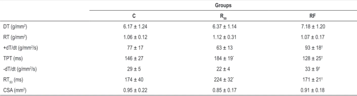
Fasting/refeeding cycles did not cause functional impairment (Tables 3 and 4). Nevertheless, the isotonic [-dL/dt, TPS, and RT50)] and isometric parameters (TPT, +dT/dt, -dT/ dt, RT50)were significantly elevated in RF rats compared to those in the R50 group, indicating that fasting/refeeding cycles preserves the contraction and relaxation phase of cardiac function. Furthermore, the R50 rats presented
cardiac damage in relation to the C group for isotonic and isometric variables. In addition, the papillary muscle CSA showed no difference among groups.
Calcium stimulation
After baseline condition, the increases in extracellular Ca2+
concentrations from 0.625 to 5.2 mM resulted in a positive inotropic effect in myocytes from all groups (Figures 2A-F). However, the results shown in Figures 2B, C and E indicate that extracellular Ca2+ (1.25 and 2.5 mM) induced a greater
response in +dT/dt (RF: 99.1 ± 23.6; 132.1 ± 36.2 g/mm2/svs.
R50: 63.2 ± 12.8; 91.5 ± 22.0 g/mm2/s; p < 0.05, respectively),
-dT/dt (RF: 30.6 ± 5.9; 35.9 ± 5.8 g/mm2/svs. R
50: 22.0 ± 4.4;
28.5 ± 6.1 g/mm2/s; p < 0.05, respectively) and -dL/dt
(RF: 2.19 ± 0.45; 2.77 ± 0.51 ML/svs. R50: 1.47 ± 0.24;
1.99 ± 0.31 ML/s; p < 0.05, respectively) in the RF rats than in the R50 rats. In addition, -dT/dt and -dL/dt were significantly
diminished in the R50 myocardium at Ca2+ concentration
of 5.2 mM when compared to those in the RF group. When submitted to inotropic maneuvers, DT, PS and +dL/dt were similar between RF and R50. In relation to the cardiac function of C rats after Ca2+stimulation, the fasting/refeeding
Figure 1 – Changes in body weight after 90 days of treatment. Control (C; closed squares, n = 7), animals with food restriction of 50% (R50; closed triangles, n = 7) and
animals with alternation between food restriction of 50% and refeeding (RF; open circles, n = 7). Values are means ± SD; * significant at p < 0.05, R50vs. C; † p < 0.05,
RF vs. R50;
#p < 0.05, C vs. RF. Repeated measures two-way ANOVA; post hoc Bonferroni’s test. Source: Research team.
500
400
300
200
Body weight (g)
Weeks
0 1 2 3 4 5 6 7 8 9 10 11 12
C R50 RF
#
# #
# # # #
*† * *†
*† *† *† *† *† *† *† *†
Ca2+ concentration (5.2 mM); -dL/dt was significantly lower in
R50 than in C group (C: 2.66 ± 0.35 vs. R50: 2.18 ± 0.33 ML/s, p < 0.05) (Figure 2E).
Isoproterenol stimulation
The fasting/refeeding cycles increased +dT/dt, -dT/dt and -dL/dt at the highest isoproterenol concentration (1 µM) compared to those in the R50 group, indicating a positive inotropic effect in myocytes. In contrast, the RF grouppromoted a reduction in +dL/dt than the R50 group at the same isoproterenol concentration (Figure 3F). In addition, the similar effects were noted in +dT/dt and -dT/dt at 1µM isoproterenol of the C group when compared to the R50 (Figures 3B and C). Furthermore, RF rats presented
higher +dL/dt at baseline and isoproterenol concentrations (0.01 µM) in comparison to C group (Figure 3F). There were no significant differences in mechanical data (DT and PS) under inotropic stimulation with isoproterenol among the groups (Figures 3A and D).
Myocardial morphology
The C group rats showed normal morphological characteristics, with myofibrils filling the entire sarcoplasm, well-defined sarcomeres, mitochondria with lamellar cristae, sarcoplasmic membranes with regular aspect, sarcoplasmic reticulum among myofibrils and nuclei with uncondensed chromatin (Figures 4A and B). The R50 group presented focal changes, including disorganization or absence of myofibrils, some polymorphic mitochondria with a decreased number of cristae and areas of sarcoplasmic reticulum dilation (Figures 4C, D and E). In RF rats, the only change observed was a loss of mitochondrial cristae in some organelles. Most of the fibers had normal morphology (Figures 4F and G).
Discussion
Interestingly, little information is available on the relationship between cardiac function and morphology during fasting/refeeding in SHR hypertrophied hearts. Within this context, this dietary regimen has become the subject of Table 3 – Isotonic contraction of groups at baseline condition
Groups
C R50 RF
PS (%) 19 ± 3 18 ± 3 20 ± 2
-dL/dt (ML/s) 1.89 ± 0.40 1.60 ± 0.36 2.19 ± 0.45†
TPS (ms) 168 ± 26 205 ± 14* 161 ± 14†
+dL/dt (ML/s) 4.28 ± 1.26 4.13 ± 1.11 4.79 ± 0.86
RT50 (ms) 58 ± 10 76 ± 12
* 53 ± 9†
CSA (mm2) 0.95 ± 0.22 0.85 ± 0.17 0.91 ± 0.18
C: control group; R50: animals with food restriction of 50%; RF: animals with alternation between food restriction of 50% and refeeding; PS: percentage of shortening;
-dL/dt: maximum shortening velocity; TPS: time to peak shortening; + dL/dt: maximum relaxation velocity; RT50: time from peak tension to 50% relaxation;
CSA - muscle cross-sectional area. Values are means ± SD (n = 7) at basal calcium concentration (1.25 mM); * significant at p < 0.05 vs. C; † p < 0.05 vs. R
50. One-way
Figure 2 –Effects of increased extracellular calcium on myocardial isotonic and isometric parameters in papillary muscles from control (C = black bars), animals with food restriction of 50% (R50 = gray bars) and animals with alternation between food restriction of 50% and refeeding (RF = white bars). Extracellular calcium experiment:
7 animals each group. Isometric parameters: A: DT (peak developed tension normalized per cross-sectional area); B: +dT/dt (peak isometric tension development rate normalized per cross-sectional area); C: -dT/dt, g/mm2/s (maximum tension decline rate normalized per cross-sectional area). Isotonic parameters: D: PS
(percentage of shortening); E: -dL/dT (maximum shortening velocity); F: +dL/dT (maximum relaxation velocity). Lmax: muscle length at peak DT. Values are means ± SD; * significant at p < 0.05 vs. C; † p < 0.05 vs. R
50. Repeated measures two-way ANOVA and post hoc Tukey’s test. Source: Research team.
A B
D E F
C
10
8
6
4
2
0
DT (g/mm
2)
+dT/dt (g/mm
2/s)
–dT/dt (g/mm
2/s)
PS (%)
–dL/dT (ML/s) +dL/dT (ML/s)
200
100 150
50
0
50
40
30
20
10
0
30
20
10
0
4
3
2
1
0
8
6
4
2
0
0.625 1.25 2.5 5.2 0.625 1.25 2.5 5.2 0.625 1.25 2.5 5.2
0.625 1.25 2.5 5.2
0.625 1.25 2.5 5.2
0.625 1.25 2.5 5.2
calcium concentration (mM) calcium concentration (mM) calcium concentration (mM)
calcium concentration (mM) calcium concentration (mM) calcium concentration (mM) C R50 RF
† †
†
† †
* †
† †
considerable scientific interest for weight loss and improving cardiometabolic health. Thus, the main finding of this study was that fasting/refeeding attenuated the damage caused by CR. The results reveal that fasting/refeeding showed increased isotonic and isometric parameters at baseline, as well as improved the myocardial inotropic response to calcium and
isoproterenol. In addition, fasting/refeeding prevented cardiac atrophy and morphological injuries.
Less body weight gain was observed in the RF group than in the C group (Table 1, Figure 1), but more body weight gain than in the R50 group. According to literature, body weight
reduces approximately 13% when the animals are submitted Table 4 – Isometric contraction of groups at baseline condition
Groups
C R50 RF
DT (g/mm2) 6.17 ± 1.24 6.37 ± 1.14 7.18 ± 1.20
RT (g/mm2) 1.06 ± 0.12 1.12 ± 0.31 1.07 ± 0.17
+dT/dt (g/mm2/s) 77 ± 17 63 ± 13 93 ± 18†
TPT (ms) 146 ± 27 184 ± 19* 128 ± 25†
-dT/dt (g/mm2/s) 29 ± 5 22 ± 4 33 ± 9†
RT50 (ms) 174 ± 40 224 ± 32
* 171 ± 21†
CSA (mm2) 0.95 ± 0.22 0.85 ± 0.17 0.91 ± 0.18
C: control group; R50: animals with food restriction of 50%; RF: animals with alternation between food restriction of 50% and refeeding; DT: peak developed
tension; RT: resting tension; TPT: time to peak tension; +dT/dt: maximum tension development rate; -dT/dt: maximum tension decline rate; RT50: time from peak
tension to 50% relaxation; CSA: muscle cross-sectional area. Values are means ± SD (n = 7) at basal calcium concentration (1.25 mM); * significant at p < 0.05 vs. C; † p < 0.05 vs. R
to 48 hours of fasting.25 This result appears to be mediated
by hormones, such as leptin that acts regulating appetite and weight gain. A rapid inhibition of ob gene expression in the white adipose tissue occurs in fasting, and this effect can be reversed by refeeding.25,26
Cardiac hypertrophy, a major pathological process involved in cardiac remodeling, initially serves as a compensatory mechanism to preserve cardiac output.27 Cardiac remodeling
may be regarded as a first step in the sequence of adaptive responses of the heart to stress caused by a large number of physiological and pathological conditions, such as changes in volume and pressure loads and/or metabolic alterations.28
Current study revealed that fasting/refeeding induced cardiac atrophy visualized by reduced total heart and left ventricle, as well as in the LVW/FBW. A decrease in left ventricle weight relative to body weight is very common in small animals submitted to food restriction22 and fasting/refeeding.29
Inhibition of myocardial protein synthesis and reduction in average protein half-lives are possible explanations for reduced cardiac mass under starvation.30 Protein synthesis,
an anabolic process, is required for cardiac hypertrophy. Two major pathways regulating protein synthesis are inhibited by AMPK, a primary regulator of metabolic pathways, which plays an essential role in a wide variety of cellular processes to protect against cardiac hypertrophy.31 Therefore, cardiac
atrophy could be regulated by the common signaling pathway of AMPK in the hypothalamus.
In the ultrastructural analysis, food restriction caused focal morphological damage in most papillary muscle fibers. The same alterations were less intense in the intermittent refeeding condition. Intermittent refeeding seems to aid in the attenuation of the mechanisms responsible for this damage and seems to act by enhancing protein anabolism and retarding protein degradation. Recent findings suggest that the beneficial effects of refeeding result from a reduction in oxidative injury and an increase in cellular stress resistance.2,32 One possible
mechanism for our result may be linked to the expression of atrogin-1, an E3 ubiquitin ligase also known as muscle atrophy F-box (MAFbx). E3-ligases are part of the ubiquitin proteasome pathway utilized for protein degradation during muscle atrophy. The literature has shown that atrogin-1/MAFbx expression results in muscle atrophy during catabolic condition.33 In cardiac muscle, atrogin-1/MAFbx expression
increases during heart failure and pressure overload.33,34
The isolated papillary muscle analysis showed that food restriction promotes cardiac dysfunction, but refeeding condition prevents the state. These stimuli provide evidence that the improvement of myocardial function assigned to fasting/refeeding cycles was related to changes in intracellular Ca2+ handling, mainly in the recapture and/or extrusion of
Figure 3 –Effects of isoproterenol stimulation on myocardial function in papillary muscles from control (C = black bars), animals with food restriction of 50% (R50 = gray
bars) and animals with alternation between food restriction of 50% and refeeding (RF = white bars). Isoproterenol stimulation experiment: 7 animals each group. Isometric parameters: A: DT (peak developed tension normalized per cross-sectional area); B: +dT/dt (peak isometric tension development rate normalized per cross-sectional area); C: -dT/dt, g/mm2/s (maximum tension decline rate normalized per cross-sectional area). Isotonic parameters: D: PS (percentage of shortening); E: -dL/dt
(maximum shortening velocity at Lmax); F: +dL/dt (maximum relaxation velocity at Lmax). Lmax: muscle length at peak DT. Values are means ± SD; * significant at p < 0.05 vs. C; † p < 0.05 vs. R
50.Repeated measures two-way ANOVA and post hoc Tukey’s test. Source: Research team.
10.0
7.5
5.0
2.5
0.0
basal 0.01 0.1 1
Isoproterenol (µM)
basal 0.01 0.1 1
Isoproterenol (µM)
basal 0.01 0.1 1
Isoproterenol (µM)
basal 0.01 0.1 1
Isoproterenol (µM)
basal 0.01 0.1 1
Isoproterenol (µM)
basal 0.01 0.1 1
Isoproterenol (µM) DT (g/mm 2) +dT/dt (g/mm 2/s) +dL/dT (ML/s) –dT/dt (g/mm 2/s) –dL/dT (ML/s) 200 150 100 50 0 60 30 45 15 0 PS (%) 30 20 10 0 4 3 2 1 0 6.0 4.5 3.0 1.5 0.0 † † * * † * * * † *† †
C R50 RF
A B
D E F
Figure 4 –Ultrastructural study of LV papillary muscle (n = 3 per group). Photographs A and B correspond to the control group, photographs C, D and E to the food-restriction (R50) group and photographs F and G to the refeeding group (RF) group. The control group showed preserved ultrastructure with normal myofibrils (M),
sarcoplasmic reticulum (arrowhead), mitochondria (mi), nuclear membrane (N) and plasma membrane (arrow). Food restriction rats showed cellular changes, including polymorphic mitochondria (*), myofibril disorganization (**), and infolding of the plasma membrane (arrow). The papillary muscle during refeeding showed preserved myofibrils (M), mitochondria (mi), and plasma membranes (arrow), polymorphic mitochondria (*) and capillary (C). Source: Research team.
cytosolic Ca2+, and β-adrenergic system. Nevertheless, the
lower response of food restricted rats to the increase of extracellular Ca2+ concentration can be related to changes
in the general mechanisms involved in Ca2+ cycling such
as sarcolemmal Na+/Ca2+ exchanger, sarcolemmal L-type
channel, sarcoplasmic reticulum (SR), ryanodine receptor, SR Ca2+ uptake pump, and the myofilament Ca2+ sensitivity.35
In relation to RF, this process may be faster and more balanced, but no study was found to support this statement and show the activity and protein expression of Ca2+ handling
regulatory proteins.
Another explanation could be related to the role of cytokine in intermittent fasting mediated cardioprotection. The influx of inflammatory cells and production of pro-inflammatory mediators contribute to myocardial injury.36 Nevertheless, adiponectin can protect myocardial
cells against ischemic injury by activating the cyclic AMP-dependent protein kinase - Akt pathway, being the latter mediated, in part, by caloric restriction.37 Thus, the
beneficial effects of fasting/refeeding may function through anti-inflammatory cytokine pathways.
Few studies have evaluated the β-adrenergic components in experimental models of fasting/refeeding.25,35 Some studies
have shown that cardiac function impairment is related to β-adrenergic system changes,35 while other researchers have
not reported reduced β-adrenergic response.25 The literature
shows that a decrease in cardiac β-receptor number has been reported in several hypertensive models known to be associated with an increase in sympathetic nerve activity, including SHR.38 Thus, the association between increased
sympathetic activity and cardiac β-receptor downregulation is sufficiently close to suggest that the finding of decreased β-receptor number after starvation and refeeding is indicative of persistently elevated cardiac sympathetic drive. However, in the current study, there is no damage of β-system in the RF rats, since the cardiac function was similar to that of the C group. The present data tend to support the hypothesis that isoproterenol stimulation reveals that the β-adrenergic system and cAMP phosphorylation of proteins related to Ca2+
handling were preserved in refeeding rats.
The beneficial effects of the intermittent fasting result from at least two mechanisms: the oxidative stress and the stress resistance hypothesis.39 According to literature, during the
intermittent fasting, there are fewer free radicals produced in the mitochondria of cells and, therefore, less oxidative damage to the cells.39 Another hypothesis is the resistance
to stress that is associated with increased resistance of cells in many different tissues to injury induced by oxidative, genotoxic and metabolic insults. The conservation of stress resistance responses to intermittent fasting across a range of species provides strong evidence that this mechanism contributes to the lifespan-extending action of dietary restriction.39
It is worth noting that according to studies in rodents and humans, intermittent food restriction is capable of promoting weight loss and/or favorably influence an array of cardiometabolic health indices, with equal or greater efficacy than conventional continuous energy restriction approaches, such as food restriction.29 Fasting/refeeding
cycles increase cardiac tolerance to ischemic injury and can affect the development of cardiovascular disease, preventing postinfarct cardiac remodeling, and impending chronic heart failure.29 Comparing the two dietary approaches, studies
show that caloric restriction may exert its beneficial effects primarily by reducing oxidative stress, whereas RF may act primarily by a stress resistance mechanism,40 which can have
a cardioprotective effect.
Study limitations
The study did not investigate the activity and protein expression of Ca2+ handling regulatory proteins known to
affect myocardial contraction and relaxation. In addition, the current study did not evaluate the involvement of anti-inflammatory cytokines, free-radical production and cellular stress response, which could help and consolidate the beneficial effects of intermittent fasting.
Conclusion
We demonstrated that fasting/refeeding promotes cardiac beneficial effects and attenuates myocardial injury caused by CR in SHR rats, contributing to the reduction of cardiovascular risk profile and morphological injuries. Furthermore, RF promotes mild improvement in the Ca2+ handling and β-adrenergic system.
Author contributions
Conception and design of the research: Pinotti MF, Cicogna AC, Leopoldo AS; Acquisition of data: Pinotti MF, Matias AM, Sugizaki MM, Nascimento AF, Pai MD, Leopoldo APL; Analysis and interpretation of the data: Pinotti MF, Sugizaki MM, Nascimento AF, Pai MD, Leopoldo APL; Statistical analysis: Sugizaki MM, Nascimento AF, Pai MD, Leopoldo APL; Obtaining financing: Cicogna AC; Writing of the manuscript: Pinotti MF, Matias AM, Cicogna AC, Leopoldo AS; Critical revision of the manuscript for intellectual content: Matias AM, Pai MD, Leopoldo APL, Cicogna AC, Leopoldo AS.
Potential Conflict of Interest
No potential conflict of interest relevant to this article was reported.
Sources of Funding
This study was funded by FAPESP number 04/04654-6.
Study Association
This study is not associated with any thesis or dissertation work.
Ethics approval and consent to participate
This study was approved by the Ethics Committee on Animal Experiments of the Faculdade de Medicina de Botucatu, UNESP under the protocol number 439/2004.
1. World Health Organization. (WHO). Noncommunicable doenças WHO. [Cited in 2016 Sep 26]. Available from: http://www.who.int/mediacentre/ factsheets/fs355/en/
2. Han X, Turdi S, Hu N, Guo R, Zhang Y, Ren J. Influence of long-term caloric restriction on miocardial and cardiomyocyte contractile function and autophagy in mice. J Nutr Biochem. 2012;23(12):1592-9.
3. Martin B, Mattson MP, Maudsley S. Caloric restriction and intermittent fasting: two potential diets for successful brain aging. Ageing Res Rev. 2006;5(3):332-53.
4. Dolinsky VW, Morton JS, Oka T, Robillard-Frayne I, Bagdan M, Lopaschuk GD, et al. Calorie restriction prevents hypertension and cardiac hypertrophy in the spontaneously hipertensive rat. Hypertension. 2010;56(3):412-21.
5. Keenan KP, Laroque P, Ballam GC, Ballam GC, Soper KA, Dixit R, et al. The effects of diet, ad libitum overfeeding, and moderate dietary restriction on the rodent bioassay: the uncontrolled variable in safety assessment. Toxicol Pathol. 1996;24(6):757-68.
6. Cicogna AC, Padovani CR, Okoshi K, Aragon FF, Okoshi MP. Myocardial function during chronic food restriction in isolated hypertrophied cardiac muscle.. Am J Med Sci. 2000;320(4):244-8.
7. Haddad F, Bodel PW, McCue SA, Herrick PE, Baldwin KM. Food restriction-induced transformations in cardiac functional and biochemical properties in rats. J Appl Physiol (1985). 1993;74(2):606-12.
8. Melo DS, Costa-Pereira LV, Santos CS, Mendes BF, Costa KB, Santos CF, et al. Severe calorie restriction reduces cardiometabolic risk factors and protects rat hearts from ischemia/reperfusion injury. Front Physiol. 2016 Apr 8;7:106.
9. Okoshi K, Matsubara LS, Okoshi MP, Cicogna AC, Fioretto JR, Padovani CR, et al. Food restriction-induced myocardial disfunction demonstrated by the combination of in vivo and in vitro studies. Nutr Res. 2002;22(11):1353-64.
10. Cicogna AC, Padovani CR, Okoshi K, Matsubara LS, Aragon FF, Okoshi MP. The influence of temporal food restriction on the performance of isolated cardiac muscle. Nutr Res. 2001;21(4):639-48.
11. Gut AL, Okoshi MP, Padovani CR, Aragon FF, Cicogna AC. Myocardial dysfunction induced by food restriction is related to calcium cycling and beta-adrenergic system changes. Nutr Res. 2003;23(7):911-9.
12. Okoshi MP, Okoshi K, Pai VD, Pai-Silva MD, Matsubara LS, Cicogna AC. Mechanical, biochemical, and morphological changes in the heart from chronic food-restricted rats. Can J Physiol Pharmacol. 2001;79(9):754-60.
13. Ahmet I, Wan R, Mattson M, Lakatta EG, Talan MI. Chronic alternate-day fasting results in reduced diastolic compliance and diminished systolic reserve in rats. J Card Fail. 2010;16(10): 843-53.
14. Longo VD, Mattson PM. Fasting: molecular mechanisms and clinical applications. Cell Metab. 2014;19(2):181-92.
15. Fann DY, Ng GY, Poh L, Arumugam TV. Positive effects of intermittent fasting in ischemic stroke. Exp Gerontol. 2017 Mar;89:93-102.
16. Snorek M, Hodyc D, Sedivý V, Durišová J, Skoumalová A, Wilhelm J, et al. Short-term fasting reduces the extent of myocardial infarction and incidence of reperfusion arrhythmias in rats. Physiol Res. 2012;61(6):567-74.
17. Wiggins JE, Goyal M, Sanden SK, Wharram BL, Shedden KA, Misek DE, et al. Podocyte hypertrophy, “adaptation,” and “decompensation” associated with glomerular enlargement and glomerulosclerosis in the aging rat: prevention by calorie restriction. J Am Soc Nephrol. 2005;16(10):2953-66.
18. Gouw AM, Efe G, Barakat R, Preecha A, Mehdizadeh M, Garan AS, et al. Roles of estrogen receptor-alpha in mediating life span: the hypothalamic deregulation hypothesis. Physiol Genomics. 2017;49(2):88-95.
19. Winter TA, O’Keefe SJ, Callanan M, Marks T. The effect of severe undernutrition and subsequent refeeding on the whole-body metabolism and protein synthesis in human subjects. JPEN J Parenter Enteral Nutr. 2005;29:221-8.
20. Ševela S, Novák F, Kazda A, Brodská H. [Refeeding syndrome]. Cas Lek Cesk. 2016;155(2):34-40.
21. Young EA, Cantu TL, Harris MM. Gastrointestinal and cardiac response to refeeding after low-calorie semistarvation. Am J Clin Nutr. 1989;50(5):922-9.
22. Ameredes BT, Daood MJ, Watchko JF. Refeeding reverses cardiac myosin shifts induced by undernutrition in aged rats: modulation by growth hormone. J Mol Cell Cardiol. 1998;30(8):1525-33.
23. Freund HR, Holroyde J. Cardiac function during protein malnutrition and refeeding in the isolated rat heart. JPEN J Parenter Enteral Nutr. 1986;10(5):470-3.
24. Pinotti MF, Leopoldo AS, Silva MD, Sugizaki MM, do Nascimento AF, Lima-Leopoldo AP, et al. A comparative study of myocardial function and morphology during fasting/refeeding and food restriction in rats. Cardiovasc Pathol. 2010;19(5):175-82.
25. Pinotti MF, Silva MD, Sugizaki MM, Diniz YS, Sant’Ana LS, Aragon FF, et al. Effect of unsaturated fatty acids on myocardial performance, metabolism and morphology. Braz J Med Biol Res. 2006;39(2):305-12.
26. Trayhurn P, Thomas ME, Duncan JS, Rayner DV. Effects of fasting and refeeding on ob gene-expression in white adipose-tissue of lean and obese (ob/ob) mice. FEBS Lett. 1995;368(3):488-90.
27. Tham YK, Bernardo BC, Ooi JY, Weeks KL, McMullen, JR. Pathophysiology of cardiac hypertrophy and heart failure: signaling pathways and novel therapeutic targets. Arch Toxicol. 2015;89(9):1401-38.
28. Wu QQ, Xiao Y, Yuan Y, Ma ZG, Liao HH, Liu C, et al. Mechanisms contributing to cardiac remodelling. Clin Sci (Lond). 2017;131(18):2319-45.
29. Lee SR, Ko TH, Kim HK, Marquez J, Ko KS, Rhee BD, et al. Influence of starvation on heart contractility and corticosterone level in rats. Pflugers Arch. 2015;467(11):2351-60.
30. Samarel AM, Parmacek MS, Magid NM, Decker RS, Lesch M. Protein synthesis and degradation during starvation-induced cardiac atrophy in rabbits. Circ Res. 1987;60(6):933-41.
31. Feng Y, Zhang Y, Xiao H. AMPK and cardiac remodelling. Sci China Life Sci. 2018;61(1):14-23.
32. Mattson MP, Longo VD, Harvie M. Impact of intermittent fasting on health and disease processes. Ageing Res Rev. 2017 Oct;39:46-58.
33. Carvalho RF, Castan EP, Coelho CA, Lopes FS, Almeida FL, Michelin A, et al. Heart failure increases atrogin-1 and MuRF1 gene expression in skeletal muscle with fiber type-specific atrophy. J Mol Histol. 2010;41(1):81-7.
34. Razeghi P, Baskin KK, Sharma S, Young ME, Stepkowski S, Essop MF, et al. Atrophy, hypertrophy, and hypoxemia induce transcriptional regulators of the ubiquitin proteasome system in the rat heart. Biochem Biophys Res Commun. 2006;342(2):361-4.
35. Gut AL, Sugizaki MM, Okoshi MP, Carvalho RF, Pai-Silva MD, Aragon FF, et al. Food restriction impairs myocardial inotropic response to calcium and beta-adrenergic stimulation in spontaneously hipertensive rats. Nutr Res. 2008;28(10):722-7.
36. Souza Junior AL, Malfitano C, Figueroa D, de Souza LE, Ignotti E, Irigoyen MC, et al. Effect of fasting and refeeding on the consequences of myocardial infarction in rats. Integr Mol Med. 2015;31(1):478-83.
37. Dogan S, Ray A, Cleary MP. The influence of different calorie restriction protocols on serum pro-inflammatory cytokines, adipokines and IGF-I levels in female C57BL6 mice: short term and long term diet effects. Meta Gene. 2017 Jun;12:22-32.
38. Stiles GL, Lefkowitz RJ. Cardiac adrenergic receptors. Ann Rev Med. 1984;35:149-64.
39. Sohal RS, Weindruch R. Oxidative stress, caloric restriction, and aging. Science. 1996;273(5271):59-63.