DIGITAL RECTAL EXAMINATION (DRE) DOES NOT INFLUENCE
TOTAL SERUM LEVELS OF PROSTATE SPECIFIC ANTIGEN (TPSA), IN
INDIVIDUALS WITHOUT PROSTATE PATHOLOGY
MARIA DE F. FIGUEIRÊDO, GALENO T. LOPES, TALAPALA G. NAIDU
Department of Pathology and Legal Medicine, Medicine School, Federal University of Ceará, Fortaleza, Ceará, Brazil
ABSTRACT
Objective: To evaluate if the digital rectal examination (DRE) performed before determina-tion of total serum prostate specific antigen (tPSA) influences the levels of this protein.
Materials and Methods: Forty-eight men without a diagnosis of prostate pathology were assessed for tPSA levels, before and 30 minutes after DRE examination. Values of tPSA in the indi-viduals’ serum were measured by the electrochemoluminescence (ECLIA), in Roche’s Elecys 1010 analyzer.
Results: DRE examination induced a modest elevation in tPSA values in 34 of the 48 men, with a variation in mean elevation from 2.19% in the age range ≥ 70 years to 11.96% in the age range of 60-69 years. Additionally, moderate decreases in values were detected in 11 individuals and 3 did not present any alteration following the procedure. Differences in mean values of tPSA, pre- and post-DRE were not statistically significant, neither in the total sample of individuals or in the age range groups.
Conclusion: DRE examination does not significantly influence the tPSA values in individu-als under study.
Key words: prostate; prostate-specific antigen; diagnosis Int Braz J Urol. 2003; 29: 423-7
INTRODUCTION
In many institutions the diagnosis of prostate pathologies, such as benign prostatic hyperplasia (BPH) and prostate cancer (PCa), is made through clinical and laboratory procedures: digital rectal ex-amination (DRE), assessment of serum total PSA (tPSA), ultrasonography and histopathologic exami-nation in biopsies of the gland, usually in that order. The evidence of an altered structure of the gland through DRE and/or tPSA value above 4 ng/mL, gen-erally leads to the performance of a prostate biopsy for definitive diagnosis.
signifi-cance. In a more recent study, Lechevallier et al. re-ported that DRE induced a significant increment of total PSA in the peripheral bloodstream, mainly due to the elevation of free PSA. Conjugated PSA seemed to be less sensitive to the procedure (4).
Though DRE and tPSA examinations do not individually have a diagnostic value for prostate pa-thology, the association of both parameters was rec-ommended as the most effective way for “screening” the risk population (5,6).
Routine application of DRE before blood collection for dosing tPSA is performed in many cen-ters that are specialized in prostate diseases. The avail-ability of relatively few studies in literature about the effects of DRE on serum PSA and the lack of find-ings in literature about such assessments in our envi-ronment, led to the present study about the potential effects of digital rectal examination on serum total PSA levels (tPSA).
MATERIALS AND METHODS
During the period from June to September 2001, 48 men aged over 39 years were selected for this study. All men were asymptomatic concerning the prostate, and none of them had a history of sys-temic inflammatory disease. Men were grouped by the following age ranges: a) 39 - 49 years (n = 18); b) 50 - 59 (n = 18); c) 60 - 69 (n = 8); d) ≥ 70 (n = 3).
Determination of tPSA
Patients had their venous blood collected in sterile Vacutainer tubes without anticoagulant and cen-trifuged for serum separation. Quantitative dosing of tPSA was performed by the electrochemoluminescence immunoassay technique (ECLIA), using Roche’s Elecsys 1010 analyzer. The first dosing was performed before DRE, and the second one within a 30-minute interval of this examination. Values were estimated in ng/mL and expressed in means ± standard deviation and medians. Results were analyzed for potential dif-ferences between median values, before and after DRE examination in the whole group of 48 individuals, as well as in each of the age ranges under study.
Differences between median values before and after DRE examination were assessed by
McNemar’s test for comparing the medians of paired samples (7), as well as the difference in pre- and post-DRE values in each age range to the significance grade of p ≤ 0.05.
RESULTS
The DRE examination did not find detect-able abnormalities in none of the 48 individuals, in-cluding the 3 men aged ≥ 70 years, 2 of whom pre-sented tPSA values above the values regarded as nor-mal of 4.00 ng/mL.
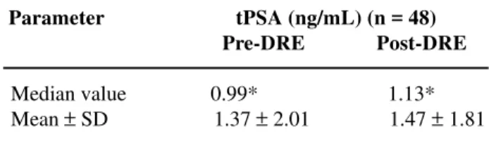
Before the DRE examination the individuals revealed a tPSA value of 1.37 ± 2.01 ng/mL with the median values established in 0.99 ng/mL (Table-1). Two individuals from the investigational group, both above 70 years old, had values above 4.00 ng/mL, one with 4.36 and other with 14.00 ng/mL, which contributed to the high value of standard deviation around the mean evidenced in this group. Following the post-DRE 30-minute interval, the group presented values for mean ± standard deviation and median, respectively, of 1.47 ± 1.81 and 1.13 ng/mL. The dif-ference between median values of tPSA, before and after the examination, was minimal and non signifi-cant (p > 0.05). In a new tPSA assay after DRE ex-amination the same 2 individual that had revealed values above 4 ng/mL, maintained values above this threshold (4.35 and 13.00 ng/mL, respectively) con-tributing, once more to elevating the standard devia-tion around the mean (Table-1).
The group’s mean values of tPSA were ap-proximately 37% higher than the medians, both be-fore and after DRE, proving that the median reflected the value distribution of the group in a more reliable way, without being excessively influenced by indi-vidual values that are largely different from the aver-age. The expressive majority of individuals shows a trend of having presented some elevation in tPSA values following DRE (34 dos 48), and 3 maintained themselves at the same threshold, while 11 demon-strated a slight decrease in the levels of this serum protein, including the 2 individuals with values above 4.00 ng/mL.
re-Table 1 – Pre- and post-digital rectal examination (DRE) values of total PSA (tPSA) in individuals without prostate pathology.
Parameter tPSA (ng/mL) (n = 48) Pre-DRE Post-DRE
Median value 0.99* 1.13* Mean ± SD 1.37 ± 2.01 1.47 ± 1.81
* Difference between the values was not statistically significant
(p > 0.05). tPSA values increased in 34 individual, decreased in
11 and remained unchanged in 3. veal that mean values of tPSA progressively increase
from the lowest age range to the range ≥ 70 years. The group ≥ 70 years evidenced the highest mean value of all four age ranges, influenced by 2 indi-viduals’ high values.
Median values of the age ranges also recorded a progressive elevation with increasing age, except for the group from 50 - 59 years whose median value, below the other ranges, was maintained after the DRE examination. The difference in pre- and post-DRE median values was not statistically significant in any age group (p > 0.05).
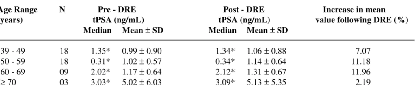
The percentage of variation in the tPSA mean levels, before and after DRE examination in the age groups are presented in Table-2. DRE pro-cedure caused the lowest variation for levels higher than 2.19% in the group ≥ 70 years, and the highest increase of 11.96% in individuals between 60-69 years. A progressive increase in the percentage of elevation in tPSA mean values was found in the first 3 age groups (7.07; 11.18 and 11.96, for the 39 - 49, 50 - 59 e 60 - 69 years ranges, respec-tively), with a drop to 2,19% being recorded then in the group ≥ 70 years.
DISCUSSION
DRE and determination of serum PSA are regarded as highly valuable for “screening” male population in ages that are more susceptible to pros-tate pathologies (8-11). In comparative terms, the in-crease in serum PSA levels seem to better correlated to the evolution of prostate pathologies (12), but this protein can be high in other conditions unrelated to the prostate (13,14), which renders difficult the use of this marker as an exclusive parameter for diagno-sis of PCa and BPH. Even though rectal examination allows a direct assessment of structural changes of the prostate and surrounding tissues, DRE examina-tion is considered less sensitive than PSA determina-tion, particularly for detecting diseases in an early stage of development. Such considerations have largely contributed for recommending the use of both procedures in combination for routinely “screening” the male population for detection of prostate patholo-gies (5,6).
When DRE examination is performed before the dosing of serum PSA, the use of post-rectal pal-pation PSA values as reliable markers of true values for the individual could be compromised, in principle, if such examination induces significant changes in the serum protein. Such possibility was investigated in some studies, mainly in the USA (1-3) and in France (4), with observations suggesting that DRE induced, in a general way, minimal changes in serum levels of PSA, which did not significantly compromise the use of this parameter. However, doubts seem to persist on this respect (4).
For the present study, the urology team of the institution determined the 30-minute interval between DRE examination and the new dosing of tPSA. Pre-vious works used intervals between DRE and the as-sessment of PSA, ranging from a few minutes and up to 90 minutes following digital rectal examination (1,2), however without finding significant differences in values that could be attributed to the post-DRE measuring time. The interval employed in this work is the same one used in the study by Lechevalier et al. (4).
Present data reveal that DRE resulted, almost always, in increase in the tPSA value, of about 8 to 13% (Tables-1 and 2). This observation is in accor-dance, in general terms, to reports by Stamey et al. (15) and by Brawer et al. (16), during the 80s, and in subsequent studies (1-4).
Table 2 – Pre- and post-digital rectal examination (DRE) values of total PSA (tPSA) in individuals without prostate pathology, according to age range.
Age Range N Pre - DRE Post - DRE Increase in mean (years) tPSA (ng/mL) tPSA (ng/mL) value following DRE (%)
Median Mean ± SD Median Mean ± SD
39 - 49 18 1.35* 0.99 ± 0.90 1.34* 1.06 ± 0.88 7.07 50 - 59 18 0.31* 1.02 ± 0.57 0.34* 1.14 ± 0.64 11.18 60 - 69 09 2.02* 1.17 ± 0.64 2.12* 1.31 ± 0.67 11.96
≥70 03 3.03* 5.02 ± 6.03 3.09* 5.13 ± 5.35 2.19
* There was no statistically significant difference between pre- and post-DRE values in none of the age ranges under study.
reasonable to assume that he effects of DRE exami-nation are not long lasting to the point of maintaining PSA alteration for a long time. The modest rise in mean and median values of tPSA following the per-formance of the DRE was not shown to be statisti-cally significant, which is in accordance to other re-ports in the literature (1-16), however it does not cor-roborate the observations of studies that found a sig-nificant increase in serum PSA values following digi-tal recdigi-tal examination (2-4,15). The increase in post-DRE PSA, of about 1.5 to 2.0 times above the initial values as reported by Stamey et al. (15), was not cor-roborated in none of the subsequent studies, includ-ing the present work.
In this study, in addition to moderate increases in the majority of individuals assessed, DRE has also caused a decrease in tPSA values in 11 of the 48 indi-viduals, with other 3 that did not present any change in their levels. This finding seems to suggest that DRE does not contribute, in fact, for a significant change in mean or median profile of tPSA in this group of individuals (men with no diagnosed prostate pathol-ogy). In this aspect, this work, which was performed with a group of only 48 healthy individuals, directly and favorably parallels with the conclusion by Crawford et al. (2), that DRE did not result in signifi-cant changes in the PSA level.
Data from Table-2 reveal that the percentage of DRE-induced increase in mean tPSA values re-lates to the increasing age of the male population, in a crescent way, from the age range of 39 - 49 years to the range of 60 - 69 years. Such data is in accordance,
in a general way, to the well-known elevation of PSA with increasing age. Despite this effect of age over tPSA in older individuals, the differences found in values of age ranges were not significant.
Finally, we observed that the group of indi-viduals aged ≥ 70 years had a decrease in tPSA value following the DRE procedure. Considering that there were only 3 individuals in this age range, the obser-vation becomes little relevant. Even if we disregard the tPSA values of the group ≥ 70 years, it is evident that the digital rectal examination induced some al-teration for higher tPSA values in approximately 72% of the individuals assessed. However, this modest el-evation does not seem to be significant so to nega-tively influence the use of post-DRE tPSA values as reliable markers.
Maria de F. Figueiredo had a scholarship from CNPq - Brazil
REFERENCES
1. Yuan JJ, Coplen DE, Petros JA, Figenshau RS, Ratliff TL, Smith DS, et al.: Effects of rectal examination, prostatic massage, ultrasonography and needle biopsy on serum prostate-specific antigen levels. J Urol. 1992; 147 (Pt 2): 810-4.
2. Crawford ED, Schutz MJ, Clejan S, Grago J, Resnick MI, Chodak GW, et al.: The effect of digital rectal examination on prostate-specific antigen levels. JAMA. 1992; 267: 2227-8.
ef-fect of digital rectal examination on the serum pros-tate-specific antigen concentration: results of a ran-domized study. J Urol. 1992; 148: 83-6.
4. Lechevallier E, Eghazarian C, Ortega JC, Roux F, Coulange C: Effect of digital rectal examination on serum complexed and free prostate-specific antigen and percentage of free prostate-specific antigen. Urology. 1999; 54: 857-61.
5. Vieira JGH, Nishida SK, Pereira AB, Arraes RF, Verreschi TN: Serum levels of prostate-specific anti-gen in normal boys throught puberty. J Clin Endocrinol Metabol. 1994; 78: 1185-7.
6. Garzotto M, Hudson RG, Peters L, Hsieh YC, Barrera E, Mori M, et al.: Predictive modeling for the pres-ence of prostate carcinoma using clinical, laboratory, and ultrasound parameters in patients with prostate specific antigen levels < or = 10 ng/mL. Cancer. 2003; 98: 1417-22.
7. Sachs L: Estatística Aplicada. Barcelona, Editorial Labor. 1978.
8. Galic J, Karner I, Cenan L, Tucak A, Hegedus I, Pasini J, et al.: Comparison of digital rectal examination and prostate specific antigen in early detection of prostate cancer. Coll Antropol. 2003; 27 (Suppl 1): 61-6. 9. Miller DC, Hafez KS, Stewart A, Montie JE, Wei JT:
Prostate carcinoma presentation, diagnosis, and stag-ing: an update form the National Cancer Data Base. Cancer. 2003; 98: 1169-78.
10. Becciolini A, Porciani S, Lanini A: Marker determi-nation for response monitoring: radiotherapy and dis-appearance curves. Int J Biol Markers. 1994; 9: 38-42. 11. Carter HB, Pearson JD, Metter EJ, Brant LJ, Chan DW, Andres R, et al.: Longitudinal evaluation of prostate-specific antigen levels in men with and without pros-tate cancer. JAMA. 1992; 267: 2215-20.
12. Roehrborn CG, Gregory A, McConnell JD, Sagalowsky AI, Wians FH Jr.: Comparison of three assays for total serum prostate-specific antigen and percentage of free prostate-specific antigen in predict-ing prostate histology. Urology. 1996; 48 (6A Suppl): 23-32.
13. Rhoden EL, Riedner CE, Maffesson R, Gobbi D, Teloken C, Souto CAV: Free to total prostate-specific antigen ratio for the diagnosis of prostate cancer. Int Braz J Urol. 2001; 27: 454-60.
14. Maciel LMZ, Martins ACP, Falconi RA, Cologna AJ, Suaid HJA: Comparative study of assays for prostate-specific antigen (PSA) determination. Int Braz J Urol. 2001; 27: 542-8.
15. Stamey TA, Yang N, Hay AR: Prostate-specific anti-gen as a serum marker for adenocarcinoma of the pros-tate. N Engl J Med. 1987; 317: 909-16.
16. Brawer MK, Schifman RB, Ahmann FR, Ahmann ME, Coulis KM: The effect of digital rectal examination on serum levels of prostate-specific antigen. Arch Pathol Lab Med. 1988; 112: 1110-2.
Correspondence address:
Dr. Talapala G. Naidu
Rua Vilebaldo Aguiar, 95 / 903 Fortaleza, CE, 60190-780, Brazil E-mail: [email protected]