PRIAPISM – ETIOLOGY, PATHOPHYSIOLOGY AND MANAGEMENT
C. VAN DER HORST, HENRIK STUEBINGER, CHRISTOPH SEIF, DIETHILD MELCHIOR,
F.J. MARTÍNEZ-PORTILLO, K.P. JUENEMANN
Department of Urology, University Hospital Schleswig Holstein, Campus Kiel, Germany
ABSTRACT
The understanding of erectile physiology has improved the prompt diagnosis and treatment of priapism. Priapism is defined as prolonged and persistent erection of the penis without sexual stimulation and failure to subside despite orgasm. Numerous etiologies of this condition are consid-ered. Among others a disturbed detumescence mechanism, which may due to excess release of con-tractile neurotransmitters, obstruction of draining venules, malfunction of the intrinsic detumescence mechanism or prolonged relaxation of intracavernosal smooth muscle are postulated. Treatment of priapism varies from a conservative medical to a drastic surgical approach. Two main types of pri-apism; veno-occlusive low flow (ischemic) and arterial high flow (non-ischemic), must be distin-guished to choose the correct treatment option for each type. Patient history, physical examination, penile hemodynamics and corporeal metabolic blood quality provides distinction between a static or dynamic pathology. Priapism can be treated effectively with intracavernous vasoconstrictive agents or surgical shunting. Alternative options, such as intracavernous injection of methylene blue (MB) or selective penile arterial embolization (SPEA), for the management of high and low flow priapism are described and a survey on current treatment modalities is given.
Key words: priapism; classification; pathophysiology; etiology; treatment; pharmacologic therapy;
surgery
Int Braz J Urol. 2003; 29: 391-400
INTRODUCTION
Definition
Priapism is a persisting erection caused by disturbances in the mechanism controlling penile de-tumescence and the maintenance of penile flaccidity. However, priapism will either start de novo, as a re-sult of a prolonged nocturnal erection, or may persist after sexual intercourse. During priapism, blood con-tinues to accumulate in the cavernous sinusoids and, in time, results in painful erection. The corpora cavernosa become rigid and painful, whereas the cor-pus spongiosum and the glans penis remain soft and uninvolved.
endot-392 PRIAPISM
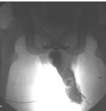
helial and trabecula destruction of the erectile tissue and subsequently in permanent erectile dysfunction (2) (Figure-1). After 48h widespread smooth muscle ne-crosis will take place (3). The natural squealer of un-treated ischemic priapism is penile fibrosis and impo-tence (4). On their long-term follow-up (mean 66 months) El-Bahnasawy and co-workers reported only a 43% rate of preserved erectile function after a long lasting priapism (median range of duration 48h) (5).
Erectile dysfunction is the most outstanding late complication. The incidence of this latter is di-rectly related to the duration of priapism and neces-sarily used aggressive treatment methods. As a result of priapism the overall rate of erectile dysfunction can be as high as 59%. High-flow arterial priapism shows a lower incidence of erectile dysfunction up to 20% (6). To preserve erectile function immediate uro-logical consultation must be provided.
The duration time of a normal erection before it is classifiable as priapism is still controversial. On-going penile erections for more than 6 hours can be classified as priapism owing to studies analyzing blood gases revealing ischemia and acidosis after 4 to 6 hours that could lead to potential damage (7,8).
Physiology of Penile Erection
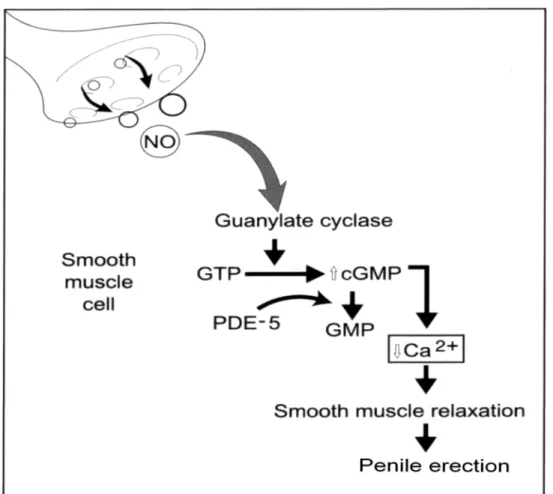
Penile erection is a complex neurovascular event involving the interaction of three physiological systems: the central nervous system (CNS), the pe-ripheral nervous system and the penile arterial and trabecula smooth muscle (9). The penile corpora, a specialized vascular tissue, consisting of endothelial-lined sinusoidal spaces supported by a framework of smooth muscle, collagen, nerves, nutritive arterioles and capillaries (4). Normal erectile function is a com-plex interaction of both, the nervous and vascular system. Erection requires relaxation of trabecula smooth muscle that results in an increased compli-ance of the sinusoids and arterial wall as well as a dilatation of the arterioles and arteries. As a result of arterial engorgement that occurs through smooth muscle dilatation, a three step mechanism providing full erection follows. Expanding sinusoids are sam-pling blood passively. Resulting in compression of the subtunical venous plexuses sited into the trabe-cula network between the tunica albuginea and the peripheral sinusoids. This reduces venous outflow. Stretching of the tunica albuginea to its capacity en-closing the emissary veins between the tunica layers leading to a decreased venous outflow (4). The smooth muscle relaxation during erection depends upon the promotion of Ca2+ efflux. This relaxation of smooth muscle cells is mediated mainly by nitric oxide, which activates the enzyme guanylate cyclase. This cyto-plasmic enzyme increases formation of the second messenger, cGMP. Elevated levels of peripheral cGMP in turn promote the efflux of Ca2+ ions from the cavernosa smooth muscle cells. This induces muscle relaxation, facilitates blood flow into the cor-pora cavernosa, and thereby helps to obtain and main-tain penile erection (10) (see also Figure-2). Under physiological conditions the process of penile detu-mescence, mediated by efferent sympathetic path-ways, follows the tumescence phase. Adrenergic sym-pathetic nerves release norepinephrine, which acts on adrenoceptors in penile smooth muscle. This result in reduced arterial inflow diminished lacunar space volume and accelerated corporeal venous outflow (11,12). The flaccid state of the penis is maintained by contraction of penile smooth muscle cells medi-Figure 1 – Corporeal cavernosography demonstrating
ated by the intracellular accumulation of Ca2+ ions, which is mainly effected by noradrenergic stimula-tion of α1-adrenergic receptors (9,13).
Despite the fact that the metabolic rate of corporal smooth muscles has not been reported yet, the penis as an external organ supplies a decreased temperature compared to the mean warmth of the central body. Therefore its energy requirements can be met at very low blood flow rates. During sexual excitement, the helicine arteries dilate and straighten which in turn allows blood to enter directly into the sinusoidal spaces. At that time, there is a 5-10 fold increase in blood flow to the penis. Intracavernal needle aspiration of blood during normal erection has a pO2 of approximately 40 mmHg, a pCO2 of 40 mmHg and a pH of 7.4 (4,14).
ETIOLOGY AND PATHOPHYSIOLOGY
Over the last years, improved comprehension of the physiology and pharmacology of erectile func-tion has led to a wider knowledge on etiology, patho-physiology and treatment of priapism (15-17). As a result, the treatment of priapism has evolved from predominantly surgical management to less invasive pharmacological therapies (18). For this reason, it is important to distinguish between arterial and veno-occlusive priapism as the choice of treatment depends on the underlying pathophysiology. Meanwhile, the diagnosis and therapeutically management of arterial priapism still remains controversial (19), whereas the diagnosis and treatment of veno-occlusive priapism has become well established.
394 PRIAPISM
The cause of priapism can be primary, sec-ondary or idiopathic. Priapism with primary etiology is not accompanied by a disorder responsible for a prolonged erection, e.g. of physical or psychological origin (20). Secondary priapism is induced by fac-tors directly or indirectly affecting the penile erec-tion (Table-1). It has been suggested that the increas-ing frequency of priapism is related to the risincreas-ing wide-spread use of intracavernous injection therapy (CCIT) for erectile dysfunction (2). Between 1.1% (for PGE1) and 7.7% (combination of papaverine and phentola-mine) of patients treated with CCIT develop a pro-longed erection (21). The cause of an idiopathic pri-apism cannot be traced and no pathological condi-tions are obvious. Its incidence is estimated between 50% and 60% (4,6,15,22).
During initial assessment, depending from the underlying hemodynamic pathology, the physician must distinguish between the 2 basic types of priapism – high flow (non-ischemic) and low flow (ischemic) – as the methods of treatment and prognosis differ accordingly (1,4,14,23). The etiology of arterial high-flow priapism remains unclear, although pharmaco-logical, traumatic and neurological diseases have been proposed (24,25). High-flow priapism results from unregulated, continuous arterial inflow into the lacu-nar spaces (an arterial-laculacu-nar fistula), usually sec-ondary to a lacerated cavernosal artery from blunt or penetrating trauma (25). The increase arterial flow is not regulated by helicine arteries and does not acti-vate the veno-occlusive mechanism (4). The paucity of cases reported in the literature implies that arterial priapism is a rare urological disorder (6,26). Initially CCIT induced high-flow priapism as an increase in arterial flow is evident. With time the veno-occlusive mechanism becomes activated changing into a pain-ful, ischemic low-flow priapism (15).
The more common low-flow or veno-occlu-sive priapism results from persistent obstruction of venous outflow from the lacunar spaces (27). 80% to 90% of clinically presented priapisms are low flow disorders (6,15).
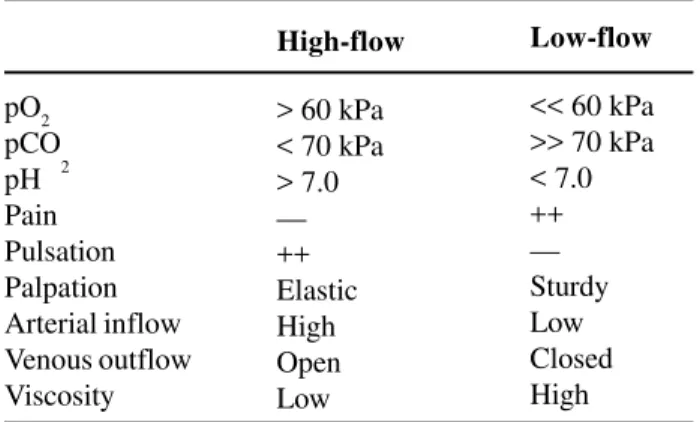
In order to specify the type of priapism pre-cisely, the assessment of history, physical examina-tion, penile hemodynamics and corporeal metabolic blood quality (Tables-2 and 3) is essential. In ischemic
low flow priapism, venous outflow is not revealed by cavernosonography and pulsation is not palpable. One of the main pathologies of low flow priapism is blood stasis in the corpora cavernosa resulting in low pO2 and high pCO2. The pH of corporeal blood drops be-low 7.0 (acidosis). Erection then becomes painful and irreversible corporeal fibrosis can develop (Figure-1). Pain is associated with tissue hypoxia and acido-sis. Urgent therapeutic intervention with irrigation and corporeal blood aspiration of up to 150 ml to 200 ml is necessary.
In color-flow duplex sonography, the often painless, non-ischemic high-flow priapism shows a high arterial inflow and cavernosonography reveals normal venous outflow. The pH rises above 7.0. The blood gas level is similar to that of arterial blood. Color-flow duplex sonography was proposed to be a less invasive diagnostic examination method as op-posed to cavernosonography (15,19).
CONSERVATIVE TREATMENT FOR
PRIAPISM
pa-395
PRIAPISM
- sickle cell disease
- leukemia
- thalassemia
- polycythemia
- thrombocytasthenia
- coagulopathologies
- hemophilia
- corpus cavernosal thrombosis
- congenital dyserythropoietic anemia
Surgical
- spinal cord injury
- penile trauma
- perineal trauma
- pelvic trauma
- arterial anastomosis to corpus
- scorpion and snake venom
- herniated disc (lumbar)
- ruptured cerebral aneurysm
- spinal cord tumor or metastasis
- multiple sclerosis
- spinal tumor with compression
- spinal anesthesia
- cycling
- prostatitis
- urethritis
- phimosis
- mumps
- syphilis
- malaria
- rabies
- diabetes mellitus
- atypical pneumonia
- acute appendicitis
- papaverine
- theophylline
- prostaglandin E
- nitroglycerine
- verapramil
- imipramine
- haloperidol
- phenelzine
- cannabis
- doxozosine - cocaine - clozapine - prazosine - trazodone - vancomycine - quetiapine - chlorpromazine - heparin - alcohol - primary sarcoma
- metastasis
- myeloma
- prostate cancer
- penis cancer
- lymphoma
- bladder carcinoma
- paraneoplastic
396 PRIAPISM
tient expeditiously by increasing the outflow of cav-ernous blood. This procedure is followed by intracavernous injection of vasoconstrictive agents and finally progressing to surgical procedures de-signed to shunt blood from the corpora cavernosa to the corpus spongiosum or to the vena saphena (22,24,29).
Pharmacological intervention comprises intracavernous injection of metaraminol or alpha-adr-energic agonists such as phenylephrine, norepineph-rine, ethylephrine and epinephrine – all with similar
pO2 pCO
2
pH Pain Pulsation Palpation Arterial inflow Venous outflow Viscosity
High-flow
> 60 kPa < 70 kPa > 7.0 — ++ Elastic High Open Low
Low-flow
<< 60 kPa >> 70 kPa < 7.0 ++ — Sturdy Low Closed High
Table 2 – Metabolic and hemodynamic classification of priapism (intracavernal blood)
Table 3 – Oral and intracorporeal agents for the therapy of priapism
Intracavernous Application
Drug Dose
Epinephrine 0.03 – 0.05 mg Etylephrine 2 - 20 mg Phenylephrine 0.1 - 1 mg Norepinephrine 0.01 – 0.02 mg Methylene blue 50 mg
Intravenous / Oral application
Drug Dose
Dopamine 2 - 4 µg / kg i.v. Terbutaline sulfate 5 mg oral
Ketamine hydrochloride 1 mg / kg i.v. or i.m.
modes of action – and oral or intravenous adminis-tration of sympathomimetica such as dopamine or terbutaline (12,16,20,30,31) (Table-3). Alpha-agonist agents exert a vasoconstrictive effect on smooth muscle. Intracavernous injection of these drugs can cause pain, hematoma, infection and fibrosis of the penis (20). In order to avoid systemic complications, it is of outstanding importance to inject the agent into the fully erected corporeal cavernosa only (32). As no blood is retained in the penis, local administration of these agents may lead to such systemic complica-tions as sustained hypertension up to 200 mmHg or cerebral bleeding (26,33).
Alpha-agonists for oral administration may be taken into consideration, but hypertension and car-diac arrhythmia could assert a limiting effect (20). This report considered the successful oral use of terbutaline (2.5 to 5 mg), a ß2-adrenergic-agonist, for recurrent idiopathic priapism. In 5 patients the pri-apism resolved completely within 30 minutes after oral administration of 5 mg terbutaline. ß2-agonists should be used with caution in patients with severe arterial coronary disease, suspected excessive intra-vascular fluid volume or low potassium because this latter can cause tachycardia and pulmonary edema.
effects and is rapidly excreted by the kidneys into the urine. The routine use of intracavernous MB in the treatment of priapism is appealing, since its toxicity and systemic side effects could prove slighter than those of commonly used alpha-adrenergic agents (18). Whilst treating 11 patients for priapism, deHoll et al. aspirated up to 200 ml corporeal blood and then intracavernosal injection of 50 mg MB for a 3 to 5 minutes period (34). The MB was then also aspirated and the penis was lightly compressed for 5 minutes. Immediate detumescence was the response in 67% of these patients. There was a 100% response in pa-tients suffering priapism after intracavernous injec-tion therapy (prostaglandin E1). Alpha-agonists of-fered no benefit in cases where MB was unsuccess-ful. These results confirmed that MB is effective in the treatment of pharmacologically-induced priapism. A temporary blue discoloration of the penis was noted subsequent to treatment with this substance. Further to this, after injection of MB, patients complained of a burning sensation in the penis that fortunately sub-sided within a short time (35). Despite this, penile necrosis has been observed after intracavernous ad-ministration of MB. Necrosis was attributed to cav-ernous fibrosis in one patient with a history of 5 years intermittent priapism and after performing an inef-fective shunt surgery (38). In our opinion, MB alone provides favorable results if the existing endothelium is in good condition (35). With respect to these ob-servations and in accordance to Mejean et al. (38) we do not recommend the administration of MB in cases of suspected corporeal fibrosis, e.g. in patients with a long history of recurrent priapism. Fibrosis possibly inhibits the endothelial action of MB and thus even-tually eliminates any positive effect (38). It is there-fore of eminent importance to aspirate the injected MB. In consequence, to exclude any possible exist-ence of fibrosis, MB should only be administered during the first hours of priapism. No cutaneous ne-crosis was observed at the injection site in our pa-tient cohort.
The sickle cell mediated priapism is normally treated with pharmacotherapy. The main step of therapy is hydration, alkalization, analgesia and he-modilution, which is used to increase the hemoglo-bin concentration. If conservative measures are
inef-fective, then the former described therapy options must be attempted (4). Gonadotropin-releasing hor-mone (GnRH) analogues have been tried for the chronic treatment of recurrent priapism. Antiandrogens like flutamide can be administered as well (27).
INVASIVE TREATMENT FOR PRIAPISM
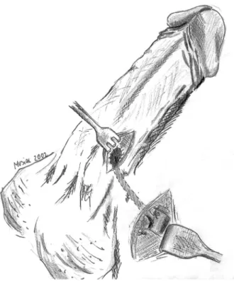
Despite widespread administration of phar-macological substances in the treatment of priapism, there is still a call for surgical intervention if all at-tempts of conservative treatment fail. A wide range of surgical procedures is established for this type of treatment. This includes proximal ligation of the in-ternal pudendal artery, anastomosis between the cor-pus cavernosum and the saphenous vein (Grayhack et al. shunt) see Figure-3 (39), cavernous spongiosum shunt (Winter shunt), see Figure-4 (22), cavernosal dorsal vein shunt, incision, irrigation and drainage of corporeal sinuses, percutaneous aspiration of the cor-pus cavernosum, division of the pudendal nerves and ischiocavernosum muscle, amputation of the penis if associated with malignant disease of the genitalia, and other procedures (20).
The Winter shunt is the most common proce-dure (22). With this technique, a fistula is created between the glans penis and the corpora cavernosa. The success rate of these procedures ranges between 50% and 65% (40). Possible postoperative compli-cations are infection of the corpora cavernosa with abscess formation, urethral injury leading to stricture or urethrocutaneous fistula and penile hematoma with or without penile thrombosis resulting in erectile dys-function (6).
The problem with these surgical procedures is the high rate of erectile dysfunction (50%). There-fore they should only be used after failed conserva-tive treatment (29). If erectile dysfunction is present the best treatment is implantation of a penile prosthe-sis to enable the patient to sexual intercourse (41).
398 PRIAPISM
Bastuba et al. to evaluate the use of selective penile arterial embolization (SPAE) (26). Candidates for SPAE underwent diagnostic arteriography of the pel-vis with bilateral selective injections of the internal pudendal arteries that revealed a unilateral arterial-lacunar fistula in all cases first. A catheter was ad-vanced up to the level of the ipsilateral common pe-nile artery. Approximately 3 ml autologous clot were either unilaterally or bilaterally injected until com-plete stasis was achieved, producing a transient in-terruption of arterial flow. This resulted in immedi-ate pain relief with subsequent detumescence in all the patients treated by SPAE. Post-embolization ar-teriogram studies documented the occlusion of the cavernous artery. Normal erectile function, with a delay of up to 5 months because of clot lysis, was restored in 86% of the patients. This group of inves-tigators propagated SPAE as a successful treatment option for high-flow priapism attributed to trauma or A-V fistulas. Potential complications after
emboliza-tion include penile gangrene, persistent erectile dys-function, gluteal ischemia, perineal abscess or migra-tion of embolizamigra-tion material (4,18).
CONCLUSIONS
Priapism is a considered urologic emergency and should be treated prompt and consequent. The treatment options for all types of priapism are ini-tially conservative but surgical therapy must be avail-able when applicavail-able. MB is an effective and safe treatment for priapism. This approach is most suit-able for drug-mediated high-flow priapism (corpus cavernosum injection therapy, CCIT) of short dura-tion. Alpha-agonists offered no benefit in cases where MB was unsuccessful. Apparently, either MB or al-pha-agonists can be initially administered. However, if one of these substances fails to take effect, no addi-tional benefit is likely to be gained from the other. When mechanical compression and pharmacological
Figure 4 – Winter-shunt (percutaneous glandulocavernous shunt with a True-Cut-Needle)
treatment after the aspiration of cavernous blood do not take effect, then the alternatives are either a sur-gical shunt procedure or selective arterial emboliza-tion. Embolization is a less invasive option for re-fractory priapism. The restoration of erectile func-tion with MB treatment and unilateral common pe-nile arterial embolization is possible.
REFERENCES
1. Hauri D, Spycher M, Bruhlmann W: Erection and pri-apism - a new pathophysiological concept. Urol Int. 1983; 88: 138-45.
2. Hashmat AI, Rehman JU: Priapism. In: Hashmat AI, Das S (eds.), The Penis. Philadelphia, Lea & Febiger. 1993; pp 219-43.
3. Spycher MA, Hauri D: The ultrastructure of the erec-tile tissue in priapism. J Urol. 1986; 135: 142-7. 4. Melman A, Serels S: Priapism. Int J Imp Res. 2000; 12
(Suppl 4): 133-9.
5. El-Bahnasawy MS, Dawood A, Farouk A: Low-flow priapism: risk factors for erectile dysfunction. BJU Int. 2002; 89: 285-90.
6. Parivar F, Lue TF: Priapism. In: Hellstrom WJG (ed.), Male Infertility and Sexual Dysfunction. Berlin, Springer-Verlag. 1997; 401-8.
7. Broderick GA, Gordon D, Hypolite J, Levin RM: An-oxia and corporeal smooth muscle dysfunction: a model for ischemic priapism. J Urol. 1994; 151: 259-62.
8. Juenemann KP, Lue TF, Abozeid M, Hellstrom WJG, Tanagho EA: Blood gas analysis in drug induced pe-nile erections. Urol Int. 1986; 41: 207-11.
9. Wagner G, Mulhall J: Pathophysiology and diagnosis of male erectile dysfunction. Int J Imp Res. 2001; 88 (Suppl 3): 3-10.
10. Burnett AL: Role of nitric oxide in the physiology of erection. Biol Reprod. 1995; 52: 485-9.
11. Diederichs W, Stief CG, Lue TF, Tanagho EA: Nore-pinephrine involvement in penile detumescence. J Urol. 1990; 143: 1264-6.
12. Koga S, Shiraishi K, Saito Y: Post-traumatic priapism treated with metaraminol bitartate: case report. J Trauma. 1990; 30: 1591-3.
13. Chuang AT, Steers WD: Neurophysiology of Penile Erection. In Carson CC, Kirby RS, Goldstein I (eds.), Textbook of Erectile Dysfunction. Oxford, ISIS Medi-cal Media. 1999; pp 59-72.
14. Martínez Portillo FJ, Juenemann KP: New aspects in
the treatment of priapism. Andrologia. 1999; 31 (Suppl): 53-8.
15. Juenemann KP: Priapismus. In: Thüroff JW (ed.), Urologische Differentialdiagnose. Stuttgart, Georg Thieme Verlag. 1995; pp 301-8.
16. Lue TF, Tanagho EA: Physiology of erection and phar-macological management of impotence. J Urol. 1987; 137: 829-36.
17. Pohl J, Pott B, Kleinhans G: Priapism - A three-phase concept of management according to etiology and prog-nosis. Br J Urol. 1986; 58: 113-8.
18. Steers WD, Selby JB: Use of methylene blue and se-lective embolization of the pudendal artery for high flow priapism refractory to medical and surgical treat-ment. J Urol. 1991; 146: 1361-3.
19. Brock G, Breza J, Lue TF, Tanagho EA: High flow priapism: a spectrum of disease. J Urol. 1993; 150: 968-71.
20. Shanta TR, Finnerty DP, Rodriguez AP: Treatment of persistent penile erection and priapism using terbutaline. J Urol. 1989; 141: 1427-9.
21. Spahn M, Manning M, Juenemann KP: Intracavernosal Therapy. In: Carson CC, Kirby RS, Goldstein I (eds.), Textbook of Erectile Dysfunction. Oxford, ISIS Medi-cal Media. 1999: pp 345-53.
22. Winter CC: Priapism treated by modification of cre-ation of fistulas between glans penis and corpora cavernosa. J Urol. 1979; 121: 743-4.
23. Hormon WJ, Nehra A: Priapism. In: Hashmat AI, Das S (eds.) The Penis. Philadelphia, Lea & Febiger. 1997; pp 219-43.
24. Lue TF, Hellstrom WJG, McAninch JW, Tanagho EA: Priapism: a refined approach to diagnosis and treat-ment. J Urol. 1986; 136: 104-8.
25. Witt MA, Goldstein I, Saenz de Tejada I, Greenfield A, Krane RJ: Traumatic laceration of intracarvenosal arteries: the pathophysiology of non-ischemic, high flow arterial priapism. J Urol. 1990; 143: 129-32. 26. Bastuba MD, Saenz de Tejada I, Dinlenc CZ, Sarazen
A, Krane RJ, Goldstein I: Arterial priapism: diagno-sis, treatment and long-term followup. J Urol. 1994; 151: 1231-7.
27. Levine LA, Guss SP: Gonadotropine-releasing hor-mone analogues in the treatment of sickle cell anemia-associated priapism. J Urol. 1993; 150: 475-7. 28. Lue TF: Editorial Comment. J Urol. 1988; 139: 737. 29. Bertram RA, Webster GD, Carson CC: Priapism:
eti-ology, treatment and results in series of 35 presenta-tions. Urology. 1985; 26: 229-32.
400 PRIAPISM
terbutaline and pseudoephedrine in management of prostaglandine E1-induced prolonged erections. Urol-ogy. 1993; 42: 51-3.
31. Sidi AA: Vasoactive intracavernous pharmacotherapy. Urol Clin North Am. 1988; 15: 95-101.
32. Potempa D, Juenemann KP, Schuller A, Lšbelenz M, Rassweiler J, Alken P: Die Therapie der prolongierten Erektion. Akt Urol. 1991; 143: 933-5.
33. Rösener M, Wechsel HW, Dichgans J: Intrazerebrale Massenblutung nach intrakavernöser Metaraminol-Behandlung einer prolongierten Erektion. Akt Urol. 1995; 26: 427-30.
34. deHoll JD, Shin PA, Angle JF, Steers WD: Alternative approaches to the management of priapism. Int J Imp Res. 1998; 10: 11-4.
35. Martínez Portillo FJ, Hoang-Böhm J, Weiss J, Alken P, Juenemann KP: Methylene blue as a successful treat-ment alternative for pharmacologically-induced pri-apism. Eur Urol. 2001; 39: 20-3.
36. Bush PA, Aronson WJ, Buga GM, Rajfer J, Ignarro LJ: Nitric oxide is a potent relaxant of human and rab-bit corpus cavernosum. J Urol. 1992; 147: 1650-5. 37. Furchgott RF: The role of endothelium in the responses
of vascular smooth muscle to drugs. Ann Rev Pharmacol Toxicol. 1984; 24: 175-97.
38. Mejean A, Marc B, Rigot JM, Mazeman E: Letter to the editor. J Urol. 1993; 149:1149.
39. Grayhack JT, McCullough W, O´Connor VJ: Venous bypass to control priapism. Invest Urol. 1964; 1: 509-13.
40. Winter CC, McDowell G: Experience with 105 patients with priapism: update review of all aspects. J Urol. 1988; 140: 980-3.
41. Kabalin JN: Corporeal fibrosis as a result of priapism prohibiting function of self-contained inflatable penile prosthesis. Urology. 1994; 43: 401-3.
Correspondence address:
Dr. Francisco José Martínez-Portillo Department of Urology, University Hospital Schleswig-Holstein Campus Kiel, Germany Arnold-Heller-Str. 7 D-24105 Kiel, Germany Fax: + 49 431 597-1957
E-mail: [email protected]