266
Pure Conventional Laparoscopic Radical Nephrectomy with
Level II Vena Cava Tumor Thrombectomy
_______________________________________________
Mingshuai Wang, Hao Ping, Yinong Niu, Junhui Zhang, Nianzeng Xing
Department of Urology, Beijing Chao-yang Hospital, Beijing Chaoyang, Beijing, China
ABSTRACT
ARTICLE
INFO
______________________________________________________________ ______________________
The surgical management with laparoscopic technique for renal cell carcinoma with inferior vena cava tumor thrombus (IVTT) remains challenging and technically de-manding in urological oncology. We present two patients with level II IVTT that were managed with pure conventional laparoscopic radical nephrectomy and thrombectomy. Two patients were diagnosed with a renal tumor with level II IVTT from December 2011 to January 2012. They both underwent pure conventional laparoscopic radical nephrectomy with thrombectomy. During these operations, intraoperative laparosco-pic ultrasonography was used to detect the thrombus and ensure complete removal. Two patients were operated through retroperitoneal approach for right renal tumor and transperitoneal approach for left renal tumor respectively. The demographics, periopera-tive and follow-up data were recorded for the study. Both operations were successfully performed without conversion. They both had no radiographic evidence of recurrence during follow-up. It is concluded that it is feasible to manage renal cell carcinoma with level II IVTT through pure conventional laparoscopic approach in carefully se-lected patients, which might expand the indication for laparoscopic surgery. The pure laparoscopic approach in the treatment of renal cell carcinoma with level II vena cava tumor thrombus is challenging and requires advanced laparoscopic skills. Multicenter prospective randomized control trials are needed to prove the benefits of this approach.
Key words:
Carcinoma, Renal Cell; Thrombosis; Laparoscopy
Int Braz J Urol. 2014; 40: 266-73
_____________________
Submitted for publication: July 23, 2013
_____________________
Accepted after revision: December 03, 2013
INTRODUCTION
Renal cell carcinoma (RCC) has a natural tendency for formation of tumor thrombus, which can extend into the inferior vena cava (IVC) in 4% to 10% of cases (1). The surgical management for RCC with IVC tumor thrombus (IVTT) is ex-tremely challenging and technically demanding. Open surgery is still the preferred method to ma-nage RCC with IVTT (2). With continued advances in laparoscopic surgical techniques and increase experiences among urologists, the indications of laparoscopic techniques have expanded to more complex renal tumors.
In the present study, we aim to demons-trate two cases of renal tumor with level II IVTT managed by pure LRN and thrombectomy.
MATERIAL AND METHODS
From January 2011 to February 2012, a total of 75 radical nephrectomies (RN) were performed in our department of which one patient underwent open RN with level II tumor thrombectomy and three pa-tient received LRN and tumor thrombectomy. Among the three patients, two patients underwent LRN with level II tumor thrombectomy. With Institutional Re-view Board approval, two patients undergoing LRN and IVC tumor thrombectomy for RCC with level II IVTT were identified and reviewed. Both patients had clinically localized disease without metastasis. The renal tumor thrombus was classified according to the Mayo classification (7). The RCC was classified based on American Joint Committee on Cancer 2010 TNM staging criteria, Fuhrman grading system and 2004 WHO classification. One surgical team leaded by a laparoscopic surgeon (N.Z.X) with high volume ex-periences performed both procedures.
Data including demographics, periopera-tive data, pathologic data and oncological outco-mes were collected and analyzed. The comorbidity was classified according to Charlson’s index (CI) (8). Perioperative data involved tumor’s compu-ted tomography-scan size, side and location, IVTT length (evaluated by abdominal vascular magne-tic resonance imaging), operative time, estimated blood loss, and intraoperative and postoperative complications (Figure-1). Postoperative complica-tions within 30 days were classified according to Clavien’s system (9).
SURGICAL TECHNIQUE
The transperitoneal laparoscopic approach was used for the patient 1 with left renal tumor and IVTT, while the retroperitoneal approach was used for the patient 2 with right renal tumor and IVTT.
Following general anesthesia and Foley catheter placement, patient 1 was positioned in a 60º modified lateral decubitus position. Five trocars were placed: one 10mm trocar at the umbilicus for the camera, one 5mm trocar lateral to the
umbili-Figure 1 - Preoperative CT and MRI scan. IVTT, inferior vena caval tumor thrombus; RT, renal tumor.
A
C
B
268
cus at the left anterior axillary line and one 12mm trocar in the midline between the xiphoid and the umbilicus for dissection, and one 10mm trocar subxiphoid trocar and one 10mm trocar lateral to the umbilicus at the right anterior axillary line for clamping the IVC.The colon was reflected medially and the renal hilum was identified. The left renal artery was clipped with Hem-o-lok clip and divided by Liga-Sure. After the left renal vein was mobilized the IVC was dissected circumferentially above the left renal vein as proximal as possible (Figure-2A). The intraoperative laparoscopic ultrasound probe (UST-5536-7.5 Ultrasound; Aloka) was utilized to identi-fy the extent of the tumor thrombus. The right renal vein was identified and any lumbar tributaries were isolated and ligated. A laparoscopic Satinsky clamp was used to block the IVC distal to the thrombus. Since the dissection of IVC can not reach the tail of the tumor thrombus, we didn’t clamp the IVC above the thrombus. The left renal vein was
inci-sed circumferentially at its junction with the IVC, and the tumor thrombus was immediately extracted entirely (Figure-2B). Then another prepared lapa-roscopic Satinsky clamp was immediately introdu-ced to clamp the IVC above the renal vein through the subxiphoid trocar. The IVC was closed with 5-0 polypropylene suture. The rest of the kidney and adrenal gland were dissected outside of Gerota’s fascia and were removed integrally with tumor thrombus using a retrieval bag (Figure-2C).
The patient 2 was firmly secured to the operating table in the lateral position. The right kidney bridge was elevated moderately, and the operating table was flexed somewhat to increa-se the space between the lowermost rib and the iliac crest. During LRN, four trocars were placed. In brief, the first 2cm incision was performed under the 12th rib in the posterior axillary line. A retro-peritoneal space was created with the index finger followed by a balloon dilator inflated with 1000mL air. A 5mm port was placed in the subcostal region
Figure 2 - A) The IVC was recognized and carefully dissected. B) After clamping the IVC, the IVC was incised and the IVTT was identified. C) The tumor was 13*10*8 cm in size with 8cm tumor thrombus in length. IVC, inferior vena cava; IVTT, inferior vena caval tumor thrombus.
A
C
B
on anterior axillary line guided by the forefinger. Another two 10mm port were positioned on the midaxillary line above the iliac crest and 3cm an-terior at the same level as this point respectively.
Gerota’s fascia was incised longitudinally in the general area of the renal hilum. Lifting the kidney, blunt and sharp dissection in this area was performed to identify renal arterial pulsations, which could indicate the location of the renal ar-tery. After complete circumferential mobilization of the artery Hem-o-lok clips were applied and the artery was transected directly by LigaSure. Dissec-tion continued toward the renal vein, which was lying anterior to the renal artery. As the renal vein was dilated by thrombus, it was easy to identify and dissect the renal vein, contralateral renal vein and IVC. Any luminal veins were clipped and se-vered. Then dissection continued upwards towards the adrenal gland as far as the diaphragm. The adrenal gland was isolated and the adrenal vein was clip-ligated and divided. After the kidney was fully mobilized, the IVC could be exposed with
only connecting to renal vein in renal hilum area. After the IVC was isolated, intraoperative ultraso-nography was performed to identify the extent of IVC thrombus. Tourniquet loops were placed and tightened around the IVC above and below the IVC tumor thrombus and the contralateral renal vein was clamped by bulldog clamp (Figure-3A). Then the renal vein was incised circumferentially at its junction with the IVC, and the tumor throm-bus was extracted entirely (Figure-3B and C). The IVC was stitched with a running 5-0 polypropyle-ne suture (Figure-3D). No tumor thrombus pieces were dislodged detected by intraoperative ultraso-nography. No bleeding was noted when the tour-niquet loops were removed.
RESULTS
Table-1 shows patient characteristics and perioperative data. Pure LRN with IVC thrombec-tomy was successfully performed in both patients with no conversion.
A
C
B
D
270
Table 1 - Demographics, clinical/pathologic data, and perioperative variables.
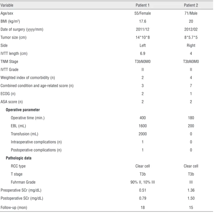
Variable Patient 1 Patient 2
Age/sex 55/Female 71/Male
BMI (kg/m2) 17.6 20
Date of surgery (yyyy/mm) 2011/12 2012/02
Tumor size (cm) 14*10*8 8*5.7*5
Side Left Right
IVTT length (cm) 6.9 4
TNM Stage T3bN0M0 T3bN0M0
IVTT Grade II II
Weighted index of comorbidity (n) 2 4
Combined condition and age-related score (n) 3 7
ECOG (n) 2 1
ASA score (n) 2 2
Operative parameter
Operative time (min.) 400 180
EBL (mL) 1600 200
Transfusion (mL) 2000 0
Intraoperative complications (n) 1 0
Postoperative complications (n) 1 0
Pathologic data
RCC type Clear cell Clear cell
T stage T3b T3b
Fuhrman Grade 90% II, 10% III III
Preoperative SCr (mg/dL) 0.51 1.36
Postoperative SCr (mg/dL) 0.79 1.50
Follow-up (mon) 18 15
BMI = body mass index; IVTT = inferior vena cava tumor thrombus; ECOG = Eastern Cooperative Oncology Group Performance Status; ASA = American Society of Anesthesiologists score; EBL = estimated blood loss; RCC = renal cell carcinoma; SCr = serum creatinine.
The first patient was a 55 years old wo-man, whose BMI was 17.6kg/m2. She presented with the symptoms of anemia. Preoperatively, computed tomography (CT) indicated the tumor was left sided measuring 14*10*8cm (Figure-1A). Magnetic resonance imaging (MRI) were used to evaluate the extent of the tumor thrombus,
(ECOG) was 2 and the weighted index of comorbi-dity and the combined condition and age-related scores were 2 and 3 according to Charlson’s in-dex. She underwent angioembolization 24 hours before surgery.
The estimated blood loss was 1600mL, and the operative time was 400 min. She received blood transfusion (2000mL) intraoperatively. The postoperative serum creatinine was 0.79mg/dL. A small amount of oral intake began at postopera-tive day 2 and she got out of bed that afternoon. The drainage tube was removed at postoperative day 8. Her final pathology was 13*10*8cm, 90% Fuhrman grade II and 10% grade III, clear cell carcinoma with tumor thrombus protruding 8cm into the IVC with negative surgical margin and no invasion of renal vein and IVC walls. The patient was discharged from the hospital on postoperati-ve day 15. One week after discharge, the patient went to our department again because of two time convulsion at home. After laboratory and imaging examinations she was diagnosed with hypokale-mia due to bad diet. Her electrolyte was corrected and intravenous nutrition therapy was given to improve her condition. The surgical complication was grade II according to Clavien classification system. The patient returned to full activity two months postoperatively and had no radiographic evidence of recurrence at 18 months follow-up.
Our second patient was a 71 years old man with BMI 20kg/m2. CT-scan indicated the renal tumor was right sided measuring 8*5.7*5cm and abdominal vascular MRI demonstrated the length of the tumor thrombus in IVC was 4cm, which was also a level II tumor thrombus (Fi-gure 1C and D). The TNM staging of this renal tumor was also T3bN0M0. The patient had mul-tiple comorbidities, including chronic obstructi-ve pulmonary disease for 6 years, hypertension for 16 years and type II diabetes mellitus for 2 years, which placed the patient in the high risk category for surgical intervention. The ASA score was 2, the ECOG was 1 and the weighted index of comorbidity and the combined condition and age-related scores were 4 and 7.
The estimated blood loss was 200mL, and the operative time was 180 min. No serious
in-occurred. The postoperative serum creatinine was 1.50mg/dL. He got out of bed and oral in-take began at postoperative day 1. The drainage tube was withdrawn at postoperative day 7. He was discharged from the hospital on postopera-tive day 10. The final pathology was 7.5*6*4cm, Fuhrman grade III, clear cell carcinoma with tu-mor thrombus protruding 4cm into the IVC with surgical margins free of tumor and without renal vein and IVC walls invasion. The patient returned to full activity about one month postoperatively and had no radiographic evidence of recurrence at 15 months follow-up.
DISCUSSION
Since the first introduction of LRN in 1990, the indications for LRN have expanded (10,11). RCC with IVTT was once considered a relative contraindication to LRN (12). In 1996, McDougall et al. presented the first successful LRN with level I thrombectomy (3). During the following years, many case series studies involving LRN with le-vel I thrombectomy were reported proving that the procedure was feasible (4,5). In this procedure, the tumor thrombus was “milked back” into the renal vein so as to block the vein with vascular clips or an endoscopic stapling device (13). Before Romero et al. (6) reported the first case of pure LRN with level II tumor thrombectomy, Fergany et al. (14) successfully performed laparoscopic ra-dical nephrectomy with level II thrombectomy in a survival porcine model, which showed clinical application of this technique appeared possible. The report of conventional pure LRN with level II tumor thrombectomy was very rare.
272
thrombus which was similar to ours. They pla-ced vessel loops around the IVC above and below the tumor thrombus and around the contralateral renal vein. Although the favorable outcomes re-ported in this first series, case-control studies are needed to evaluate whether a robotic approach is superior to pure laparoscopic and open technique.Currently most authors agree that the pre-sence of the thrombus itself has no specific prog-nostic significance if it can be removed success-fully (16). With advances in immunotherapy and molecular targeted therapy with such agents as interferon and Sorafenib, control of distant metas-tases in patients with RCC extending into the IVC can be achieved, thus survival of these patients may increase if aggressive surgery including tu-mor thrombectomy is combined with immunothe-rapy and molecular targeted theimmunothe-rapy. It appears worthwhile to perform thrombectomy even in pa-tients in whom RCC thrombus extends to the level of the right atrium or the pulmonary artery (17).
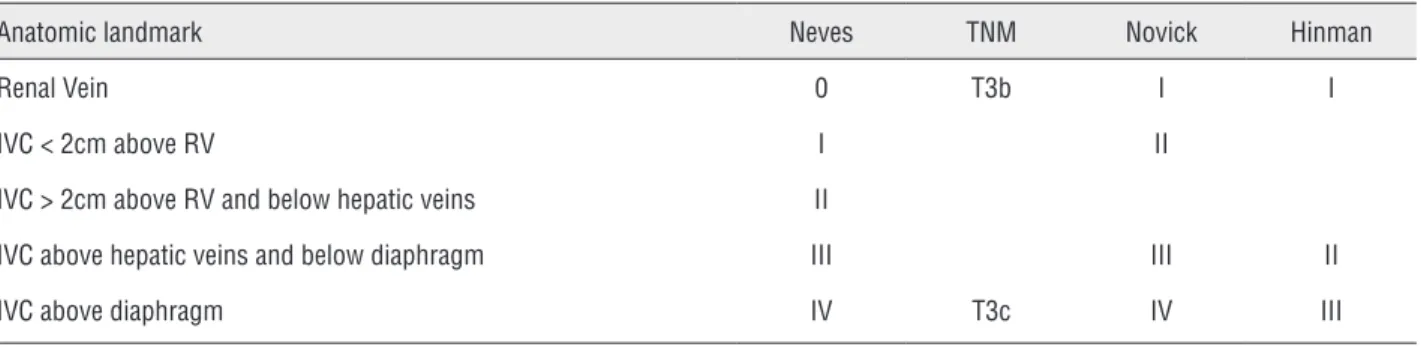
In our study, we employed Neves system to surgically stage tumor thrombus. Four staging systems were proposed for RCC with thrombus in the literature, TNM (18), Neves (7), Novick (19) and Hinman (20) (Table-2). The Neves and Novick are currently the most widely used. However, di-fferences should be noted among several staging systems. Since the surgical approach depends mainly on the tumor thrombus level, so a consis-tent surgical staging system is of utmost impor-tant for preoperative planning.
Similar to open surgery, pure LRN with le-vel II thrombectomy has several important steps. (1) dissection and ligation of the renal artery;
(2) vena cava, contralateral and ipsilateral renal vein exposure; (3) occlusion of the IVC under the thrombus, contralateral renal vein, and the IVC above the thrombus; (4) opening of the vena cava and thrombectomy; (5) stitching the IVC and rele-ase of the vascular clamps; (6) nephrectomy. Com-pletely control of the IVC is a vital important step. In our first case, we didn’t clamp the IVC above the thrombus because the dissection of IVC could not reach the tail of the tumor thrombus. However, this late clamping technique has several drawba-cks, mainly intraoperative massive hemorrhage. As the IVC above the thrombus wasn’t clamped, intraoperative massive hemorrhage occurred when we incised the IVC wall and drew out the tumor thrombus. However, after the tumor thrombus was dragged out, the IVC was immediately clamped by a prepared laparoscopic Satinsky clamp, which could avoid fatal hemorrhage. Secondly, pulmo-nary embolism. Patients may develop pulmopulmo-nary embolism secondary to tumor thrombus detach-ment. Our first patient was stable intraoperatively and continued to have no evidence of disease.
In our cases, we employed flexible intra-operative ultrasonography to detect the extent of IVC thrombus. The tumor thrombus can’t be dis-lodged if we find the right place to block the IVC. Another important function of intraoperative ultra-sonography is identification whether the IVC wall was invaded by the tumor thrombus. Conversion is required if the IVC wall is invaded by the thrombus.
With the present study, we demonstrated another report that pure LRN with level II IVTT thrombectomy might be safe for highly selecti-ve patients. This study had seselecti-veral shortcomings:
Table 2 - Different staging systems of IVC tumor thrombus in literature.
Anatomic landmark Neves TNM Novick Hinman
Renal Vein 0 T3b I I
IVC < 2cm above RV I II
IVC > 2cm above RV and below hepatic veins II
IVC above hepatic veins and below diaphragm III III II
retrospective study design, the small number of patients, and relatively short length of follow-up. Only two cases were involved in our study. Addi-tional reports and randomized studies are needed. Long-term oncologic outcomes are required to elu-cidate the safety and efficiency of this approach.
CONCLUSIONS
Pure LRN with level II IVC thrombectomy is feasible and can be performed in carefully se-lected patients. However, the laparoscopic appro-ach in the treatment of RCC with level II IVTT is challenging and technically demanding.
ABBREVIATIONS
RCC = renal cell carcinoma
IVC = inferior vena cava
IVTT = inferior vena cava tumor thrombus
RN = radical nephrectomy
LRN = laparoscopic radical nephrectomy
CONFLICT OF INTEREST
None declared.
REFERENCES
1. Marshall FF, Dietrick DD, Baumgartner WA, Reitz BA: Surgical management of renal cell carcinoma with intracaval neoplastic extension above the hepatic veins. J Urol. 1988; 139: 1166-72.
2. Ali AS, Vasdev N, Shanmuganathan S, Paez E, Dark JH, Manas D, et al.: The surgical management and prognosis of renal cell cancer with IVC tumor thrombus: 15-years of experience using a multi-specialty approach at a single UK referral center. Urol Oncol. 2013; 31: 1298-304.
3. McDougall E, Clayman RV, Elashry OM: Laparoscopic radical nephrectomy for renal tumor: the Washington University experience. J Urol. 1996; 155: 1180-5.
4. Martin GL, Castle EP, Martin AD, Desai PJ, Lallas CD, Ferrigni RG, et al.: Outcomes of laparoscopic radical nephrectomy in the setting of vena caval and renal vein thrombus: seven-year experience. J Endourol. 2008; 22: 1681-5.
5. Kovac JR, Luke PP: Hand-assisted laparoscopic radical nephrectomy in the treatment of a renal cell carcinoma
6. Romero FR, Muntener M, Bagga HS, Brito FA, Sulman A, Jarrett TW: Pure laparoscopic radical nephrectomy with level II vena caval thrombectomy. Urology. 2006; 68: 1112-4. 7. Neves RJ, Zincke H: Surgical treatment of renal cancer with
vena cava extension. Br J Urol. 1987; 59: 390-5.
8. Nuttall M, van der Meulen J, Emberton M: Charlson scores based on ICD-10 administrative data were valid in assessing comorbidity in patients undergoing urological cancer surgery. J Clin Epidemiol. 2006; 59: 265-73.
9. Dindo D, Demartines N, Clavien PA: Classification of surgical complications: a new proposal with evaluation in a cohort of 6336 patients and results of a survey. Ann Surg. 2004; 240: 205-13.
10. Clayman RV, Kavoussi LR, Soper NJ, Dierks SM, Meretyk S, Darcy MD, et al.: Laparoscopic nephrectomy: initial case report. J Urol. 1991; 146: 278-82.
11. Fenn NJ, Gill IS: The expanding indications for laparoscopic radical nephrectomy. BJU Int. 2004; 94: 761-5.
12. Eisenberg MS, Meng MV, Master VA, Stoller ML, Rini BI, Carroll PR, et al.: Laparoscopic versus open cytoreductive nephrectomy in advanced renal-cell carcinoma. J Endourol. 2006; 20: 504-8.
13. Henderson A, Murphy D, Jaganathan K, Roberts WW, Wolf JS Jr, Rané A: Hand-assisted laparoscopic nephrectomy for renal cell cancer with renal vein tumor thrombus. Urology. 2008; 72: 268-72.
14. Fergany AF, Gill IS, Schweizer DK, Kaouk JH, ElFettouh HA, Cherullo EE, et al.: Laparoscopic radical nephrectomy with level II vena caval thrombectomy: survival porcine study. J Urol. 2002; 168: 2629-31.
15. Abaza R: Initial series of robotic radical nephrectomy with vena caval tumor thrombectomy. Eur Urol. 2011; 59: 652-6. 16. Bachmann A, Seitz M, Graser A, Reiser MF, Schäfers HJ, Löhe
F, et al.: Tumour nephrectomy with vena cava thrombus. BJU Int. 2005; 95: 1373-84.
17. Glazer AA, Novick AC: Long-term followup after surgical treatment for renal cell carcinoma extending into the right atrium. J Urol. 1996; 155: 448-50.
18. Sobin LH and Wittekind C: TNM Classification of Malignant Tumours, 6th ed. New York, WileyLiss. 2002; pp. 239. 19. Novick AC, Streem SB and Pontes E: Stewart’s Operative
Urology, 2nd ed. Philadelphia, Williams & Wilkins. 1989. 20. Hinman F: Atlas of Urologic Surgery, 2nd ed. Philadelphia,
WB Saunders. 1998; pp. 1172.