Figueiredo JA, Carvalho GM, Mota RT, Castro VM, Meyer MMMDE, Barragat AZ. Laparoscopic total pelvic exenteration and perineal amputation with wet colostomy. A case report. J Coloproctol, 2011;32(2): 175-179.
ABSTRACT: Advanced rectal tumors can be treated with curative intent by surgical resection of the rectum including other pelvic organs. The reconstruction of the urinary and gastrointestinal tracts depends on the distance between the tumor and the anus, the patient’s status and the experience of the surgical team. This is a case of a male patient with a locally advanced low rectal tumor that underwent a laparoscopic pelvic exenteration. The anus and the tumor and other organs were excised by peritoneal approach. The uretero-colic anastomosis was performed extra-abdominally. The patient was discharged on the 14th postoperative day and remains healthy six months after the surgery. This approach
has shown to be feasible and safe. The aesthetical result was well accepted by the patient. The laparoscopic route should be considered as an alternative approach to pelvic exenteration in the treatment of locally advanced low rectal tumors that demand perineal amputation.
Keywords: rectal neoplasm; pelvic exenteration; laparoscopy.
RESUMO: O tumor de reto localmente avançado pode ser tratado com intenção curativa com uma operação ampliada que inclua outros órgãos da pelve. A reconstrução do trânsito urinário e do trânsito intestinal dependerá da distância do tumor em relação à margem do ânus, da experiência da equipe de cirurgiões, assim como das morbidades associadas do paciente. Apresentou-se neste artigo o caso de um paciente do sexo masculino, com tumor de reto baixo localmente avançado que foi submetido à exenteração pélvica por laparoscopia. Houve indicação para ressecção do ânus e a peça cirúrgica foi retirada por via perineal. A anastomose uretero-colônica foi confeccionada de maneira extracor-pórea. O paciente recebeu alta hospitalar após 14 dias e encontra-se com seis meses pós-operatórios. O método se mostrou factível e seguro. O resultado estético foi bem aceito pelo paciente. A via de acesso laparoscópica pode ser considerada uma alternativa para a exenteração pélvica no tratamento do tumor de reto baixo avançado que necessita de amputação anoperineal.
Palavras-chave: neoplasia de reto; exenteração pélvica; laroscopia.
Laparoscopic total pelvic exenteration and perineal amputation
with wet colostomy. A case report
Juliano Alves Figueiredo1, Gustavo Mareli de Carvalho2, Rafael Turano da Mota3, Vivian Monteiro de Castro3, Matheus Matta Machado Duque Estrada Meyer3, André Zucollo Barragat3
1Member of the Brazilian Society of Coloproctology and the Brazilian Society of Digestive Endoscopy; in Doctor’s
Degree Program in Surgery, School of Medical Sciences, Universidade Federal de Minas Gerais (UFMG) – Belo Horizonte (MG), Brazil. 2Urologist at the Hospital da Baleia – Belo Horizonte (MG), Brazil. 3Resident in General
Surgery, Hospital da Baleia – Belo Horizonte (MG), Brazil.
Study carried out at the Hospital da Baleia – Belo Horizonte (MG), Brazil. Financing source: none.
Conlict of interest: nothing to declare.
Submitted on: 05/04/2010 Approved on: 05/19/2010
INTRODUCTION
A locally advanced rectal tumors is a challenging situation in the clinical practice. Only complete sur-gical resection can offer the possibility of long-term disease control.
of pelvic exenteration, it is possible to have good
sur-vival rates within ive years2,3.
Laparoscopy has shown to be an alternative to the treatment of locally advanced neoplasms within the pelvis, with some reports and series of cases published in the world literature4,5,6.
The purpose of this study was to describe, with emphasis on the surgical approach, the case of a patient with locally advanced rectal tumor, treated with pelvic exenteration and wet colostomy by laparoscopy.
CASE REPORT
A 43-year-old male patient, with body mass in-dex of 19, presented hematochezia and tenesmus three
months ago. Low rectal cancer was conirmed near the
pectineal line, as well as moderately differentiated
ad-enocarcinoma, with istula directed to the anus margin.
The computed tomography showed invasion of pros-tate. Tomographic exams showed no signs of hepatic or pulmonary metastasis. The neoadjuvant treatment with chemotherapy was indicated (5-Fluorouracil 675 mg and Leucovorin 50 mg) and radiotherapy. The preoperative bowel preparation was performed with polyethylene glycol.
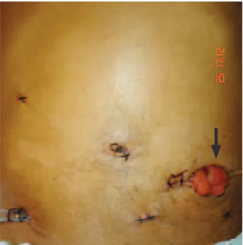
Total pelvic exenteration was performed using six trocars: umbilical (10 mm), right hypochondri-um (5 mm), right iliac fossa (12 mm), two in left iliac fossa (5 mm) and hypogastric (5 mm) trocars (Figure 1). The patient remained in dorsal decubi-tus position, with lower limbs extended, during the laparoscopic exams of rectosigmoidectomy and cys-toprostatectomy. The patient’s lower limbs were placed on stirrups only during the perineal surgical time. The colorectal surgery was performed before the bladder and prostate surgery. A double-barrel stroma was placed in the left iliac fossa trocar inci-sion (Figure 1). As the patient had the preoperative stoma marking, it was used for one of the trocars. A monopolar cautery was coupled to the laparoscopic curved scissors for the surgery and the 400 clips were used for hemostasia of mesenteric, vesical and prostat-ic vessels. The vesprostat-icoprostatprostat-ic dissection started with the Retzius space opening. The vascular pedicles were posterolaterally connected with the 400 clips
and Hemolock. The ureters were distally identiied,
released above the iliac vessels. The prostate
dissec-tion started with the endopelvic fascia opening, with the venous complex controlled with 2-0 silk suture, incision of the urethra and rectourethralis near the pelvic musculature. Denovilliers’ (rectoprostatic) fascia was not opened, and the single vesicoprostat-ic specimen was laterally dissected until the pelvvesicoprostat-ic
loor. A pelvic drain was placed through the right ili
-ac fossa trocar incision. The greater omentum, keep-ing the vascular nutrition through the left
gastroepi-ploic artery, was placed in the pelvis to ill the empty
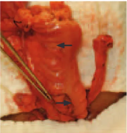
space and between the ureters after the wet colosto-my placement. No metastatic lesion was observed in the surgery. No lymphadenectomy of internal iliac vessels or obturator fossa was performed. The surgi-cal specimen was extracted through the perineum by two surgeons, at the same time, and part of the team performed the anastomosis of ureters with the distal segment of the descending colon. That was an extra-corporeal anastomosis (Figure 2).
The patient remained hospitalized for 14 days after the surgery. No supplementary nutrition was required, as the patient did not tolerate feeding for the maximum period of four days. No blood trans-fusion was required. Leucocytosis occurred after
the sixth postoperative day, secondary to a left il-iac fossa wall collection near the colostomy. This collection was treated with percutaneous drainage guided by ultrasound and use of Vancomycin and Meropenem. Histology showed an adenocarcinoma that invaded the muscularis propria, and the lymph
nodes were free of neoplasm. In the prostate, ibrot
-ic tissue was identiied, but no signs of adenocar
-cinoma after the adjuvant treatment. The margins were not affected (R0). Four postoperative chemo-therapy cycles were scheduled.
The total surgical time was seven hours and
thir-ty minutes, with ive hours and thirthir-ty minutes only for
the laparoscopic procedure. The procedure was well
tolerated by the patient, without any signiicant reduc
-tion of hemoglobin or blood transfusion. He remained 14 days hospitalized after the surgery due to a puru-lent collection near the stoma. He is in the 10-month follow-up. There is no evidence of recurrent hydro-nephrosis and no sign of metastatic disease.
DISCUSSION
Pelvic exenteration for the treatment of ad-vanced pelvic tumors alleviates symptoms of refrac-tory pain, lower limb edema, urinary sepsis and re-current hemorrhage7. It is estimated that 6 to 10% of the rectal tumors invade adjacent organs2. The prostate involvement changes the primary treatment into a total pelvic exenteration1,2,7. The survival rate
within ive years after the total pelvic exenteration
for primary rectal cancer is between 28 and 64%1,2,7. Pelvic oncologic surgery is also performed for the treatment of advanced cervical cancer, with radical hysterectomy and aortic and pelvic lymphadenecto-my8. Anterior pelvic exenterations can be performed with the urinary tract reconstruction, using a ureter-sigmoid anastomosis4. Surgeries such as radical cys-tectomy and prostacys-tectomy, via laparoscopic route, are performed by trained surgeons9.
In this study, the urologist had already conclude the learning curve in laparoscopic urologic surgery. The colorectal surgeon had already performed more than 40 colorectal surgeries via laparoscopy and par-ticipated in a number of cases with wet colostomy via conventional access10.
In this case report, the option of pelvic exen-teration via laparoscopic route was considered due to the possibility of complete extraction of the surgical specimen through the perineum, as it is, according to the tomographic exams, a tumor close to the pectineal line and that invaded the prostate. The urology team performed the rectal touch examination and cystos-copy and kept the indication of pelvic exenteration.
It is desirable to have the deinition about the pelvic
exenteration before the surgery, although it is known
that some cases are only deined during the surgery3.
The survival of patients with locally advanced rectal tumor without lymph nodes is better than when metastatic lymph nodes are present, and it is an in-dependent variable for survival2. In locally advanced rectal tumors, the prostate is the second most frequent-ly involved organ3. The neoadjuvant treatment is well accepted in the treatment of locally advanced rectal cancer with indication of pelvic exenteration (R0)11, although some renowned authors prefer not to use neoadjuvant chemoradiotherapy. There is also some debate on the use of lymphadenectomy near the inter-nal iliac vessels in cases of pelvic exenteration either via laparotomy12 or laparoscopy6. The authors of this study preferred to use preoperative chemotherapy and radiotherapy and did not use lymphadenectomy near the internal iliac vessels during the surgery.
Possible advantages of the laparoscopic sur-gery are: reduced blood loss, reduced postoperative pain and better cosmetic effect without affecting the oncologic radicality13. Pelvic exenteration via lapa-roscopic route should not affect the oncologic
REFERENCES
1. Koda K, Tobe T, Takiguchi N, Oda K, Ito H, Miyazaki M. Pelvic exenteration for advanced colorectal cancer with reconstruction of urinary and sphincter functions. Brit J Surg 2002;89(10):1286-9.
2. Costa SRP, Teixeira ACP, Lupinacci RA. A exenteração pélvica para o câncer de reto: Avaliação dos fatores prognósticos de sobrevida de 27 pacientes operados. Rev Bras Coloproct 2008;28(1):7-18.
3. Moriya Y, Akasu T, Fujita S, Yamamoto S. Aggressive surgical treatment for patients with T4 rectal cancer. Colorectal Dis 2003;5(5):427-31.
4. Puntambekar S, Kudchadkar R J, Gurjar AM, Sathe RM, Chaudhari YC, Agarwal GA, et al. Laparoscopic pelvic exenteration for advanced pelvic cancer: a review of 16 cases. Gynecol Oncol 2006;102(3):513-6.
5. Ferron G, Querleu D, Martel P, Chopin N, Soulié M. Laparocopy-assisted vaginal pelvic exenteration. Gynecol Obst Fertil 2006;34(12):1134-6.
6. Lin MY, Fan EW, Chiu AW, Tian YF, WU MP, Liao AC. Laparoscopy-assisted transvaginal total exenteration for locally advanced cervical cancer with bladder invasion after radiotherapy. J Endourol 2004;18(10):867-70.
7. Kecmanovic D M, Pavlov M J, Kovacevic P A, Sepetkovski A V, Ceranic M S, Stamenkovic A B. Management of advanced pelvic cancer by exenteration. Eur J Surg Oncol 2003;29(9):743-6.
8. Spirtos NM, Schlaerth JB, Kimball RE, Leiphart VM, Ballon SC. Laparoscopic radical hysterectomy (type III) with aortic and pelvic lymphadenectomy. Am J Obstet Gynecol 1996;174(6):1763-8.
9. Gupta NP, Gill IS Fergany A, Nabi G. Laparoscopic radical
cystectomy with intracorporeal ileal conduit diversion: ive
cases with a 2-year follow-up. BJU Int 2002;90(4):391-6. 10. Queiroz FL, Barbosa-Silva T, Costa LMP, Werneck-Cortes
BJ, Figueiredo JA, Guerra F, et al. Double-barrelled wet colostomy with simultaneous urinary e faecal diversion: results in 9 patients and review of the literature. Colorectal Disease 2006;8(4):353-9.
11. Bedrosian I, Giacco G, Penderson L, Rodrigues-Bigas MA, Feig B, Hunt KK et al. Outcome after curative resection for locally recurrent rectal cancer. Dis Colon Rectum 2006;49(2):175-82.
12. Costa SR, Antunes RC, Paula RP, Pedroso MA, Farah JF, Lupinacci RA. A exenteração pélvica no tratamento do câncer de reto estádio T4: A experiência de 15 casos operados. Arq Gastroenterol 2007;44(4):284-8.
cality and the complete excision of the tumor, and lymphadenectomy should follow the same parame-ters of the open technique.
Urinary derivation, combined with total pelvic exenteration, affects the patient’s quality of life, and some options could be the Bricker procedure or dou-ble-barrel wet colostomy14,15.
Double-barrel wet colostomy is an option for patients that require simultaneous urinary and fecal derivation16. It presents two derivations that drain to a single stoma17,18. It is considered a technique of low complexity, without intestinal anastomosis, involving reduced surgical time and acceptable quality of life10.
The published series about urinary derivations via laparoscopic route have few case of wet
colos-tomy, due to the dificult production of the reservoir
and increased surgical time4,6,8. The extracorpore-al production described in this case report had the
double-barrel coniguration, using a larger incision
in one of the trocars in the left lower quadrant of
the abdomen; thus, promoting reduced surgical time without increasing surgical morbidity. The authors know only few cases in the international literature with total pelvic exenteration combined with perine-al amputation and ureterocolonic anastomosis for the treatment of advanced rectal tumor. The laparoscopic procedure, combined with wet colostomy, was fea-sible and safe and it enabled reduced blood loss and prevented abdominal incision. However, a greater number of patients is required, as well as a longer postoperative follow-up, for a better acceptance of this access route in the treatment of locally advanced rectal tumor.
CONCLUSION
13. Leroy J, Forbes L, Jamali F, Smith M, Rubino F, Mutter D, Marescaux J et al. Laparoscopic total mesorectal excision (TME) for rectal câncer surgery long-term outcomes. Surg Endosc 2004;18(2):281-9.
14. Takada H, Yoshioka K, Boku T, Yoshida R, Nakagawa K, Matsuda T et al. Double-barreled wet colostomy. A simple method of urinary diversion for patients undergoing pelvic exenteration. Dis Col Rectum 1995;38(12);1325-6.
15. Díez AB, Rosado EF, Castelo LA, Rodrígues-Losada JS, Abal VC, Castro SN et al. Colostomía húmeda em doble barra: análisis de una derivación. Actas Urol Esp 2003;27(8):611-7.
16. Gullón AO, Oca J, Costea MAL, Virgili J, Ramos E, Rio C, et al. Double-barreled wet colostomy: a safe and simple method after pelvic exenteration. Int J Colorectal Dis 1997;12(1):37-41.
17. Carter MF, Dalton DP, Garnett J.E. Simultaneous diversion of the urinary and fecal streams utilizing a single abdominal stoma: the double-barreled wet colostomy. J Urol 1989;141(5):1189-91.
18. Guimarães GC, Ferreira FO, Rossi BM, Aguiar SJ, Zequi SC, Bachega W et al. Double-barreled wet colostomy is a safe option for simultaneous urinary and fecal diversion. Analysis of 56 procedures from a single institution. J Surg Oncol 2006;93(3):206-11.
Correspondence to: Juliano Alves Figueiredo
Rua Barcelona, 226, apto. 304 – Santa Lúcia CEP: 30360-260 – Belo Horizonte (MG), Brazil