Ex vivo lung reconditioning:
a new era for lung transplantation*
Recondicionamento pulmonar ex vivo: uma nova era para o transplante pulmonar
Alessandro Wasum Mariani, Paulo Manuel Pêgo-Fernandes, Luis Gustavo Abdalla, Fabio Biscegli Jatene
Abstract
Lung transplantation has come to be viewed as the best treatment option for various end-stage lung diseases. The low number of viable donors continues to be a major obstacle to increasing the number of lung transplants, resulting in high mortality among patients on the waiting list. Unlike transplantation of other solid organs, lung transplantation is primarily limited not by the absolute number of donors but by the viability of the donor lungs, which can be damaged by brain death and by treatments given in the ICU. There are various proposals of ways to increase the number of lung donors: intensification of donation campaigns, use of non-heart-beating donors, living lobar lung transplantation, and adoption of more flexible criteria for donors. However, the proposal that has attracted the most attention from lung transplant groups is ex vivo lung perfusion, especially due to the prospect of reconditioning previously discarded lungs. This system consists of perfusion and ventilation of the isolated heart-lung block using a modified cardiopulmonary bypass circuit. Various authors have been studying this technique due to the satisfactory results obtained and the prospect of an increase in the number of organs suitable for transplantation. Researchers in Sweden, Canada, Austria, England, Spain, and Brazil have extensive experience with the method and have introduced modifications to it. The objective of this article was to review the development of, state of the art in, and future prospects for the ex vivo model of lung perfusion and reconditioning.
Keywords: Lung transplantation; Transplantation conditioning; Perfusion; Organ preservation.
Resumo
O transplante pulmonar consolidou-se como a melhor opção terapêutica para diversas pneumopatias terminais. O baixo número de doadores viáveis ainda persiste como uma grande limitação ao aumento do número de transplantes de pulmão, causando alta mortalidade na lista de espera. Diferentemente do transplante de outros órgãos sólidos, a maior limitação do transplante pulmonar não é o número absoluto de doadores e sim a viabilidade desses órgãos, que é reduzida devido às agressões ao pulmão ocasionadas pela morte encefálica e aos cuidados na UTI. Diversas são as propostas para o aumento do número de doadores: intensificação das campanhas de doação, o uso de doadores com coração parado, transplante pulmonar lobar intervivos e maior flexibilidade dos critérios para aceitação de doadores de pulmão. Todavia, a proposta que atrai a atenção de diversos grupos de transplante pulmonar é a perfusão pulmonar ex vivo, principalmente pela perspectiva de recuperação de pulmões inicialmente descartados. Esse sistema consiste na reperfusão e ventilação do bloco pulmonar isolado em um circuito de circulação extracorpórea modificado. Devido aos bons resultados apresentados e à perspectiva de aumento no número de órgãos aptos a transplante, diversos grupos têm estudado a técnica. Pesquisadores na Suécia, Canadá, Áustria, Inglaterra, Espanha e Brasil já possuem experiência sólida com o método e introduziram algumas variações. O objetivo deste artigo foi revisar o desenvolvimento, o estado da arte e as perspectivas futuras do modelo ex vivo de perfusão e recondicionamento pulmonar.
Descritores: Transplante de pulmão; Condicionamento pré-transplante; Perfusão; Preservação de órgãos.
* Study carried out at the Instituto do Coração, Hospital das Clínicas, Faculdade de Medicina da Universidade de São Paulo – InCor/ HC-FMUSP, Heart Institute/University of São Paulo School of Medicine Hospital das Clínicas – São Paulo, Brazil.
Correspondence to: Paulo Manuel Pêgo Fernandes. Avenida Dr. Enéas de Carvalho Aguiar, 44, 2º andar, bloco II, sala 9, Cerqueira César, CEP 05403-900, São Paulo, SP, Brasil.
Tel. 55 11 2661-5248. E-mail: [email protected] Financial support: None.
Ex vivo lung reconditioning: a new era for lung transplantation 777
of increasing the number of organs suitable for transplantation.(7) Steen et al. proposed a circuit that allowed an objective assessment of lung function in those patients.
Lung perfusion in mechanical circuits is not a new concept, being widely used in studies of pulmonary physiology in small- and medium-sized animals.(8,9) However, one major technical limitation to ex vivo perfusion of human lungs was the impossibility of maintaining the integrity of the alveolar-capillary membrane, leading to increased vascular resistance and edema formation, which inevitably led to loss of lung function.(7)
By studying pigs, that group of researchers developed a perfusion solution that prevented edema formation and loss of lung function. The solution was designated Steen Solution® (Vitrolife; Gothenburg, Sweden). The Steen Solution is an extracellular solution for lung preservation, being composed of electrolytes, dextran, and albumin. A noteworthy feature of the solution is its high oncotic pressure.(10)
The first clinical use of EVLP was for the evaluation of the lungs from a non-heart-beating donor. The results of the ex vivo evaluation were satisfactory, the lungs being therefore transplanted into the recipient, who had been on the Lund University waiting list.(11) The donor was a 54-year-old man who had suffered cardiac arrest due to acute myocardial infarction. The transplantation was successful, the recipient being a patient with pulmonary emphysema.
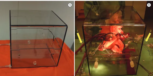
The EVLP system originally described by Steen et al. consists of a rigid, transparent rectangular box (Figure 1) to support the heart-lung block (with inlets for connecting the cardiopulmonary bypass circuit tubes), a centrifugal pump, a heat exchanger, and a membrane oxygenator, as well as a pressure transducer, a flow meter, and a thermometer (and their respective monitors) for EVLP monitoring. The perfusate consists of 1,500 mL of Steen Solution and a variable amount of packed red blood cells, the hematocrit level of the perfusate being approximately 15%. A gas mixture (of nitrogen, oxygen, and carbon dioxide) is used in order to “deoxygenate” the perfusate through the gas exchange membrane, gas flow being adjusted so that the gas concentration in the perfusate is similar to that in the venous blood. According to the Lund University EVLP protocol, flow and temperature should be gradually increased.
Introduction
Lung transplantation has come to be viewed as the best treatment option for various end-stage lung diseases. This heterogeneous group comprises diseases that are highly disabling and that have high mortality rates, despite considerable advances in the pharmacological treatment options that are currently available.
The low number of viable donors is currently the major obstacle to increasing the number of lung transplants, resulting in a long waiting time for transplantation and high mortality among lung transplant candidates on waiting lists.(1) Unlike transplantation of other solid organs, such as the liver and the kidneys, lung transplantation is primarily limited not by the absolute number of donors but by the viability of the donor lungs. Factors that can damage the donor lung and therefore make it unsuitable for transplantation include those related to brain death (including bronchial aspiration, pulmonary edema, and chest trauma) and those related to ICU treatment (including hypervolemia, barotrauma, and ventilator-associated pneumonia). The reported rates of donor lung use are low, i.e., approximately 15% (6.1-27.1%).(2) In Brazil, these rates are even lower. A study conducted in 2006 and analyzing data from the São Paulo State Department of Health Transplant Center showed that only 4.9% of all donor lungs were effectively transplanted.(3)
There have been various proposals of ways to increase the number of actual lung donors, including intensification of donation campaigns, living donor transplantation,(4) use of non-heart-beating donors,(5) and adoption of donor criteria that are more flexible.(6) However, the proposal that has attracted the most attention from lung transplant groups is ex vivo lung perfusion (EVLP), especially due to the prospect of reconditioning previously discarded lungs.
The objective of the present article was to review the development of, state of the art in, and future prospects for the ex vivo model of lung perfusion and reconditioning.
Development of the EVLP system
• The EVLP system allows the removal of clots in the pulmonary circulation.
• The high oncotic pressure of the perfusate reduces pulmonary edema.
• The EVLP system allows the removal of inflammatory cells, dextran reducing the degree of lung inflammation and improving the microvasculature.
This gave rise to what is currently known as ex vivo lung reconditioning, whereby donor lungs initially deemed unsuitable for transplantation can be “reconditioned” by EVLP.(12)
In 2005, after the good results of the experimental studies in pigs, the Lund University lung transplant group performed the first transplantation of an initially rejected lung submitted to ex vivo lung reconditioning.(14) The donor lung had been considered unsuitable for transplantation because of bilateral lung contusion (as seen on chest X-ray) and low pre-harvesting PaO2 (as revealed by arterial blood gas analysis; last measurement, 67 mmHg, with an FiO2 of 0.7). The researchers then harvested the organ for ex vivo evaluation rather than for transplantation. The organ underwent EVLP for approximately 1 h, when a new arterial blood gas analysis was performed. The analysis revealed a PaO2 of 391.5 mmHg and an FiO2 of 100%. Macroscopic evaluation revealed an apparently good compliance, despite the presence of hemorrhagic stippling on the lung surface. The organ was then deemed Ventilation is initiated when the temperature
reaches 32°C, and the maximum flow should never exceed values that will cause pulmonary artery pressure to be greater than 20 mmHg. The most important evaluation is blood gas analysis of the perfusate collected from the pulmonary veins when the temperature reaches 37°C.(11)
After having established the EVLP model, Steen et al. hypothesized that it had uses other than the evaluation of non-heart-beating donor lungs. On the basis of the hypothesis that many of the lungs deemed unsuitable for transplantation could be used if additional evaluation guaranteed their viability, the authors proposed the use of EVLP for evaluating “marginal donors”. In a study published in 2006, that group of authors evaluated six initially rejected donor lungs and found that EVLP increased the oxygenation capacity of the lungs.(12) In another study, also published in 2006, one group of researchers at the University of North Carolina at Chapel Hill, in Chapel Hill, NC, obtained satisfactory results using a similar methodology.(13) However, during the evaluation of those marginal donor lungs, the major finding was that EVLP apparently “improved” lung function. This was attributed to the following characteristics of the EVLP system:
• The EVLP system allows the use of alveolar recruitment maneuvers.
• The EVLP system allows more effective clearance of bronchial secretions.
Figure 1 - Ex vivo lung perfusion model used by the lung transplant group at Lund University, Lund,
Ex vivo lung reconditioning: a new era for lung transplantation 779
with prolonging the duration of perfusion. To that end, the Canadian group modified the Swedish EVLP protocol, as follows:
• use of an acellular perfusate, the perfusate consisting only of Steen Solution (i.e., no red blood cells)
• use of a maximum perfusion flow of approximately 40% of the estimated cardiac output
• use of a pulmonary artery pressure of 10-15 mmHg
The use of an acellular perfusate facilitates the logistics of EVLP by reducing the costs and preventing ethical conflicts arising from the use of blood products in studies of organs that may or may not be used clinically. The disadvantages of using blood in the perfusate include hemolysis caused by mechanical trauma to red blood cells and the proinflammatory response typically elicited by the transfusion. The modifications were successful in prolonging the duration of EVLP (to up to 12 h).(15) The absence of red blood cells did not affect the assessment of lung function. The degree of oxygen diffusion in the Steen Solution perfusate is high and therefore allows reliable blood gas analysis. The Canadian group also developed a new (round) containment box (Figure 2) and specific cannulae (Figure 3) that allow a better connection of the pulmonary artery and left suitable for transplantation, and the recipient
was summoned.
At the end of EVLP, in order to preserve the organ, the Lund University lung transplant group opted to place the lung on topical extracorporeal membrane oxygenation, which was achieved by reducing the temperature of the perfusate to 25°C and by stopping ventilation and perfusion. The semi-inflated organ was immersed in the same solution that was used for perfusion, which was oxygenated and kept in recirculation at a controlled temperature of 8°C for approximately 10 h until the arrival of the recipient and the performance of the transplantation. The patient underwent single (left) lung transplantation. There were no intraoperative or immediate postoperative complications, and there were no late postoperative complications. At postoperative month 11, the patient died from septic shock unrelated to EVLP.
The Canadian EVLP model
Encouraged by the good results obtained by the Lund University lung transplant group, the lung transplant group at the University of Toronto, in Toronto, Canada, conducted EVLP studies with the objective of reconditioning previously discarded lungs. A distinguishing characteristic of those studies was the concern
atrial stump (pulmonary venous connection) to the circuit. In addition, the cannulae have built-in pressure catheters. The atrial cannula allows closed perfusion (Figure 4) and the atrial pressure to be maintained at 3-5 mmHg. Although the Swedish EVLP protocol recommends 1 h of normothermic perfusion, the Canadian EVLP protocol recommends at least 4 h of normothermic perfusion with stable parameters for the organ to be considered suitable for transplantation. Chart 1 shows a comparison between the Lund University EVLP protocol and the University of Toronto EVLP protocol.
The experience in Brazil
In Brazil, Pêgo-Fernandes et al. began to study EVLP in 2008. In a study published in the Brazilian Journal of Pulmonology in 2009, the authors described a modification of the EVLP circuit aimed at reducing the amount of solution needed for EVLP.(16) The authors
Figure 3 - Cannulae for connecting the lung to the ex vivo lung perfusion circuit (Canadian model). In A, cannula for pulmonary venous return. Note lateral outlet for air removal and sample collection (arrow 1). Note also probes for pressure measurement (arrow 2) and the tip to be attached to the main pulmonary artery (arrow 3). In B, pulmonary artery cannula. Note the silicone cone to be sutured to the edge of the left atrium (arrow 4).
Figure 4 - Venous cannula being sutured to the atrial
Ex vivo lung reconditioning: a new era for lung transplantation 781
described an EVLP system comprising a set of pediatric tubes, a pediatric venous reservoir, and a pediatric membrane oxygenator (Figure 5). The system also comprised a heat exchanger and a centrifugal pump (Braile Biomédica, São José do Rio Preto, Brazil). The heart-lung block remained in a rigid, transparent box (Vitrolife), an orotracheal tube being inserted into the trachea and a perfusion cannula (Vitrolife) being inserted into the main pulmonary artery. The solution returning through the pulmonary veins flowed directly into the containment box, being drained into the venous reservoir by the force of gravity. For lung reconditioning and clinical use, the protocol included closed cannulation of the atrium with special cannulae, as described by the University of Toronto lung transplant group.(15) The system was filled with 1,500 mL of Steen Solution (Vitrolife). In that protocol, EVLP was initiated with the solution at 20°C and an initial flow of 10% of the calculated value, the
Chart 1 - Comparison between the Lund University ex vivo lung perfusion protocol and the University of
Toronto ex vivo lung perfusion protocol. CO: cardiac output; PAP: pulmonary artery pressure; and LAP: left atrial pressure.
Figure 5 - Ex vivo lung perfusion system used at the
University of São Paulo School of Medicine Hospital
Other transplant groups have published studies reporting their experience with the clinical use of EVLP. Of those studies, the most recent is a study conducted by an Austrian group and published in 2012. The authors reported that 9 of 13 lungs submitted to ex vivo evaluation showed improvement in PaO2 and were therefore transplanted. According to the authors, the postoperative outcome was similar to that in patients receiving standard lung transplants during the observation period.(23)
In England, Zych et al. evaluated 13 heart-lung blocks; of those, 6 had good parameters and were therefore transplanted, short-term (three-month) survival being excellent (100%).(24)
Moradiellos et al. evaluated 8 non-heart-beating donor lungs submitted to EVLP; of those, 4 showed good lung function and were therefore transplanted. Although two patients died before the end of post-transplantation year 1, the causes of death were unrelated to EVLP.(25)
Experimental studies of EVLP
Because EVLP has uses other than clinical use, various groups have used EVLP in experimental studies. Frank et al. used EVLP in order to investigate pathophysiological mechanisms of lung function, including edema formation.(26) Sakuma et al. studied drugs having an effect on lung function and demonstrated the role of endotracheal adrenaline and the use of beta-adrenergic stimulation in reducing pulmonary edema.(27) Inci et al. studied the addition of urokinase to the EVLP perfusate and found that it improved lung function and reduced pulmonary vascular resistance.(28) The same group of authors used an EVLP model in order to study the effect of surfactant lavage on lungs injured by gastric acid aspiration.(29)
Mariani et al. used human donor lungs deemed unacceptable for transplantation in order to develop a variation of the ex vivo model of lung evaluation. The technique consisted of separating the lung block into right and left blocks and subsequently reconnecting those two blocks, being designated the split lung block technique.(30)
Ex vivo animal model
A variation of the EVLP model can also be used for experimental studies in small animals, facilitating the logistics of such studies. Several temperature and flow being gradually increased.
When the temperature reached 32°C (20 min after EVLP initiation), ventilation was initiated and a gas (7% CO2 + 93% N2) was released into the system through the membrane oxygenator in order to “deoxygenate” the perfusate coming from the pulmonary veins so that the solution that enters the pulmonary artery has the same gas concentrations as those of the venous blood. The ventilator was set at a tidal volume of 6-8 mL/kg, an RR of 7 breaths/min, an FiO2 of 100%, and a positive end-expiratory pressure of 5 cmH2O. The flow and temperature were increased until a flow of 40% of the estimated cardiac output and a temperature of 37°C were reached (i.e., within approximately 40-60 min). If the pulmonary artery pressure was as high as 20 mmHg before the estimated maximum flow was reached, the maximum flow was kept at a lower value. Within 60 min after the initiation of EVLP, perfusate samples were collected from the pulmonary veins for blood gas analysis. The following parameters were analyzed: PaO2; PaCO2; pulmonary vascular resistance; and lung compliance. The first studies conducted by that group of authors focused on establishing an EVLP model suitable for use in Brazil(17) and on reconditioning lungs deemed unsuitable for transplantation.(18,19)
Clinical use
The first cases of clinical use of EVLP were published in 2009 and involved the monitoring of six lung transplant recipients who received initially rejected lungs submitted to EVLP. The results showed a three-month survival of 100% and a one-year survival of 67%, the deaths being apparently unrelated to EVLP.(20)
Ex vivo lung reconditioning: a new era for lung transplantation 783
an adenoviral vector encoding human IL-10 was used during EVLP in order to increase IL-10 levels and therefore produce potent anti-inflammatory activity in the donor lungs.(44)
Final considerations
Currently, EVLP represents a promising tool for use in the field of lung transplantation. In addition to having the potential to increase the number of donor lungs that are suitable for transplantation, EVLP can be used in studies aimed at developing new techniques to improve lung conditioning in the immediate pre-transplant period, therefore improving post-transplant outcomes.
Although the clinical use of EVLP is becoming increasingly more common in various countries, long-term studies are needed in order to understand the real impact of EVLP on post-transplant outcomes.
Acknowledgments
We would like to thank Dr. Marcos Naoyuki Samano for kindly contributing to the study by helping us with the figures and providing invaluable suggestions.
References
1. Costa da Silva F Jr, Afonso JE Jr, Pêgo-Fernandes PM, Caramori ML, Jatene FB. São Paulo lung transplantation waiting list: patient characteristics and predictors of death. Transplant Proc. 2009;41(3):927-31. PMid:19376390. http://dx.doi.org/10.1016/j.transproceed.2009.01.048 2. Punch JD, Hayes DH, LaPorte FB, McBride V, Seely MS. Organ
donation and utilization in the United States, 1996-2005. Am J Transplant. 2007;7(5 Pt 2):1327-38. PMid:17428283. http://dx.doi.org/10.1111/j.1600-6143.2007.01779.x 3. Fernandes PM, Samano MN, Junqueira JJ, Waisberg
DR, Noleto GS, Jatene FB. Lung donor profile in the State of São Paulo, Brazil, in 2006. J Bras Pneumol. 2008;34(7):497-505. PMid:18695795. http:// dx.doi.org/10.1590/S1806-37132008000700010 4. Camargo SM, Camargo Jde J, Schio SM, Sánchez LB,
Felicetti JC, Moreira Jda S, et al. Complications related to lobectomy in living lobar lung transplant donors. J Bras Pneumol. 2008;34(5):256-63. PMid:18545820. 5. Gomez-de-Antonio D, Campo-Cañaveral JL, Crowley
S, Valdivia D, Cordoba M, Moradiellos J, et al. Clinical lung transplantation from uncontrolled non-heart-beating donors revisited. J Heart Lung Transplant. 2012;31(4):349-53. PMid:22306439. http:// dx.doi.org/10.1016/j.healun.2011.12.007
6. Pêgo-Fernandes PM, Samano MN, Fiorelli AI, Fernandes LM, Camargo SM, Xavier AM, et al. Recommendations for the use of extended criteria donors in lung transplantation. Transplant Proc. 2011;43(1):216-9. PMid:21335191. http://dx.doi.org/10.1016/j.transproceed.2010.12.050
questions regarding lung transplantation can be answered that way. Pierre et al. studied rat lungs and demonstrated that slow (i.e., gradual) reperfusion is less harmful than rapid reperfusion.(31)
Silva et al. studied rat lungs submitted to EVLP and confirmed that lung preservation techniques disarrange lung architecture and lead to ischemia-reperfusion injury.(32)
Pêgo-Fernandes et al. have conducted studies using a system that is marketed under the name IL2 - Isolated Perfused Rat or Guinea Pig Lung System (Harvard Apparatus, Holliston, MA, USA; Hugo Sachs Elektronik, Hugstetten, Germany). This system allows ventilation and perfusion of the heart-lung block, as well as being capable of measuring respiratory mechanics and hemodynamic parameters.(33) There have been studies evaluating the system itself(34) and lung preservation, either by comparing preservation solutions(35,36) or by investigating the effect of additives in the solutions.(37)
Future prospects
All lung transplant groups believe that ex vivo lung reconditioning will lead to an increase in the number of lung transplants; nevertheless, studies have proposed new, ingenious uses for EVLP, including the treatment of certain conditions that prevent transplantation, such as pneumonia and acute lung injury.(38)
Another prospect is the optimization of donor lung conditioning by reducing inflammatory activity. Some researchers foresee the possibility of using EVLP to “condition” all donor lungs, including those initially deemed suitable for transplantation, in an attempt to improve post-transplant outcomes.(39)
The EVLP model including activation of lung metabolism at normothermia seems to be ideal for lung reconditioning and for monitoring the effectiveness of lung reconditioning before transplantation. In addition, side effects are minimized because the drugs used are not systemically absorbed by the recipient. Furthermore, there is the prospect of prolonging EVLP for several days, which would allow prolonged treatment of infected lungs.(40)
rejected donor lungs after reconditioning ex vivo. Ann Thorac Surg. 2009;87(1):255-60. PMid:19101308. http:// dx.doi.org/10.1016/j.athoracsur.2008.09.049
21. Cypel M, Yeung JC, de Perrot M, Karolak W, Chen F, Sato M, et al. Ex vivo lung perfusion in clinical lung transplantation – the HELP trial. J Heart Lung Transplant. 2010;29(2S):S88. http://dx.doi.org/10.1016/j. healun.2009.11.270
22. Cypel M, Yeung JC, Liu M, Anraku M, Chen F, Karolak W, et al. Normothermic ex vivo lung perfusion in clinical lung transplantation. N Engl J Med. 2011;364(15):1431-40. PMid:21488765. http://dx.doi.org/10.1056/ NEJMoa1014597
23. Aigner C, Slama A, Hötzenecker K, Scheed A, Urbanek B, Schmid W, et al. Clinical ex vivo lung perfusion--pushing the limits. Am J Transplant. 2012;12(7):1839-47. PMid:22458511. http:// dx.doi.org/10.1111/j.1600-6143.2012.04027.x 24. Zych B, Popov AF, Stavri G, Bashford A, Bahrami
T, Amrani M, et al. Early outcomes of bilateral sequential single lung transplantation after ex-vivo lung evaluation and reconditioning. J Heart Lung Transplant. 2012;31(3):274-81. PMid:22088786. http:// dx.doi.org/10.1016/j.healun.2011.10.008
25. Moradiellos FJ, Naranjo JM, Córdoba C, Salas MC, Gómez D, Campo-Cañaveral JL, et al. Clinical lung transplantation after ex vivo evaluation of uncontrolled non heart-beating donors lungs: initial experience [abstract 90]. J Heart Lung Transplant. 2011;30(Suppl 4):S38. http:// dx.doi.org/10.1016/j.healun.2011.01.097
26. Frank JA, Briot R, Lee JW, Ishizaka A, Uchida T, Matthay MA. Physiological and biochemical markers of alveolar epithelial barrier dysfunction in perfused human lungs. Am J Physiol Lung Cell Mol Physiol. 2007;293(1):L52-9. PMid:17351061 PMCid:2764531. http://dx.doi.org/10.1152/ ajplung.00256.2006
27. Sakuma T, Gu X, Wang Z, Maeda S, Sugita M, Sagawa M, et al. Stimulation of alveolar epithelial fluid clearance in human lungs by exogenous epinephrine. Crit Care Med. 2006;34(3):676-81. PMid:16505652 PMCid:2765117. http://dx.doi.org/10.1097/01.CCM.0000201403.70636.0F 28. Inci I, Zhai W, Arni S, Inci D, Hillinger S, Lardinois
D, et al. Fibrinolytic treatment improves the quality of lungs retrieved from non-heart-beating donors. J Heart Lung Transplant. 2007;26(10):1054-60. PMid:17919627. http://dx.doi.org/10.1016/j.healun.2007.07.033 29. Inci I, Ampollini L, Arni S, Jungraithmayr W, Inci D,
Hillinger S, et al. Ex vivo reconditioning of marginal donor lungs injured by acid aspiration. J Heart Lung Transplant. 2008;27(11):1229-36. PMid:18971096. http:// dx.doi.org/10.1016/j.healun.2008.07.027
30. Mariani AW, Medeiros IL, Pêgo-Fernandes PM, Fernandes FG, Unterpertinger Fdo V, Fernandes LM, et al. Ex vivo experimental model: split lung block technique. J Bras Pneumol. 2011;37(6):791-5. PMid:22241037. http:// dx.doi.org/10.1590/S1806-37132011000600013 31. Pierre AF, DeCampos KN, Liu M, Edwards V, Cutz E, Slutsky
AS, et al. Rapid reperfusion causes stress failure in ischemic rat lungs. J Thorac Cardiovasc Surg. 1998;116(6):932-42. http://dx.doi.org/10.1016/S0022-5223(98)70043-1 32. Silva CA, Carvalho RS, Cagido VR, Zin WA, Tavares P,
DeCampos KN. Influence of lung mechanical properties and alveolar architecture on the pathogenesis of ischemia-reperfusion injury. Interact Cardiovasc Thorac 7. Steen S, Ingemansson R, Budrikis A, Bolys R, Roscher R,
Sjöberg T. Successful transplantation of lungs topically cooled in the non-heart-beating donor for 6 hours. Ann Thorac Surg. 1997;63(2):345-51. http://dx.doi. org/10.1016/S0003-4975(96)01101-0
8. Wang LS, Yoshikawa K, Miyoshi S, Nakamoto K, Hsieh CM, Yamazaki F, et al. The effect of ischemic time and temperature on lung preservation in a simple ex vivo rabbit model used for functional assessment. J Thorac Cardiovasc Surg. 1989;98(3):333-42. PMid:2770316. 9. Findler M, Barak J, Einav S, Snir E, Hochauser E, Vidne BA.
The use of isolated auto-heart-lung perfusion apparatus on small animals. Transplant Proc. 1987;19(5):3792-4. PMid:3118525.
10. Steen S, Liao Q, Wierup PN, Bolys R, Pierre L, Sjöberg T. Transplantation of lungs from non-heart-beating donors after functional assessment ex vivo. Ann Thorac Surg. 2003;76(1):244-52; discussion 252. http://dx.doi. org/10.1016/S0003-4975(03)00191-7
11. Steen S, Sjöberg T, Pierre L, Liao Q, Eriksson L, Algotsson L. Transplantation of lungs from a non-heart-beating donor. Lancet. 2001;357(9259):825-9. http://dx.doi. org/10.1016/S0140-6736(00)04195-7
12. Wierup P, Haraldsson A, Nilsson F, Pierre L, Scherstén H, Silverborn M, et al. Ex vivo evaluation of nonacceptable donor lungs. Ann Thorac Surg. 2006;81(2):460-6. PMid:16427831. http://dx.doi. org/10.1016/j.athoracsur.2005.08.015
13. Egan TM, Haithcock JA, Nicotra WA, Koukoulis G, Inokawa H, Sevala M, et al. Ex vivo evaluation of human lungs for transplant suitability. Ann Thorac Surg. 2006;81(4):1205-13. PMid:16564244. http:// dx.doi.org/10.1016/j.athoracsur.2005.09.034 14. Steen S, Ingemansson R, Eriksson L, Pierre L, Algotsson
L, Wierup P, et al. First human transplantation of a nonacceptable donor lung after reconditioning ex vivo. Ann Thorac Surg. 2007;83(6):2191-4. PMid:17532422. http://dx.doi.org/10.1016/j.athoracsur.2007.01.033 15. Cypel M, Yeung JC, Hirayama S, Rubacha M,
Fischer S, Anraku M, et al. Technique for prolonged normothermic ex vivo lung perfusion. J Heart Lung Transplant. 2008;27(12):1319-25. PMid:19059112. http:// dx.doi.org/10.1016/j.healun.2008.09.003
16. Pêgo-Fernandes PM, Medeiros IL, Mariani AW, Fernandes FG, Unterpertinger Fdo V, Samano MN, et al. Ex vivo lung perfusion: initial Brazilian experience. J Bras Pneumol. 2009;35(11):1107-11. PMid:20011846. 17. Pêgo-Fernandes PM, de Medeiros IL, Mariani AW, Fernandes
FG, Unterpertinger FD, Samano MN, et al. Ex vivo lung perfusion: early report of Brazilian experience. Transplant Proc. 2010;42(2):440-3. PMid:20304159. http://dx.doi. org/10.1016/j.transproceed.2010.01.015
18. Pêgo-Fernandes PM, Mariani AW, Medeiros IL, Pereira AE, Fernandes FG, Valle Unterpertinger F, et al. Ex vivo lung evaluation and reconditioning. Rev Bras Cir Cardiovasc. 2010;25(4):441-6. PMid:21340372. http:// dx.doi.org/10.1590/S0102-76382010000400006 19. Medeiros IL, Pêgo-Fernandes PM, Mariani AW,
Fernandes FG, do Vale Unterpertinger F, Canzian M, et al. Histologic and functional evaluation of lungs reconditioned by ex vivo lung perfusion. J Heart Lung Transplant. 2012;31(3):305-9. PMid:22133788. http:// dx.doi.org/10.1016/j.healun.2011.10.005
Ex vivo lung reconditioning: a new era for lung transplantation 785
38. Lee JW, Fang X, Gupta N, Serikov V, Matthay MA. Allogeneic human mesenchymal stem cells for treatment of E. coli endotoxin-induced acute lung injury in the ex vivo perfused human lung. Proc Natl Acad Sci U S A. 2009;106(38):16357-62. PMid:19721001 PMCid:2735560. http://dx.doi.org/10.1073/ pnas.0907996106
39. Sanchez PG, Bittle GJ, Burdorf L, Pierson RN 3rd, Griffith BP. State of art: clinical ex vivo lung perfusion: rationale, current status, and future directions. J Heart Lung Transplant. 2012;31(4):339-48. PMid:22423980. http://dx.doi.org/10.1016/j.healun.2012.01.866 40. Karamanou DM, Perry J, Walden HR, Simpson AJ, Corris
P, Gould K, et al. The effect of ex-vivo perfusion on the microbiological profile of the donor lung [abstract 279]. J Heart Lung Transplant. 2010;29(2):S94. http://dx.doi. org/10.1016/j.healun.2009.11.291
41. Hirayama S, Cypel M, Sato M, Anraku M, Liaw PC, Liu M, et al. Activated protein C in ischemia-reperfusion injury after experimental lung transplantation. J Heart Lung Transplant. 2009;28(11):1180-4. PMid:19782612. http://dx.doi.org/10.1016/j.healun.2009.06.026 42. Nita I, Hollander C, Westin U, Janciauskiene SM. Prolastin,
a pharmaceutical preparation of purified human alpha1-antitrypsin, blocks endotoxin-mediated cytokine release. Respir Res. 2005;6:12. PMid:15683545 PMCid:549028. http://dx.doi.org/10.1186/1465-9921-6-12
43. Martins S, de Perrot M, Imai Y, Yamane M, Quadri SM, Segall L, et al. Transbronchial administration of adenoviral-mediated interleukin-10 gene to the donor improves function in a pig lung transplant model. Gene Ther. 2004;11(24):1786-96. PMid:15470481. http:// dx.doi.org/10.1038/sj.gt.3302357
44. Cypel M, Liu M, Rubacha M, Yeung JC, Hirayama S, Anraku M, et al. Functional repair of human donor lungs by IL-10 gene therapy. Sci Transl Med. 2009;1(4):4ra9. Surg. 2010;11(1):46-51. PMid:20378696. http://dx.doi.
org/10.1510/icvts.2009.222018
33. Pêgo-Fernandes PM, Werebe E, Cardoso PF, Pazetti R, de Oliveira KA, Soares PR, et al. Experimental model of isolated lung perfusion in rats: first Brazilian experience using the IL-2 isolated perfused rat or guinea pig lung system. Transplant Proc. 2010;42(2):444-7. PMid:20304160. http://dx.doi.org/10.1016/j.transproceed.2010.01.016 34. Pêgo-Fernandes PM, Werebe Ede C, Cardoso PF,
Pazetti R, Oliveira KA, Soares PR, et al. Experimental model of isolated lung perfusion in rats: technique and application in lung preservation studies. J Bras Pneumol. 2010;36(4):490-3. PMid:20835597. http:// dx.doi.org/10.1590/S1806-37132010000400015 35. Soares PR, Braga KA, Nepomuceno NA, Pazetti R,
Correia AT, Cardoso PF, et al. Comparison between Perfadex and locally manufactured low-potassium dextran solution for pulmonary preservation in an ex vivo isolated lung perfusion model. Transplant Proc. 2011;43(1):84-8. PMid:21335161. http://dx.doi. org/10.1016/j.transproceed.2010.12.005
36. Simões EA, Pêgo-Fernandes PM, Cardoso PF, Pazetti R, Werebe E, de Oliveira Braga KA, et al. Comparing the performance of rat lungs preserved for 6 or 12 hours after perfusion with low-potassium dextran or histidine-tryptophan-ketoglutarate. Transplant Proc. 2011;43(5):1520-4. PMid:21693228. http://dx.doi. org/10.1016/j.transproceed.2010.12.001
37. Cardoso PF, Pazetti R, Moriya HT, Pêgo-Fernandes PM, Almeida FM, Correia AT, et al. An experimental rat model of ex vivo lung perfusion for the assessment of lungs after prostacyclin administration: inhaled versus parenteral routes. J Bras Pneumol. 2011;37(5):589-97. PMid:22042390. http://dx.doi.org/10.1590/ S1806-37132011000500005
About the authors
Alessandro Wasum Mariani
Attending Physician. Department of Thoracic Surgery, Instituto do Coração, Hospital das Clínicas, Faculdade de Medicina da Universidade de São Paulo – InCor/HC-FMUSP, Heart Institute/University of São Paulo School of Medicine Hospital das Clínicas – São Paulo, Brazil.
Paulo Manuel Pêgo-Fernandes
Associate Professor. Department of Cardiorespiratory Diseases, Faculdade de Medicina da Universidade de São Paulo – FMUSP, University of São Paulo School of Medicine – São Paulo, Brazil.
Luis Gustavo Abdalla
Attending Physician. Lung Transplant Group, Instituto do Coração, Hospital das Clínicas, Faculdade de Medicina da Universidade de São Paulo – InCor/HC-FMUSP, Heart Institute/University of São Paulo School of Medicine Hospital das Clínicas – São Paulo, Brazil.
Fabio Biscegli Jatene