269
Primeira submissão em 14/06/11 Última submissão em 04/04/12 Aceito para publicação em 31/05/12 Publicado em 20/08/12Sclerosing thymoma: case report
Timoma esclerosante: relato de caso
Gesine Gregorio Siqueira1; Lisley Calixto de Araujo2; Guilherme Cançado Rezende3;
Nuno Ferreira de Lima4; Larissa Cardoso Marinho5
J Bras Patol Med Lab • v. 48 • n. 4 • p. 269-272 • agosto 2012
RELATO DE CASO CASE REPORT
We present a rare case of thymoma in a 36-year old woman, who was initially diagnosed with severe myasthenia gravis and subsequently undergone surgical resection. During surgery tumor was found at the anterior mediastinum, tightly attached to the phrenic nerve, pleura and pericardium. Histological assessment showed large areas of sclerosis and ibrous collagenous tissue as well as islands of epithelial and lymphoid cells. Sclerosing thymoma, which is a rare subtype of thymoma (< 1%), was diagnosed, thus conirming the irst report in Brazil. The patient showed partial improvement of symptoms associated with myasthenia gravis.
abstract
key words
Thymoma
Sclerosis
Myasthenia gravis
resumo
Relatamos um caso raro de timoma em uma mulher de 36 anos de idade, com clínica e diagnóstico de miastenia gravis de difícil controle clínico, submetida à ressecção cirúrgica. No intraoperatório, observou-se tumor no mediastino anterior, firmemente aderido ao nervo frênico, à pleura e ao pericárdio. Ao exame histológico, foram evidenciadas extensas áreas de tecido fibrocolagenoso e esclerose, assim como ilhas de células epiteliais e células linfoides. Diagnosticado timoma esclerosante, subtipo raro de timoma (< 1%), sendo este o primeiro caso relatado no Brasil. A paciente apresentou melhora parcial dos sintomas associados à miastenia gravis.unitermos
Timoma Esclerose Miastenia gravis
1. Especialista em Patologia.
2. Médica residente de Patologia do Hospital Universitário da Universidade de Brasilia (UnB).
3. Cirurgião torácico.
4. Cirurgião torácico; chefe do serviço de Cirurgia Torácica do Hospital Universitário da UnB. 5. Doutora em Patologia; médica do Hospital Universitário da UnB.
Introduction
Sclerosing thymoma is a rare tumour (< 1%) ex-hibiting the features of a conventional thymoma in terms of epithelial cell morphology and lymphocyte content, but with exuberant collagen-rich stroma(7).
Considering its uniqueness, we report a case of scle-rosing thymoma in a 36 year-old woman.
Case report
A 36 year-old female patient has, for six months, bilateral ptosis, reduced muscle strength on the right limbs and dysphagia. The clinic
di-agnosis of myasthenia gravis was made with the
270
SIQUEIRA, G. G. et al. Sclerosing thymoma: case report • J Bras Patol Med Lab • v. 48 • n. 4 • p. 269-272 • agosto 2012
pyridostigmine bromide and prednisone in high doses partially controlled the symptoms.
Chest computed tomography (CT) evidenced an
in-creased and homogeneous thymus. The patient was then indicated for a video-assisted thymectomy with cervical and thoracic approach.
During surgery, the left lobe of the thymus was evidenced irmly attached to the pericardium, pleura and the left phrenic nerve, which required sternotomy. Partial resection of the lesion was performed, including a portion of the pleura and pericardium adhered, preserving the phrenic nerve. The patient had good post operative evolution.
The surgical specimen was formalin-ixed at 10% and submitted to macroscopic evaluation. Fragments were processed through traditional technique (dehydrated in alcohol, xylene) and embedded in parafin. Histological sections of 5μm were stained with hematoxylin and eosin.
Immunohistochemical staining was performed by pretreating histological sections in antigen retrieval solu-tion (citrate buffer retrieval solusolu-tion, pH 6, Dako Labora-tories, USA) in a water bath at 98ºC for 20 minutes and subsequent cooling at room temperature. The following primary antibodies were used: a) monoclonal mouse anti-human CD3, Clone F7.2.38, (Dako Laboratories, USA), working dilution 1:50, to identify T cells; b) monoclonal mouse anti-human CD20cy, Clone L26 (Dako Laborato-ries, USA), working dilution 1:200, to identify B cells; c) monoclonal mouse anti-human Cytokeratin, clones AE1/ AE3 (Dako Laboratories, USA) working dilution 1:200 to identify epithelial cells. External control was used for validation of the reaction.
Clinical, laboratory and follow-up data were obtained from medical charts.
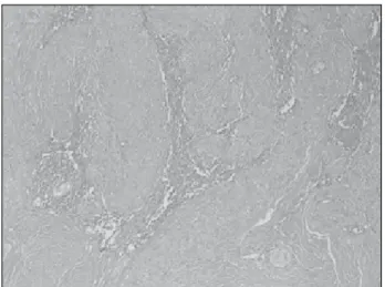
At gross examination tumor mass measured 7.5 × 3.5 × 1 cm, in pale brown color, firm con-sistence and hardened areas on the cut surface. Microscopic examination showed extensive areas of ibrocollagen tissue, with hyalinization and sclerosis sur-rounding islands of epithelial cells without atypia, with interspersed lymphoid cells (Figures 1 and 2). Between the sclerotic areas, we observed cystic degeneration, bone metaplasia, dystrophic calciication and negative images of cholesterol crystals. There were no mitoses or areas of necrosis.
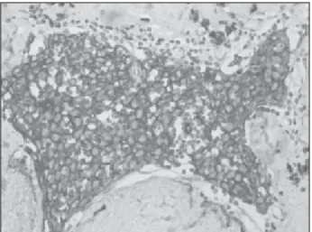
The immunohistochemal study was performed with antibodies to PanCK, CD3 and CD20, with positive for PanCK in epithelial clusters (Figure 3) and CD3 and CD20 in lymphoid clusters, showing a biphasic marking in the tumor, which lead to conirmation of thymoma, sclerosing subtype according to World Health Organization (WHO).
Discussion
Sclerosing thymoma is an extremely unusual presenta-tion of thymoma and, including this one, only 13 cases have been reported.This is the irst case reported in Brazil(3, 5).
In ive cases in the literature, patients were asymptom-atic, four were symptomatic (shortness of breath and chest pain) and three had clinical diagnosis of myasthenia gravis.
Figure 1 – Sclerosing, thymoma showing areas of fibrocollagen tissue, hialinization and sclerosis surrounding island of epithelial cells
HE 100×
HE: hematoxylin and eosin.
Figure 2 – Islands of epithelial cells without atypia, with interspersed lymphoid cells HE 400×
271
SIQUEIRA, G. G. et al. Sclerosing thymoma: case report • J Bras Patol Med Lab • v. 48 • n. 4 • p. 269-272 • agosto 2012In our case there was a correlation with clinical indings of myasthenia gravis, a fact that was consistent with indings in the report of Kuo(5) and in only one case in the series of
Moran and Suster(5) .
Grossly, lesion was poorly demarcated, unencap-sulated, pale brown colored and irm consistence with smooth and homogeneous surface. The cut surface was smooth, with areas of crackle and no macroscopic cystic areas, haemorrhage or necrosis were observed. In the literature, the described cases of sclerosing thymoma measured between 2.5 and 10 cm, well deined without bleeding or necrosis(3, 5).
Histologically, there were extensive areas of ibrocol-lagen tissue, with hyalinization and sclerosis, microscopic foci of cystic degeneration, bone metaplasia, dystrophic calciication and negative images of cholesterol crystals. In the midst of those areas we could observe islands of epi-thelial cells without atypia and lymphoid cells. In our case the complete resection was not possible for being a case of ill-deined lesion adhered to the phrenic nerve, pleura and pericardium.
In Moran and Suster review all thymoma cases had extensive areas of hyalinization or sclerosis, areas of granu-lomas with cholesterol crystals, calciication and islands of neoplastic epithelial cells. Seven cases of this review, like ours, present dual population of cells (epithelial and lym-phoid) without atypia or mitotic activity.
Cases described by Kuo(3), histology showed small
ibrotic and sclerotic nodules, containing predominance of epithelial component at one case and lymphocyte pre-dominance in another. In both, it was observed central
hemorrhagic zone, hemosiderin deposits and externally ibrous, sclerotic and dense tissue. It also had areas of cal-ciication and cholesterol granulomas.
After the morphological diagnosis, an immunohisto-chemical study was performed to highlight the two cellular components: the epithelial one, stained for panCK and T and B lymphoid one, with immunostaining for CD 3 and CD 20, respectively. Moran and Suster(5) made an
immu-nohistochemical study for cytokeratin in six of his cases highlighting the epithelial component.
Two months after surgery, patient lies with partial im-provement of symptoms and remains in clinical follow up. According to the follow up of Kuo’s cases(3), the patients
showed improvement of symptoms gradually after surgery and had remission of symptoms between two and four years after surgery. In the series of Moran and Suster(5), there was
follow-up of eight of the 10 patients reported. Six of them died from causes unrelated to thymoma, such as renal failure, congestive heart failure and pulmonary edema and two of these patients survived asymptomatic for one to six years after surgical resection.
Pathogenesis of the sclerosing thymoma is uncertain. The irst hypothesis was described by Kuo(3) and is based
on a possible spontaneous phenomenon of regression and sclerosis possibly associated with infarct through the inter-ruption of vascular low or apoptosis. Another hypothesis is that these sclerosing tumor originate from “ancient” thymoma whose pre-existing collagen bands coalesce, remaining some areas preserved.
In our view, it is a healing process induced by previous cell injury that could initiate, for example, from the in-terruption of vascular low and consequent necrosis. The necrotic cells and their contents disappear by phagocytoses and enzymatic digestion by leukocytes. The migration of ibroblasts into the injury site is guided by chemokines and their subsequent proliferation is triggered by several growth factors, macrophages being the main source of these factors(4, 8).
The number of proliferating ibroblasts and new vessels decreases, however, the ibroblasts progressively assume a more synthetic phenotype, and hence there is increased deposition of extracellular matrix and collagen synthesis by ibroblasts. As the scar matures, vascular regression eventually transforms the highly vascularized granulation tissue into a pale, largely avascular scar. Other indings, such as dystrophic calciication and bone metaplasia, also represent evidence of previous cell injury(1).
272
Nevertheless, regardless cause of these changes, it is important to consider thymomas in the differential diagnosis of hyalinized or sclerotic lesions of the anterior mediastinum. These lesions include sclerosing mediastinitis, solitary ibrous tumors, and lymphoma with prominent sclerosis(2, 6).
Sclerosing thymoma is a rare tumour exhibiting the feature of a conventional thymoma, but with exuberant collagen-rich stroma. The histopathologic features of thymomas are very important and can cause problems in diagnosis with small mediastinoscopic biopsy specimens.
References
Mailing address
Larissa Cardoso Marinho HUB – Centro de Anatomia Patológica Via L2, Norte SGAN, 604/605 Módulo C – Asa Norte CEP: 70840-050 – Brasilía-DF e-mail: [email protected]
1. DARBY, I. A.; HEWITSON, T. D. Fibroblast differentiation in wound healing and ibrosis. Int Rev Cytol, v. 257, p. 143-79, 2007.
2. HIROSHI, S.; MOTOHIRO, Y.; EISAKU, K.; SHIGEKI, S.; NORIHIRO, T. Solitary ibrous tumor of the mediastinum. Gen Thorac Cardiovasc Surg, v. 58, p. 205-8, 2010. 3. KUO T. Sclerosing thymoma: a possible phenomenon of
regression. Histopathology, v. 25, p. 289-91, 1994. 4. MARTINEZ, F. O.; SICA, A.; MANTOVANI, A.; LOCATI, M.
Macrophage activation and polarization. Front Biosci, v. 13, p. 453-61, 2008.
5. MORAN, C. A.; SUSTER, S. Ancient (sclerosing) thymomas:
a clinicopathologic study of 10 cases. Am J Clin Pathol, v. 121, p. 867-71, 2004.
6. TOSHIO, M.; MAKOTO, T.; RYOJI, Y.; RYU, N.; HIROHITO, T. Sclerosing mediastinitis mimicking anterior mediastinal tumor. Ann Thorac Surg, v. 88, p. 293-5, 2009. 7. TRAVIS, W. D.; BRAMBILLA, E.; MULLER-HERMELINK,
H. K.; HARRIS, C. C. Rare thymomas in pathology
and genetics of tumours of the lung, pleura, thymus and heart-World Health Organization classification of tumours. Lyon: IARC Press, 2004.
8. WERNER, S.; GROSE, R. Regulation of wound healing by growth factors and cytokines. Physiol Rev, v. 83, n. 3, p. 835-70, 2003.