Mailing Address: Brivaldo Markman Filho Ć $Y 9LVFRQGH GH -HTXLWLQKRQKD ă 5HFLIH 3( %UD]LO
(PDLOEULPDUN#FDUGLROEURXEULYDOGRPDUNPDQ#XROFRPEU 5HFHLYHGRQĆ$FFHSWHGRQ
Stratifying Risk in Unstable Angina with Dobutamine
Stress Echocardiography
Brivaldo Markman Filho, Maria Celita Almeida, Manuel Markman, Andrea Chaves, Miguel A. Moretti, José A. F. Ramires, Luiz A. César Serviço de Cardiologia do Hospital das Clínicas da Universidade Federal de Pernambuco e
,QVWLWXWRGR&RUDomRGR+RVSLWDOGDV&OtQLFDVă)08635HFLIH3(%UD]LO
O
BJECTIVETo evaluate the role of dobutamine stress echocardiography (DSE) in the risk stratification of low to moderate risk unstable angina (UA) patients, to predict the combined clinical outcome of cardiovascular death, myocardial infarction (MI), recurrent UA and the need of revascularization procedures in a 6 month period.
M
ETHODSMulticenter prospective study. Patients should be admitted to the hospital and asymptomatic in the last 24 hours. The exam was performed up to 72 hours from the hospital admission and no medication was stopped prior to the test.
R
ESULTSNinety-five consecutive patients were evaluated by DSE. Forty patients (42,1%) had a positive ischemic test and fifty five (57,9%) had a negative one. Clinical events occurred in twenty eight patients, twenty six of whom had a positive test. The rest of the patients (67) did not have clinical events and fifty three of them, had a negative test. The sensibility, specificity, accuracy, positive predictive value and negative predictive value of the test related to the clinical events were: 92,9%, 79,1%, 83,2%, 65% and 96,4%, respectively. Event-free survival after 6 months for pacients with a negative DSE was 96% compared to 35% for those with a positive DSE (p<0,001). The UA classification, left ventricular ejection fraction, rest and peak wall motion score index, DSE result and history of previous MI were associated with the combined end point by univariate analysis. The test result was the only independent predictor of cardiac events by multivariate analysis (p<0.001).
C
ONCLUSIONDSE has shown an excellent negative predictive value allowing for early hospital discharge without further exams. The positive test result was the only independent predictor for adverse cardiac events.
K
EY WORDSUnstable angina, risk stratification, stress-echocardiography.
Unstable angina (UA), together with non-Q wave acute myocardial infarction (AMI), in view of anatomopathological and clinical similarities, make up the so-called non-ST segment elevation unstable myocardial ischemic syndromes. Multiple physiopathological processes are involved in their genesis, in that the rupture of the coronary atherosclerotic plaque associated with non-occlusive thrombosis being the most commonly found1. Unstable
angina includes a heterogeneous group of patients, with varied prognosis for adverse clinical events2, in the short
and long term. Therefore, the stratification of risk in these patients is supposed to allow the rationalization of the best therapeutic strategy, the reduction of the hospitalization period and the consequently savings in financial resources3.
Dobutamine stress echocardiogram (DSE) has been considered as a versatile and accurate complementary method to diagnose and follow up coronary artery disease4. It has been used to stratify the risk of patients
submitted to non-cardiac surgery5, following an episode
of AMI6 and to investigate thoracic pain7. Its value in the
assessment of UA patients is limited8,9. Therefore, we
carried out a prospective study to test the importance of DSE in the risk stratification of patients admitted to hospital with a diagnosis of UA with low to moderate risk of adverse events, concerning the ability to predict combined clinical events (death from cardiovascular cause, non-fatal AMI, a new episode of UA, need of treatment with myocardial revascularization) within 6 months from the day of performance of the DSE.
M
ETHODSThis was a prospective study involving four hospitals of the metropolitan region of the city of Recife which had a cardiology unit: Procárdio Diagnósticos e Urgências Cardiológicas LTDA., Hospital das Clínicas da Universidade Federal de Pernambuco, Hospital Agamenon Magalhães and Hospital Universitário Oswaldo Cruz. The study was approved by the respective clinical research ethical committees and all the participants understood and signed the term of free and informed consent.
In order to be included in the study, the patients had to present the following characteristics: 1-be asymptomatic for 24 hours; 2- be hospitalized, 3- meet the clinical criteria of low to intermediate risk UA according to the then-current guideline10. We excluded those patients
who: had a diagnosis of high risk UA or secondary UA10
and severe limiting diseases such as neoplasias with
PHWDVWDVLV UHQDO IDLOXUH ZLWK FUHDWLQLQH PJGO
unbalanced diabetes mellitus and hepatic failure with ascitis. The DSA was performed preferably within 72 hours of hospital admission and the medication in use was not interrupted before the performance of the test.
We used a Philips Medical Systems® HDI 3000
echocardiography device, with a Kodak® integrated
stress echocardiography module. Dobutamine infusion was administered in a continuous fashion, with dose increments at regular 3-minute intervals as follows: 5,
10, 20, 30 and 40 µg/kg/min, in that the dose considered to be the peak was the 40 µg/kg/min or the prior dose if the objectives for the end of the test had been achieved. If the sub-maximum [(220- the patient’s age) x 0.85] heart rate (HR) was not reached, we administered atropine at 0.25 mg/min until a maximum dose of 2 mg11,12. At this
point, and at the discretion of the practitioner, the patients were told to compress a rubber ball with one hand (the handgrip maneuver), with the objective of increasing the positive chronotropic effect of atropine. The beginning of atropine administration could be carried out early at a dose of 20 µg/kg/min, if HR < 100 bpm13,14. The HR
and the arterial pressure were monitored continuously throughout the procedure. In order to analyze regional contractility, we used the 16-segment division model of the left ventricle (LV) as recommended by the American Society of Echocardiography15. The DSE was considered
positive for myocardial ischemia with the occurrence of a change in LV segment contractility (hypokinesia, akinesia or dyskinesia) or with the worsening of a pre-existing change in contractility. Biphasic response characterized by an improvement in the contractile pattern of an LV segment which is changed at rest, in response to low doses of dobutamine, and which later presented worsening with higher doses, indicating viability with a component of ischemia, was also considered a positive test12. Points were attributed according to the response
of the LV segment during the test, varying from 1 for normal segments to 4 for dyskinetic regions. Hypokinesia and akinesia were assigned 2 and 3 respectively. We calculated the LV wall motion score index (LVWMSI) which was considered as the sum of the points of the 16 segments of LV divided by the number of segments analyzed16,17. A uniform contractility of all the segments
of LV implied an LVWMSI equal to one. Values above these were deemed abnormal.
The follow-up period of patients was of at least six months, since the most important complications occur within this period of progression following hospital discharge18,19. Follow-up was carried out through hospital
records, telephone interview or medical assessment of the patients by the researchers or even interview with the patients’ practitioner.
Statistical analysis – The analysis of the data included two stages with different statistical procedures. In the first stage, we carried out a descriptive and comparative study of the variables, through frequency distributions (absolute and relative distributions) and descriptive measures such as means and standard deviation. The comparison between the means of continuous variables of interest, in patients with events and without events, was carried out using Student’s t test for independent samples. The tests were considered significant for p values < 0.05. The event-free survival rate was described though the Kaplan – Meier method, with the differences between the groups being compared using the log-rank test. After this, we carried out a logistic regression analysis to identify and quantify the association of the factors considered as potentially predictive of one of the outcomes. Then, after a univariate analysis, we selected those variables
which could be used to compose a multivariate logistic model. The criterium of choice was based on the value of p and, following the recommendation of Hosmer and Lemeshow20, we selected those variables whose p value,
REWDLQHGLQWKHXQLYDULDWHDQDO\VLVZDV
R
ESULTSIn the period between January 2000 and June 2002, 95 consecutive patients who met the inclusion and exclusion criteria were assessed by using the DSE. The tests were carried out in a single center (Procárdio) by two echocardiographers with experience on the method. The clinical characteristics of the patients are on table 1.
Of the 95 patients assessed, 62 (65.3%) had moderate risk UA and 33 patients (34.7%) had low risk UA. As regards the time elapsed between admission to hospital and the performance of the DSE, 70 patients (73.7%) were tested within the first 72 hours. The average dose of dobutamine administered was 29.5±6.4 µg/kg/min. Atropine was administered in association with
recurring UA. Therefore, the event-free survival rate for patients with negative DSE was 96%, as compared with 35% for patients with positive DSE (log rank 45.3; p < 0.001) as shown in figure 1.
Table 2 shows the association between possible clinical and electrocardiographic variables and events, whereas table 3 shows the association between DSE variables and events, both through univariate analysis.
We verified that the UA classification, the left ventricle ejection fraction (LVEF), the rest and peak LVWMSI, DSE result and previous AMI had a statistically significant association with the events, with p<0.05.
We then carried out the multivariate analysis and then only the variable DSE result maintained a statistically significant association with the events (p< 0.01; OR 49.2; CI-95% for OR: 10.4 to 232.8).
As regards the DSE safety profile, the side effects observed in 95 patients studied are shown on table 4.
The episode of paroxysmal atrial fibrillation (AF) occurred in the recovery phase and was reversed after the intravenous infusion of metoprolol. As regards systolic arterial hypertension (SAH), there were two episodes of systolic hypertension – 250mmHg and 240mmHg respectively and 1 episode of diastolic hypertension – 130mmHg. They occurred at the last stage of the test, at the peak of dobutamine infusion and the patients did not present symptoms. After the administration of metoprolol, blood pressure levels returned to baseline levels. Arterial hypotension was observed in one patient, at the end of the infusion protocol, and was reversed with the intravenous administration of saline solution at 0.9%. Mild precordial pain, with no contractile deficit in LV segments was not a condition to terminate the infusion protocol. There was no episode of ventricular tachycardia, AMI, ventricular fibrillation or death in the patients studied, during or immediately after the test.
D
ISCUSSIONThe concept of noninvasive risk stratification for clinically stabilized patients during the UA episode is applicable to those who present low to moderate risk of adverse events in the short and mid term, according to current guidelines10,21,22. In our study, most patients,
approximately 2/3 of the sample, had moderate risk for developing ischemic events, exactly the groups where there is higher divergence as regards the handling, through early invasive procedures or not. Low risk patients composed approximately 1/3 of the sample, and when the outcomes were compared, low risk UA, as expected, had a protective effect as regards ischemic events, since 85% of the patients of this class did not present events.
Among the clinical variables of interest, a history of previous AMI has been demonstrated to be an independent predictive risk factor for cardiac events, and also a prognostic factor of adverse events in the univariate analysis, for example, in a study which used DSE to stratify risk in unstable angina8. In our study, having a prior
dobutamine in 84% of the patients, with an average dose of 0.61±0.30 mg. The DSE was positive for myocardial ischemia in 40 patients (42.1%) and negative in 55 patients (57.9%). Throughout the observation period, 28 patients had events; of these, 26 patients had a positive DSE for myocardial ischemia. The other 67 patients did not have events and of these, 53 had a negative DSE. This way, the sensitivity, specificity, accuracy, positive predictive value and negative predictive value of the test relative to the clinical outcomes were: 92.9%, 79.1%, 83.2%, 65% and 96.4%, respectively. Only 2 patients out of the 55 who had a negative DSE presented one of the clinical events at the end of the 6-month observation period, i.e. percutaneous transluminal coronary angioplasty (PTCA) with stent implantation in both. The other events, in 26 patients, occurred in the DSE group which was compatible with myocardial ischemia, and corresponded to 11 coronary bypass graft surgeries (CABG), 9 PTCA and 6 admissions to hospital due to
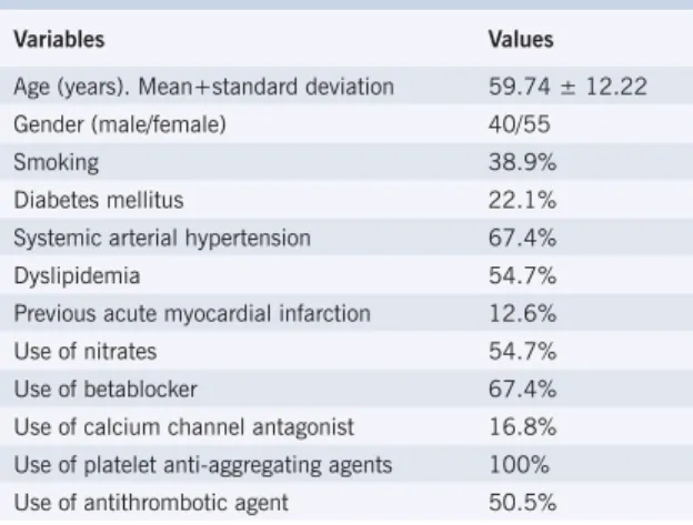
Table 1 – Clinical characteristics, history and medication in use
Variables Values
Age (years). Mean+standard deviation 59.74 ± 12.22
Gender (male/female) 40/55
Smoking 38.9%
Diabetes mellitus 22.1%
Systemic arterial hypertension 67.4%
Dyslipidemia 54.7%
Previous acute myocardial infarction 12.6%
Use of nitrates 54.7%
Use of betablocker 67.4%
Use of calcium channel antagonist 16.8%
Use of platelet anti-aggregating agents 100%
Use of antithrombotic agent 50.5%
history of AMI was a statistically significant predictive risk factor, in the univariate analysis, but was not statistically significant in the multivariate analysis, probably because the number of cases was not so large.
Considering the LV systolic function, this is a determinant factor in the prognosis of patients with cardiopathy at the time they have acute ischemia and also from the chronic point of view 23,24. LVEF is one of
the most widely used measures in clinical practice to
Table 2 – Association between clinical and electrocardiographic variables with the events
Events
Variables Yes n=28 No n=67 Total n=95 OR CI95% for OR p value
Class of unstable angina 0.031
Low risk 5 (15.2%) 28 (84.8%) 33 (100.0%) 1.0
Moderate risk 23 (37.1%) 39 (62.9%) 62 (100.0%) 3.3 1.1 a 9.7
Pre electrocardiogram 0.135
Normal 14 (21.9%) 50 (78.1%) 64 (100.0%) 1.0
Changed - anterior 7 (50.0%) 7 (50.0%) 14 (100.0%) 3.6 1.1 a 11.9
Changed - inferior 4 (4.44%) 5 (55.6%) 9 (100.0%) 2.9 0.7 a 12.1
Changed - lateral 3 (37.5%) 5 (62.5%) 8 (100.0%) 2.1 0.5 a 10.1
Previous Infarction 0.026
Yes 7 (58.3%) 5 (41.7%) 12 (100.0%) 4.1 1.2 a 14.4
No 21 (25.3%) 62 (74.7%) 83 (100.0%) 1.0
Systemic arterial hypertension 0.586
Yes 20 (31.3%) 44 (68.8%) 64 (100.0%) 1.3 0.5 a 3.4
No 8 (25.8%) 23 (74.2%) 31 (100.0%) 1.0
Diabetes mellitus 0.520
Yes 5 (23.8%) 16 (76.2%) 21 (100.0%) 0.7 0.2 a 2.1
No 23 (31.1%) 51 (68.9%) 74 (100.0%) 0.1
Smoking 0.062
Yes 15 (40.5%) 22 (59.5%) 37 (100.0%) 2.4 1.0 a 5.8
No 13 (22.4%) 45 (77.6%) 58 (100.0%) 1.0
Dyslipidemia 0.761
Yes 16 (30.8%) 36 (69.2%) 52 (100.0%) 1.1 0.5 a 2.8
No 12 (27.9%) 31 (72.1%) 43 (100.0%) 1.0
TOTAL 28 (29.5%) 67 (70.5%) 95 (100.0%)
Table 3 – Association between echocardiographic values and events.
Events
Variables Sim n=28 Não n=67 TOTAL n=95 OR CI95% for OR p value
LV ejection fraction 0.025
< 0,5 5 (71.4%) 2 (28.6%) 7 (100.0%) 7.1 1.3 a 39.0
23 (26.1%) 65 (73.9%) 88 (100.0%) 1.0
LVWMSI at rest < 0.001
= 1 14 (18.9%) 60 (81.1%) 74 (100.0%) 1,0 2.9 a 25.2
> 1 14 (66.7%) 7 (33.l3%) 21 (100.0%) 8.6
LVWMSI at peak < 0.001
= 1 2 (3.8%) 50 (96.2%) 52 (100.0%) 1,0
> 1 26 (60.5%) 17 (39.5%) 43 (100.0%) 38.2 8.2 a 178.3
DSE < 0.001
Positive 26 (65.0%) 14 (35.0%) 40 (100.0%) 49.2 10.4 a 232.8
Negative 2 (3.6%) 53 (96.4%) 55 (100.0%) 1.0
LV- left ventricle; LVWMSI-LV wall motion index; DSE- dobutamine stress echocardiogram.
quantify this function, and is extremely valuable to stratify risk after an acute event. Only seven patients (7.4%), of our sample had LVEF<0.50, with five of these presenting events. There was a statistically significant association between LVEF and the events, although the magnitude or strength of this association was affected by the small number of patients with reduced EF.
A semi-quantitative analysis of contractility was carried out based on the division of the LV in 16 segments, as recommended by the American Society
events, that would not require supplementary diagnostic investigation, and could be candidates to early discharge from hospital32. The multivariate analysis demonstrated
that a positive DSE for myocardial ischemia, among the potentially predictive factors for adverse outcomes, was the only one to reach statistical significance.
Some studies have assessed DSE safety in patients known to have coronary disease, either chronic or under investigation33,34. Even when used in a group of patients
with a higher potential for complications during and after the test, the DSE presented a very acceptable safety profile, with no record of more serious events9. In our
set of cases, there were no serious events such as death, AMI or malignant arrhythmias (ventricular fibrillation or sustained ventricular tachycardia) which corroborates the results of the study mentioned above.
Although the fact that the DSE results were made available to part of the physicians assisting the patients may have somehow biased their decision to refer patients to cinecoronariography, we must stress that this test was not included among the clinical events listed in this study. The cinecoronario graphy surely allowed the identification of severe lesions that prompted the indication of revascularization procedures. Additionally, the virtual impossibility of carrying out another complementary method with a similar accuracy, that is, myocardial
Table 4 – Side effects during DSE
Events Number of Patients Percentage(%)
Paroxysmal AF+ 1 1
Nausea 1 1
Headache 3 3
Frequent PVC++ 7 7
Frequent PSVC+++ 10 10
SAH++++ 3 3
Arterial Hypotension 1 1
Precordial pain 17 18
+Atrial Fibrillation; ++Ventricular extrasystoles; +++Supraventricular extrasystoles; ++++Systemic arterial hypertension.
Fig. 1 – Event-free survival of patients within the 6-month follow up period according to the DSE result.
E
ven
t-f
ree
su
rviva
l
(%)
Time (days)
groups DSE negative DSE positive
of Echocardiography15, through the calculation of
LVWMSI. The higher the index, the worse the myocardial contractility and an index higher than 1.6 seems to be related with worse prognosis25. During the performance
of the DSE, the LVWMSI is closely related with the development of myocardial ischemia, which is reflected in the change in segment contractility of LV. Several clinical trials have demonstrated the importance of LVWMSI as an independent risk factor for the development of adverse cardiac events26-28. Our study evidenced a strong
association between LVWMSI at rest and at peak stress with the events. Through the univariate analysis, we verified that LVWMSI equal to one was a strong protection factor against events, in the rest and peak dobutamine infusion stages.
European studies investigating patients diagnosed with low to moderate risk UA by using DSE have been recently published8,9. These studies highlighted the
importance of a positive result for myocardial ischemia as an independent predictive factor for events throughout the observation period (18 months on average). Our results are in agreement with the literature, and emphasize the good ability of stress echocardiography to stratify risk in these patients with UA8,9,29-31. The negative predictive
value (96.4%) reached in this set of cases confirms the excellent prognosis of patients who present a negative
R
EFERENCES1. Braunwald E. Unstable angina: An etiologic approach to management (editorial). Circulation 1998; 98: 2219-22.
2. Chen L, Chester MR, Redwood S, Huang J, Leatham E, Kaski JC. Angiographic stenosis progression and coronary events in patients with “stabilized” unstable angina. Circulation 1995; 91(9): 2319-24
3. Boden WE, McKay RG. Optimal treatment of acute coronary syndromes – An evolving strategy. Editorial. N Engl J Med 2001; 344: 1939-42.
4. Sawada SG, Segar DS, Ryan T et al. Echocardiography detection of coronary artery disease during dobutamine infusion Circulation 1991; 83: 1605-14.
5. Poldermans D, Fioretti PM, Foster T et al. Dobutamine stress echocardiography for assessment of perioperative cardiac risk in patients undergoing major vascular surgery. Circulation 1993; 87: 1506-12.
6. Takeuchi M, Araki M, Nakashima Y, Kuroiwa A. The detection of residual ischemia and stenosis in patients with acute myocardial infarction with dobutamine stress echocardiography. J Am Soc Echocardiograph 1994; 7: 242-52.
7. Trippi JA, Lee KS, Kopp G, Nelson DR, Yee KG, Cordell WH. Dobutamine stress tele-echocardiography for evaluation emergency department patients with chest pain. J Am Coll Cardiol 1997; 30: 627-32.
8. Sitges M, Azqueta M, Paré C et al. Dobutamine stress echocardiography and exercise eletrocardiography for risk stratification in medically treated unstable angina. J Am Soc Echocardiogr 2000; 13 (12): 1084-90.
9. Sitges M, Paré C, Azqueta M et al. Feasibility and prognostic value of dobutamine-atropine stress echocardiography early in unstable angina. Eur Heart J 2000; 21: 1063-71.
10. Nicolau JC, Cesar LAM, Timerman A, Piegas LS, Marin-Neto JA. Diretrizes da Sociedade Brasileira de Cardiologia sobre angina instável e infarto agudo do miocárdio sem supradesnível do segmento ST. Arq Bras Cardiol 2001; 77(supl II): 1-38.
11. McNeill AJ, Fioretti PM, El-Said SM, Salustri A, Foster T, Roelandt JR. Enhanced sensitivity for detection of coronary artery disease by addition of atropine to dobutamine stress echocardiography. Am J Cardiol 1992; 70(1): 41-6.
12. Usher Jr. BW, O’Brien TX. Recent advances in dobutamine stress echocardiography. Clin Cardiol 2000; 23: 560-70.
13. Hepner AM, Bach DS, Armstrong WF. Early chronotropic incompetence predicts the need for atropine during dobutamine stress echocardiography. Am J Cardiol 1997; 79: 366-71.
14. Lewandowski TJ, Armstrong WF, Bach DS. Reduced test time by early identification of patients requiring atropine during dobutamine stress echocardiography. J Am Soc Echocardiogr 1998; 11: 236-42.
15. Shiller N, Shah P, Crawford M et al. Recommendations for quantitation of the left ventricle by two-dimensional echocardiography. American Society of Echocardiography committee on standards, subcommittee on quantitation of two-dimensional echocardiograms. J Am Soc Echocardiogr 1989; 2(5): 358-67.
16. Bach DS, Armstrong WF. Dobutamine stress echocardiography. Am J Cardiol 1992; 69: 90H-6H.
17. Tsutsui JM, Mathias Jr. W. Ecocardiografia sob estresse pela dobutamina associada à atropina. Rev Bras Eco 2000; ano VII (3): 32-47.
18. Théroux P. Clinical angiographic and progression in unstable angina. From clinical observations to clinical trials. Circulation 1995; 91: 2295-8.
19. Chen L, Chester MR, Redwood S, Huang J, Leatham E, Kaski JC. Angiographic stenosis progression and coronary events in patients with “stabilized” unstable angina. Circulation 1995; 91(9): 2319-24.
20. Hosmer DW, Lemeshow S. Applied Logistic Regression. John Wiley and Sons, Inc. New York, 1989.
21. Bertrand ME, Simoons ML, Fox KA et al. Management of acute coronary syndromes: acute coronary syndromes without persistent ST segment elevation. Recommendations of the task force of the European Society of Cardiology. Eur Heart J 2000; 21(17): 1406-32.
22. Braunwald E, Antman EM, Beasley JW et al. ACC/AHA guideline update for the management of patients with unstable angina and non-ST segment elevation myocardial infarction: a report of the American College of Cardiology/American Heart Association task force on pratice guidelines (committee on the management of patients with unstable angina) 2002. Available at:http://www.acc.org/clinical/guidelines/ unstable/unstable.pdf. Acessado em 10 de Dezembro de 2002.
23. Multicenter Postinfarction Research Group: Risk stratification and survival after myocardial infarction. N Engl J Med 1983; 309(6): 331-6.
24. Poldermans D, Fioretti PM, Boersma E et al. Long-term prognostic value of dobutamine-atropine stress echocardiography in 1737 patients with known or suspected coronary artery disease. A single-center experience. Circulation 1999; 99: 757-62.
25. Cordovil A, Ferreira LDC, Machado CV, Moisés VA, Campos Filho O. O ecocardiograma em repouso na avaliação da doença arterial coronariana. Rev Bras Eco 2000; ano VII (4): 11-22.
26. Arruda-Olson AM, Juracan EM, Mahoney DW, McCully RB, Roger VL, Pellikka PA. Prognostic value of exercise echocardiography in 5,798 patients: is there a gender difference? J Am Coll Cardiol 2002; 39 (4): 625-31.
27. Stein JH, Neumann A, Preston LM et al. Improved risk stratification in unstable angina: identification of patients at low risk for in-hospital cardiac events by admission echocardiography. Clin Cardiol 1998; 21: 725-30.
28. Gigli G, Cortigiani L, Vallebona A, Orlandi S, Mariani PR, Volterrani C. Vasodilator stress echocardiography for risk stratification of medically stabilized unstable angina. Eur J Echocardiogr 2002; 3 (1): 59-66.
29. Lin SS, Lauer MS, Marwick TH. Risk stratification of patients with medically treated unstable angina using exercise echocardiography. Am J Cardiol 1998; 82: 720-4.
30. Eriksson SV, Erhardt L, Lindvall K, Melcher A, Rehnqvist N. Long-term prognostic importance of exercise echocardiography after an episode of unstable angina. Cardiology 1995; 86: 426-31.
31. Amanullah AM, Lindvall K. Predischarge exercise echocadiography in patients with unstable angina who respond to medical treatment. Clin Cardiol 1992; 15: 417-23.
32. Chuah S-C, Pellikka PA, Roger VL, McCully RB, Seward JB. Role of dobutamine stress echocardiography in predicting outcome in 860 patients with known or suspected coronary artery disease. Circulation 1998; 97: 1474-80.
33. Mertes H, Sawada SG, Ryan T et. al. Symptoms, adverse effects, and complications associated with dobutamine stress echocardiography. Experience in 1118 patients. Circulation 1993; 88(1): 15-9.
34. Mathias Jr. W, Arruda A, Santos FC et al. Safety of dobutamine –atropine stress echocardiography: a prospective experience of 4033 consecutive studies. J Am Soc Echocardiogr 1999; 12(10):785-91.
scintigraphy, in a timely manner so as to compare it with the DSE, in such a way that the DSE results were not accessed by the physician especially in the public hospitals participating in the study, would make the non-invasive stratification procedure unviable in this group of patients.
The authors thank Prof. José Natal Figueiroa for his invaluable assistance in the analysis of the statistical data of this study.
Potential Conflict of Interest
No potential conflict of interest relevant to this article was reported.