Time Domain Analysis of the Signal Averaged
Electrocardiogram to Detect Late Potentials in Heart
Failure Patients with Different Etiologies
Ernani de Sousa Grell, Rogério Silva de Paula, Nancy Maria Martins de Oliveira Tobias, Paulo Jorge Moffa, César José Grupi, Alfredo José Mansur
,QVWLWXWRGR&RUDomRGR+RVSLWDOGDV&OtQLFDVă)08636mR3DXOR63%UD]LO
O
BJECTIVETo evaluate the frequency, clinical correlations and prognosis influence of late potentials on the of heart failure patients with different etiologies using the signal averaged electrocardiogram.
METHODS
A 42 month study of the signal averaged electrocardiograms of 288 heart failure patients with different etiologies was conducted. The group of patients included 215 males (74.65%) and 73 females (25.35%) between the ages of 16 and 70 (mean 51.5, standard deviation 11.24). The heart failure etiologies were: hypertensive heart disease (78 patients, 27.1%); idiopathic dilated cardiomyopathy (73 patients, 25.4%); ischemic cardiomyopathy (65 patients, 22.6%); Chagas disease (42 patients, 14.6%); alcoholic cardiomyopathy (9 patients, 3.1%); peripartum cardiomyopathy (6 patients, 2.1%); valvular heart disease (2 patients, 4.2%) and viral myocarditis (3 patients, 1.04%). The variables included the duration of the standard QRS complex, duration of the filtered QRS complex, duration of the signal below 40µV and the root mean square of the last 40ms which were analyzed in regard to age, gender, etiology and mortality as well as the findings of the 12-lead electrocardiogram at rest, echocardiogram and ambulatory electrocardiogram. The statistical analysis tests used were: the Fisher exact probability test, Student’s t-test, Mann Whitney test, variance analysis, Log-Hank and the Kaplan-Meyer method.
R
ESULTSLate potentials were diagnosed in 90 patients (31.3%) and there was no association with the etiologies. The presence of this condition is associated with: a lower maximum oxygen uptake during the ergospirometry (p=0.001); sustained and non-sustained ventricular tachycardia during Holter monitoring (p=0.001), sudden death and mortality (p<0.05). Patients that did not present late potentials had a higher overlife rate.
M a i l i n g A d d r e s s :E r n a n i d e S o u s a G r e l l Ć 5 X D & R U L Q W R $ 6 m R 3 D X O R 6 3 % U D ] L O
(PDLOHJUHOO#FDUGLROEU 5HFHLYHGRQĆ$FFHSWHGRQ
CONCLUSION
The presence of late potentials was not associated with the etiologies and proved to be an indication of a worse prognosis.
KEY WORDS
Heart failure, signal averaged electrocardiogram, prognosis.
Late potentials are high frequency, low amplitude, microvolt-size (µV) signals at the end of the QRS complex and the beginning of the ST segment that are related to fragmented and delayed electrical activity in the ventricles, enabling the genesis of sustained ventricular tachycardia through reentry mechanism. Late potentials can be identified using a signal averaged electrocardiogram (SAECG)1. The presence of late potentials was detected
in patients with ventricular aneurysms2,3, tachycardia,
ventricular fibrillation4, and arrhythmogenic right
ventricular dysplasia5, and is associated with a higher
frequency of sudden cardiac death6.
In heart failure patients, the presence of late potentials has been investigated in small case studies that do not include patients with Chagas disease6-8.
It is admitted that 40% of the deaths in heart failure patients are sudden and caused by cardiac arrhythmias 9-12. The SAECG is a noninvasive and relatively simple
method which is a useful tool to identify patients with a higher risk of developing lethal cardiac arrhythmias. We formulated the hypothesis that the discovery of late potentials could contribute to identify heart failure patients with a higher risk of death.
We outlined the present study to evaluate the presence of late potentials in a cohort of heart failure patients with different etiologies, their clinical associations and their relation with mortality.
M
ETHODSPatients - The study included a cohort of 288
outpatients, 215 males (74.7%) and 73 females (25.4%), between the ages of 16 and 70 (51.5 ± 11.2).
The clinical evaluation included the patient’s medical history, a physical examination, chest x-ray, 12-lead electrocardiogram, ambulatory electrocardiogram, signal averaged electrocardiogram, echocardiogram and ergospirometry. The follow-up timeframe was between July 1998 and December 2001 and varied from 8 to 42 months (36.0 ± 4.5). Follow-up for each patient started on the day of the SAECG and ended on the date of the last clinical observation or death.
Two hundred and eighty-eight individuals were evaluated: 78 (27.1%) hypertensive heart disease patients; 73 (25.4%) idiopathic dilated cardiomyopathy patients; 65 (22.6%) ischemic cardiomyopathy patients; 42 (14.6%) Chagas disease patients; 12 (4.2%) valvular heart disease patients; 9 (3.1%) alcoholic cardiomyopathy patients; 6 (2.1%) peripartum cardiomyopathy patients and 3 (1.0%) viral myocarditis patients. In order to facilitate the statistical analysis the last four etiologies mentioned above were combined in a single group named
others which included 30 individuals (10.3%).
The patients were divided in two groups: Group 1 without late potentials and Group 2 with late potentials. Patient death ended the follow-up.
Diagnostic criteria – The Framingham criteria13 was
used to diagnose heart failure and was established if the patient had at least two major criteria or one major and two minor criteria.
The diagnosis of the heart failure etiology was based on criteria previously published in medical literature for patients with symptomatic heart failure or left ventricular systolic dysfunction. (World Health Organization / International Society and Federation of Cardiology, 1980, Report of 1995 World Health Organization / International Society and Federation of Cardiology Task Force on the Definition and Classification of Cardiomyopathies, 1996 and International Statistical Classification of Diseases and Related Health Problems, tenth revision – CID 10, 1993) An ischemic cardiomyopathy diagnosis was made based on patients with symptomatic heart failure and left ventricular dysfunction that had a history of myocardial infarction, percutaneous transluminal coronary angioplasty, myocardial revascularization, stable angina pectoris, an altered electrocardiogram, myocardial ischemia demonstrated by an altered exercise stress test, myocardial perfusion scintigraphy or a cineangiocardiography showing an obstruction greater than 75%.
Inclusion criteria – Patients between the ages of 16
and 70 with a diagnosis of symptomatic congestive heart failure caused by ventricular systolic dysfunction were included in the study.
Exclusion criteria – Patients with the following
conditions were excluded from the study: aortic aneurysm, artificial pacemaker, atrial fibrillation or flutter, pregnant women, recent (less than 3 months) myocardial infarction or heart surgery, unstable angina pectoris, presence of myocardial ischemia during the exercise stress test, hypertrophic cardiomyopathy, recent systemic infection, chronic obstructive pulmonary disease or neoplasias. Patients using amiodarone were not excluded.
Signal averaged electrocardiogram (SAECG) – The
signal averaged electrocardiogram was recorded using the Arrhythmia Research Technology Inc. 1200 EPX machine. The Frank orthogonal X, Y and Z electrographic lead arrangement and a broad bandwidth bidirectional filter (0.05 to 250Hz) with a frequency cut-off (lower value) of 40Hz were used. An acceptable noise level was considered as < 0.3µV with a gain of 2000/4000.
Late potential diagnosis – The presence of late potentials was detected based on the criteria of the American College of Cardiology: 1) duration of the end of the QRS complex
PVGXUDWLRQRIWKHVLJQDOEHORZ9PV URRWPHDQVTXDUHYROWDJHRIWKHODVWPV9,Q
accordance with the findings of Moraes14, only the root
PHDQVTXDUHYROWDJHLQWKHODVWPV9ZDVXVHG
for the patients with a bundle branch block.
Variables studied – The variables studied included:
a) age; b) gender; c) heart failure etiology; d) inactive area found with the 12-lead electrocardiogram; e) bundle
f) diameters of the left ventricle during the diastole and systole from the echocardiogram; g) left ventricle ejection fraction from the echocardiogram; h) maximum oxygen uptake during the ergospirometry; i) sustained ventricular tachycardia detected on the ambulatory electrocardiogram; j) duration of the standard QRS complex; k) duration of the filtered QRS complex; l) duration of the signal below 40µV; and m) the root mean square voltage of the last 40ms, on the signal averaged electrocardiogram.
Statistical analysis – Comparisons of the data between
the two groups (with and without late potentials) and within the groups were conducted with the following statistical tests: 1) chi-square test or Fisher exact probability test for comparisons between two categorical variables with two categories each; 2) Student’s t-test for comparisons between the averages of the two groups for continuous variables; 3) Mann – Whitney test (non-parametric), for comparisons between the two groups (with and without late potentials) for numerical variables (number of occurrences); 4) ANOVA (variance analysis), for comparisons between more than two groups in the case of continuous variables; 5) log-rank test to compare the survival curve; 6) Kaplan-Meier method for death related variables. The operational variables used were: sensitivity, specificity, accuracy, positive predicative value and negative predictive value.
The significance level was p<0.05. The statistical analysis was conducted using the statistical program SPSS for Windows.
Ethical aspects - The protocol was approved by the
Scientific and Ethics Commission of the Hospital das Clínicas of the University of São Paulo Medical School. The patients were briefed on the details of the study and were included in the protocol after signing the free and informed consent form.
RESULTS
Late potentials detected by the signal averaged
electrocardiogram - Out of the total number of 288
individuals included in the study, 90 (31.3%) were diagnosed with late potentials (Table 1).
The duration of the standard QRS complex varied from 80 to 242ms (124.9 ± 31.3); the duration of the filtered QRS complex varied from 76 to 254ms (131.7 ± 33.90); the duration of the signal below 40µV varied from 6 to 183ms (33.0 ± 20.57); and the root mean square voltage during the last 40ms varied from 4.20 to 267.30µV (44.0 ± 42.42).
Late potentials in relation to clinical variables – Patients with late potentials had an older mean age (54.5 ± 10 versus 50.1± 11.5); (p=0.004).
Patients with late potentials presented a higher incidence of sustained ventricular tachycardia (32/37 – 86.5% vs. 5/37 – 13.5%) during the ambulatory electrocardiogram (p=0.001). Late potential sensitivity to identify this arrhythmia was 86.5%; specificity 76.9%; accuracy 78.1%; positive predictive value 35.6% and negative predictive value 97.5%.
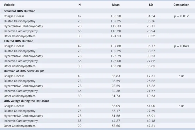
No association was found between the heart failure etiology and the presence of late potentials. However, the durations of the standard QRS complex and the filtered QRS complex revealed different mean value indexes for Chagas disease and dilated cardiomyopathy in relation to the other etiologies (Table 2).
No correlation was found between the presence of late potentials in relation to gender, heart failure etiology, the presence of electrically inactive areas during the electrocardiogram at rest, the presence of right or left bundle branch blocks or the size of the heart chambers on the echocardiogram.
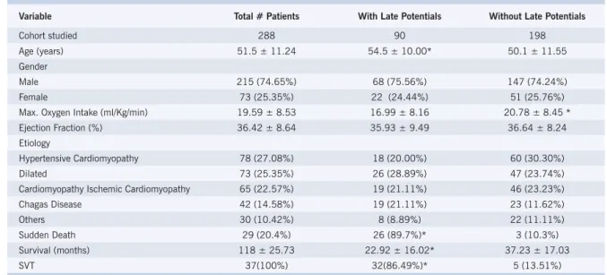
Table 1 – Clinical and Demographic Characteristics
Variable Total # Patients With Late Potentials Without Late Potentials
Cohort studied 288 90 198
Age (years) 51.5 ± 11.24 54.5 ± 10.00* 50.1 ± 11.55 Gender
Male 215 (74.65%) 68 (75.56%) 147 (74.24%) Female 73 (25.35%) 22 (24.44%) 51 (25.76%) Max. Oxygen Intake (ml/Kg/min) 19.59 ± 8.53 16.99 ± 8.16 20.78 ± 8.45 * Ejection Fraction (%) 36.42 ± 8.64 35.93 ± 9.49 36.64 ± 8.24 Etiology
Hypertensive Cardiomyopathy 78 (27.08%) 18 (20.00%) 60 (30.30%) Dilated 73 (25.35%) 26 (28.89%) 47 (23.74%) Cardiomyopathy Ischemic Cardiomyopathy 65 (22.57%) 19 (21.11%) 46 (23.23%) Chagas Disease 42 (14.58%) 19 (21.11%) 23 (11.62%) Others 30 (10.42%) 8 (8.89%) 22 (11.11%) Sudden Death 29 (20.4%) 26 (89.7%)* 3 (10.3%) Survival (months) 118 ± 25.73 22.92 ± 16.02* 37.23 ± 17.03 SVT 37(100%) 32(86.49%)* 5 (13.51%) * p<0.05; SVT- sustained ventricular tachycardia.
From the 288 patients in the study, 142 (49.3%) died; 49 (17.0%) had late potentials and 93 (32.3%) did not have late potentials.
Cause of death was classified as: heart disease evolution, sudden death and other causes not related to heart failure (pneumonia, digestive hemorrhage, stroke, etc.) Eighty patients (56.3% of the total number of deaths) died as a result of heart failure evolution. From these 80 patients, 21 (26.3%) had late potentials and 59 (73.7%) did not. Twenty-nine patients suffered sudden death (10.1%) of which 26 had late potentials (89.5%) and 3 (10.3%) did not (p<0.05). Of the 29 patients that suffered sudden death, 17 (58.6%), had late potentials and had presented a previous episode of sustained ventricular tachycardia (Table 3).
The following observations were noted in relation to the evolution of the patients: a) adverse evolution (sustained ventricular tachycardia, sudden death or death as a result
Table 2 – Descriptive Measures of the SAECG Variables according to Etiology
Variable N Mean SD Comparison
Standard QRS Duration
Chagas Disease 42 133.50 34.54 p = 0.012 Dilated Cardiomyopathy 73 132.25 36.36
Hypertensive Cardiomyopathy 78 119.33 26.11 Ischemic Cardiomyopathy 65 118.20 26.94 Other Cardiomyopathies 30 124.53 30.22 Filtered QRS Duration
Chagas Disease 42 137.88 35.77 p = 0.048 Dilated Cardiomyopathy 73 139.25 38.27
Hypertensive Cardiomyopathy 78 125.79 30.53 Ischemic Cardiomyopathy 65 125.68 27.82 Other Cardiomyopathies 30 133.20 36.85 Duration of QRS below 40 µV
Chagas Disease 42 36,83 17.31 p ns Dilated Cardiomyopathy 73 36.59 25.62
Hypertensive Cardiomyopathy 78 28.59 15.22 Ischemic Cardiomyopathy 65 32.38 21.57 Other Cardiomyopathies 30 31.73 19.53 QRS voltage during the last 40ms
Chagas Disease 42 38.09 51.00 p ns Dilated Cardiomyopathy 73 35.17 27.59
Hypertensive Cardiomyopathy 78 51.58 45.91 Ischemic Cardiomyopathy 65 44.27 42.18 Other Cardiomyopathies 29 53.66 47.21 ns= not significant
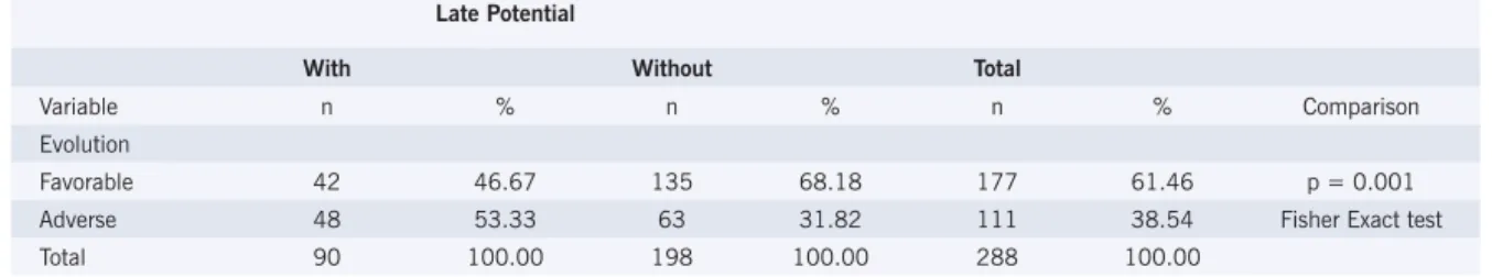
of heart failure evolution); and b) favorable evolution (none of the complications described). One hundred and seventy-seven patients (61.5%) had a favorable evolution of which 42 (46.7%) had late potentials and 135 (68.2%) did not. One hundred and eleven (38.5%) had an adverse evolution of which 48 (53.3%) had late potentials and 63 (31.8%) did not. (p=0.001) (Table 4).
The overlife rate was lower in the group with late potentials.
Late potentials in relation to echocardiograph
findings – The diameter of the left ventricle during the
diastole and systole, the diameter of the left atrium and the left ventricle ejection fraction were analyzed. The diameter of the left atrium varied from 27 to 80mm (mean 47.36; standard deviation 7.47). The diameter of the left ventricle during the diastole varied from 40 to 105mm (mean 73.98; standard deviation 9.57).
Table 3 – Presence of Late Potentials and Cause of Death
Cause of Death Number of Patients With Late Potentials Without Late Potentials Heart Failure 80 (56.4%) 21 (26.3%) 59 (73.7%) Sudden Death 29 (20.4%) 26 (89.7%) 3 (10.3%) Other Causes 33 (23.2%) 2 (6%) 31 (94.0%)
Table 4 - Distribution of the Evolution in Regard to Late Potentials
Late Potential
With Without Total
Variable n % n % n % Comparison
Evolution
Favorable 42 46.67 135 68.18 177 61.46 p = 0.001 Adverse 48 53.33 63 31.82 111 38.54 Fisher Exact test Total 90 100.00 198 100.00 288 100.00
The diameter of the left ventricle during the systole varied from 30 to 95mm (mean 63.71; standard deviation 8.64). No significant differences were found in relation to late potentials for the echocardiogram variables studied.
Late potentials in relation to the ergospirometry – The
analysis of maximum oxygen uptake varied from 4.6 to 33.7 ml/Kg/min (mean 19.59; standard deviation 8.53). In the patients that presented late potentials a value of 16.99 ml/Kg/min with a standard deviation of 8.16 was found. For the patients that did not have late potentials this value was significantly higher, that is, 20.78 ml/kg/ min with a standard deviation of 8.45 (p=0.001).
DISCUSSION
This study presented some significant observations. The most important finding was the confirmation that the presence of late potentials with congestive heart failure is related to an adverse prognosis with a higher incidence of sustained ventricular tachycardia and cardiac sudden death. More than one half of the patients (53.3%) with late potentials presented an unfavorable evolution and 26 of the 29 patients that suffered a sudden death had late potentials. The findings of this study are in agreement with those of Ohnishi et al6. These authors described
a high incidence of ventricular arrhythmias and sudden death in 54 patients with dilated cardiomyopathy and an altered signal-averaged electrocardiogram using the following criteria for detecting late potentials: duration of the filtered QRS complex >120ms or the root mean square voltage in the last 40ms < 20µV. Nevertheless there is controversy. The studies of Meinertz et al7 and
Middlekauff et al8 did not find the signal averaged
electrocardiogram to be predictive of sudden death or ventricular arrhythmia. The number of patients in both of these studies was low (30 and 22 patients, respectively) and they used different criteria to detect late potentials. Only one of the 30 patients in the non-ischemic cardiomyopathy group of Meinhertz et al7 had an altered
signal averaged electrocardiogram. Eleven patients died during the course of the study of which 5 were a result of heart failure evolution. The author concluded that the signal averaged electrocardiogram was not predictive of sudden death even though the sample was too small to make any definite conclusions.
The finding that there is a higher incidence of male individuals 215 (74.7%) with heart failure is in agreement with reliable studies such as the Framingham study. Nevertheless, the direct relationship between aging and a higher incidence of late potentials found in this study was a new fact even though aging has already been identified as a contributing factor for a worse prognosis. This was demonstrated in the Framingham study and revealed that for each decade of life mortality rates increase by 27% for males and by 61% for females.
No association was found between heart failure etiologies and late potentials. It is possible that late potentials and arrhythmia are generated by a common mechanism that is involved in the various etiologies that lead to heart failure. Interstitial fibrosis and myocardial hypertrophy are frequently found in biopsies of patients with dilated cardiomyopathy, Chagas disease, ischemic cardiomyopathy, etc. which can cause abnormal electrical conduction. It has been demonstrated that the signal averaged electrocardiogram is a useful tool to identify patients with delayed ventricular activation and consequently a substrate to trigger arrhythmia. This study demonstrated a correlation between the standard QRS complex and the filtered QRS complex in relation to dilated cardiomyopathy and Chagas disease. It is known that these pathologies lead to a sporadic fibrotic process in the heart. A specific study to enable the noninvasive investigation of the extent of myocardial fibrosis and signal averaged electrocardiogram variables was proposed by Yamada et al15. The group studied 32 patients with
dilated cardiomyopathy using a myocardial biopsy and a signal averaged electrocardiogram. They found a direct relationship between the extent of the fibrotic area and the duration of the filtered QRS complex (p<0.001), duration of the signal below 40µV (p<0.001) and the root mean square voltage in the last 40ms (p<0.005).
The presence of late potentials on the signal averaged electrocardiogram has demonstrated its predictive value for sudden death in heart failure patients16-19. The
objective of the present study was investigate similar correlations for congestive heart failure caused not only by heart failure but also by other etiologies.
R
EFERENCES1. Berbari EJ, Lazzara R, Samet P, Scherlag BJ. Noninvasive technique for detection of electrical activity during the PR segment. Circulation 1973; 48: 1005-13.
2. Wiener I, Mindich B, Pitchon R. Determinants of ventricular tachycardia in patients with ventricular aneurysms: results of intraoperative epicardial and endocardial mapping. Circulation 1982; 65: 856-61.
3. Simson MB. Clinical application of signal averaging. Cardiol Clin 1983; 1: 109-19.
4. Denes P, Santarelli P, Hauser RG, Uretz EF. Quantitative analysis of the high-frequency components of the terminal portion of the body surface QRS in normal subjects and in patients with ventricular tachycardia. Circulation 1983; 67: 1129-38.
5. Fontaine G. The use of ICD’s for the treatment of patients with Arrhythmogenic Right Ventricular Dysplasia (ARVD) J Interv Card Electrophysiol 1997; 1: 329-30.
as main heart failure capacity quantifiers - demonstrated a correlation with alterations on the signal averaged electrocardiogram. However, the maximum oxygen uptake during the ergospirometry was correlated and therefore is a good variable to analyze the peripheral component of heart failure.
Individuals with His bundle blocks are usually excluded from time domain SAECG studies, since the ventricular activation alterations can either mask or simulate late potentials. In patients with bundle branch blocks, abnormal myocardial zones generating low amplitude signals could be completely masked by the delayed activation of the normal myocardial regions. However, false positive results can be obtained when the high frequency filters are placed over the end portion of a QRS complex that has a lower than normal amplitude.
There is disagreement regarding the exclusion of patients with bundle branch blocks from studies involving time domain signal averaged electrocardiograms. According to Moffa 20, the incidence of malignant
ventricular arrhythmia in this group is higher than in individuals without conduction abnormalities (14% versus 4%). Additionally, the incidence of bundle branch blocks is relatively high in individuals with heart failure. (In this study there were 121 patients – 42.0%). Some authors have established different criteria to identify late potentials in patients with bundle branch blocks. For Nalos et al21, late potentials are considered positive in
the presence of a bundle branch block when all three variables analyzed in the time domain are abnormal, using the following reference values: filtered QRS complex
PVGXUDWLRQRIVLJQDOEHORZ9PVDQG URRWPHDQVTXDUHYROWDJHLQWKHODVWPV9)RU
Fontaine et al22, the criteria that they considered as the
best indicator was a root mean square voltage in the last
PVRI9ZLWKRUZLWKRXWDVLJQDOEHORZ9 IRUDGXUDWLRQPV)RUWKHVHDXWKRUVWKHDGGLWLRQDO
requirement of a filtered QRS complex > 180ms does not alter the total predictive accuracy.
In this study, late potentials in the patients with bundle branch blocks were identified using the criteria of Moraes14, a method used subsequently in other
studies23.
A standard QRS complex duration greater than or equal to 120ms presented a significant correlation with the prognosis (already proven in individuals with left bundle branch blocks) and 63 patients (70%) with this condition presented an adverse evolution.
The possible influence of medications is a point to be questioned. According to the experience of some authors24,25, the interpretation of the signal
averaged electrocardiogram results is jeopardized for the patients in group 1 using antiarrhythmic drugs and amiodarone. However, a prospective study26 with
27 cases of dilated cardiomyopathy was conducted to analyze the use of amiodarone and its correlation with the signal averaged electrocardiogram. This study used a series of recordings from the signal averaged and conventional electrocardiograms that were taken before the administration of amiodarone, 2 months after the administration of amiodarone and subsequently with 3 month intervals. In comparison with the initial findings there was an increase in the duration of the filtered QRS complex however there was no alteration in the QRS voltage during the last 40ms. The incidence of late potentials remained constant. In this study, 21 (7.3%) of the individuals used amiodarone and the drug was not suspended as these patients had serious heart failure and a high risk of developing cardiac arrhythmia.
CONCLUSIONS
The etiology that led to heart failure has no correlation with the incidence of late potentials detected by the time domain signal averaged electrocardiogram. The presence of late potentials is related to a higher incidence of sustained ventricular tachycardia and cardiac sudden death. Expected overlife was lower in the individuals that presented late potentials. Therefore, the presence of late potentials on the signal averaged electrocardiogram proved to be an indication of an unfavorable prognosis in individuals with congestive heart failure.
Potential Conflict of Interest
No potential conflict of interest relevant to this article was reported.
6. Ohnishi Y, Inoue T, Fukuzaki H. Value of the signal-averaged electrocardiogram as a predictor of sudden death in myocardial in the dilated cardiomyopathy. Jpn Circ J 1990; 127-36.
7. Meinertz T, Treese N, Kasper W, et al. Determinants of prognosis in idiopathic dilated cardiomyopathy as determined by programmed electrical stimulation. Am J Cardiol 1985; 56: 337-41.
8. Middlekauff HR, Stevenson WG, Woo MA, Moser DK, Stevenson LW. Comparison of frequency of late potentials in idiopathic dilated cardiomyopathy and ischemic cardiomyopathy with advanced congestive heart failure and their usefulness in predicting sudden death. Am J Cardiol 1990; 66: 1113-7.
9. Unverferth DV, Magorien RD, Moeschberger ML, Baker PB, Fetters JK, Leier CV. Factors influencing the one-year mortality of dilated cardiomyopathy. Am J Cardiol 1984; 54: 147-52.
10. Franciosa JA. Application of noninvasive techniques for measuring cardiac
11. Likoff MJ, Chandler SL, Kay HR. Clinical determinants of mortality in chronic congestive heart failure secondary to idiopathic dilated or to ischemic cardiomyopathy. Am J Cardiol 1987; 59: 634-8. 12. Simson MB, Signal-averaged electrocardiography: methods and clinical
applications, in Braunwald: Heart Disease Update. Philadelphia, WB Saunders, 1989: 145-56.
13. Mackee PA, Castelli WP, Mcnamara PM, Kannel WB. The natural history of congestive heart failure: the Framingham study. N Eng J Med 1971;285: 1441-6.
14. Moraes AP, Eletrocardiograma de alta resolução na cardiopatia chagásica crônica. Tese (Doutorado) – Faculdade de Medicina, Universidade de São Paulo. São Paulo, 1993.
15. Yamada T, Fukunami M, Ohmori M, et al. New approach to the estimation of the extent of myocardial fibrosis in patientes with dilated cardiomyopathy: Use of signal-averaged electrocardiography. Am Heart J 1993; 126: 626-31.
16. Kuthard D, Thorburn C, Sammel N. Prediction of serious arrythmic events after myocardial infarct: signal averaged electrocardiogram, holter monitoring and radionuclide ventriculography. J Am Coll Cardiol 1987;9: 531-38.
17. Breithardt G, Scwarzmaier M, Borggrefe M. Haerten K, Seipel L. Prognostic significance of late ventricular potentials after acute myocardial infarction. Eur Heart J 1983; 4: 487-95.
18. Denniss AR, Richards DA, Cody DV et al. Prognostic significance of ventricular tachycardia and fibrillation induced at programmed stimulation and delayed potentials detected on the signal-averaged electrocardiograms of survivors of acute myocardial infarction. Circulation 1986; 74: 731-45.
19. Gomes J, Winters S, Stewart D, Horowitz S, Milner M, Barreca P. A new noninvasive index to predict sustained ventricular tachycardia and sudden death in the first year after myocardial infarction based on signal averaged electrocardiogram, radionuclide ejection fraction, and Holter monitoring. J Am Coll Cardiol 1987;10: 349-57. 20. Moffa PJ. Eletrocardiograma de alta resolução - revisão da utilidade
clínica do eletrocardiograma de alta resolução e dos potencias tardios em grupos específicos de pacientes. REBRAMPA 1983; 6: 102-12. 21. Nalos PC, Gang ES, Mandel WJ, Ladenheim ML, Lass Y, Peter T. The
signal-averaged electrocardiogram as a screening test for inducibility of sustained ventricular tachycardia in high risk patients: a prospective study. J Am Coll Cardiol 1987; 9: 539-48.
22. Fontaine JM, El-Scherif N. Bundle branch block and the signal-averaged electrocardiogram. In: El-Sherif N, Turitto G. (Eds.). High Resolution Electrocardiography. New York: Futura Publishing Co. Inc., 1992: 533-67.
23. Vallejo M, Reyes K, Reyes PA, Gonzalez H, Hermosillo JA. Late potentials and variability of cardiac frequency in chronic chagasic myocardiopathy and other myocardiopathies Arch Inst Cardiol Mex 1997; 67: 485-93.
24. Greenspon AJ, Kidwell GA. The effects of antiarrhythmic drugs on the signal-averaged electrocardiogram in patients with malignant ventricular arrhythmias. Prog Cardiovasc Dis 1993; 35: 399-406. 25. Steimberg JS, Reagan A, Sciacca R, Bigger JT, Fleiss JL. Predicting
arrhytmic events after acute myocardial infartion using the signal-averaged elctrocardiogram. Am J Cardiol 1992; 69: 13-21. 26. Goedel – Meinen L, Hofmann M, Schmidt G, et al. Amiodarone
- efficacy and late potencials during long-term therapy. Int J Clin Pharmacol Ther Toxicol 1990; 28: 449-54.