Instituto da Criança do Hospital das Clínicas - FMUSP, Instituto do Coração do Hospital das Clínicas - FMUSP e Laboratório Fleury
Mailing address: Sofia Mizuho Miura Sugayama – Rua João Moura, 2381/51 Cep 05412-004 – São Paulo, SP, Brazil - E-mail: [email protected] Received 9/4/02
Accepted 10/29/02
Arq Bras Cardiol, volume 81 (nº 5), 468-73, 2003
Sofia Mizuho Miura Sugayama, Regina Lúcia Moisés, Jaqueline Wagënfur, Nana Miura Ikari,
Kikue Terada Abe, Cláudio Leone, Clóvis Artur Almeida da Silva,
Maria de Lourdes Lopes Ferrari Chauffaille, Chong Ae Kim
São Paulo, SP - Brazil
Williams-Beuren Syndrome. Cardiovascular Abnormalities in
20 Patients Diagnosed with Fluorescence in Situ Hybridization
Williams-Beuren syndrome is a rare condition of dele-tion of contiguous genes, which is characterized by elfin fa-cies, eye, teeth, cardiovascular, renal, and skeletal abnorma-lities, mental deficiency, overfriendly personality, and occa-sionally infantile hypercalcemia 1-4. Williams et al 1 in 1961
and Beuren 2 in 1962 described the syndrome independently.
The incidence is estimated at 1:13,700 to 1:25,000 live births5,6. The occurrence is generally sporadic in most
fami-lies 3,5, although reports do exist of a few cases in the same
family apparently due to dominant autosomal inheritance 7,8.
Distinctive facial features described as elfin facies in-clude bitemporal narrowness, periorbital fullness, small up-turned nose, full nasal tip, long philtrum, full cheeks, full lips, wide mouth, and small jaw 1-4. These typical facial
featu-res are usually recognizable around 4 months of age and become striking during infancy and childhood 3,4. Adult
pa-tients have dolichocephaly and thick lips 3.
Clinical diagnostic criteria are available for Williams-Beuren syndrome 3,4. Approximately 90 to 95% of the
pa-tients have micro-deletion in the long arm of chromosome 7 (7q11.23), which includes the elastin gene and is detectable by fluorescence in situ hybridization (FISH) 9,10. This test is
useful for diagnostic confirmation, because great clinical variability is present, hindering a diagnosis especially in the first year of life.
Cardiovascular abnormalities occur in almost 80% of the cases of Williams-Beuren syndrome 3,4,11-14. Aortic
su-pravalvular stenosis is the most common cardiac anomaly, present in 64% of patients 11. Other cardiopathies include
pulmonary artery stenosis, aortic hypoplasia, aortic coarc-tation, mitral valve prolapse, and septal defects 11-14. In
Williams-Beuren syndrome, located and diffuses stenosis of the thoracic and abdominal aorta 15,16 and the coronary
and cerebral arteries 17,18 may also occur.
The natural history of supravalvular aortic stenosis varies in the affected patients. It may be a progressive lesion characterized by an increase in the pressure gradient of the left ventricle due to hypoplasia of the ascending aorta12.
Objective - To evaluate the cardiovascular findings and clinical follow-up of patients with Williams-Beuren syndrome.
Methods - We studied 20 patients (11 males, mean age at diagnosis: 5.9 years old), assessed for cardiovascu-lar abnormalities with electrocardiography and Doppler echocardiography. Fluorescence in situ hybridization (FISH) was used to confirm the diagnosis of the syndrome.
Results - Elastin gene locus microdeletion was detec-ted in 17 patients (85%) (positive FISH), and in 3 patients deletion was not detected (negative FISH). Sixteen patients with a positive FISH (94%) had congenital cardiovascular disease (mean age at diagnosis: 2,3 years old). We observed isolated (2/16) supravalvular aortic stenosis and supra-valvular aortic stenosis associated (11/16) with pulmonary artery stenosis (4/11); mitral valve prolapse (3/11); bicus-pid aortic valve (3/11); aortic coarctation (2/11), thicke-ned pulmonary valve (2/11); pulmonary valvular stenosis (1/11); supravalvular pulmonary stenosis (1/11); valvular aortic stenosis (1/11); fixed subaortic stenosis (1/11); pul-monary artery stenosis (2/16) associated with pulpul-monary valvar stenosis (1/2) and with mitral valve prolapse (1/2); and isolated mitral valve prolapse (1/16). Four patients with severe supravalvular aortic stenosis underwent surge-ry (mean age: 5.7 years old), and 2 patients had normal pressure gradients (mean follow-up: 8.4 years).
Conclusion - A detailed cardiac evaluation must be performed in all patients with Williams-Beuren syndrome due to the high frequency of cardiovascular abnormalities.
This abnormality is usually diagnosed in the first 2 years of life; however, it can remain undiagnosed for years, until an increase occurs in the systolic pressure gradient. Symptoms and complications are similar to those of aortic valvar stenosis 12.
The objective of this study was to analyze the cardio-vascular findings and the clinical follow-up of cardiocardio-vascular anomalies in 20 patients with Williams-Beuren syndrome.
Methods
The cohort of this retrospective and prospective study was formed by 20 patients (11 M: 9F) with a clinical diagnosis of Williams-Beuren syndrome followed-up at the Unidade de Ge-nética do ICr-HC-FMUSP (Genetic Unit of Instituto da Criança-Hospital das Clínicas-School of Medicine) from 1991 to 2001.
Mean age during clinical diagnosis of Williams-Beuren syndrome was 5.9 years (8 months to 10.7 years). The pa-rents were healthy, and all cases were sporadic.
The Williams-Beuren syndrome diagnosis was confir-med with the FISH test. This technique enables the determi-nation of a specific deoxyribonucleic acid (DNA) sequence in a chromosomal band 19. It consists of the hybridization of a
probe fluorescently or radioactively labeled, to identify its complementary segment within a metaphase chromosome. DNA of the metaphase chromosome is denatured in the lami-na (for this reason, it is called in situ), and then hybridization is started with a labeled probe. The probe is visualized with fluorescence in the FISH technique or with an X-ray film 19.
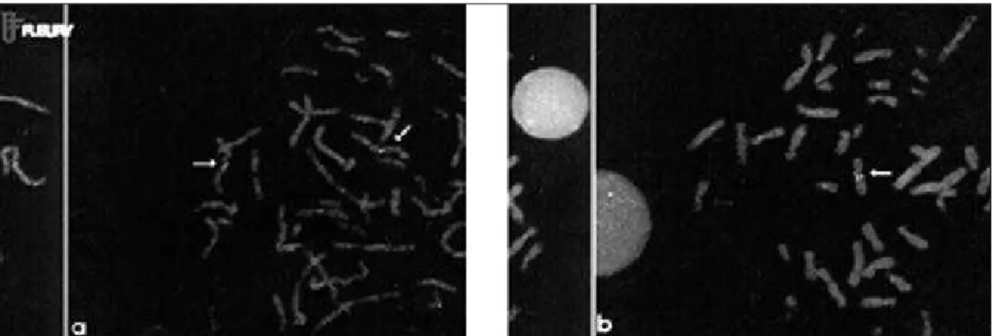
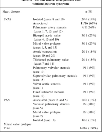
In this study, micro-deletion in the elastin gene was in-vestigated in the interphase and metaphase cells, using VYSIS® LSI Williams Syndrome Region probe (fig. 1),
follo-wing the Pinkel et al protocol 20. This is a specific locus
probe marked with Spectrum OrangeTM dye, which contains
the locus of the elastin gene, the locus of the LIMK1 gene, and the locus of D7S613, a marker of chromosome 7. The control probe marked with Spectrum GreenTM is included in
the mixture and corresponds to the loci D7S486 and D7S522
located in the spectrum 7q31. Figure 2 represents the ideo-gram of chromosome 7, showing the hybridization regions of the probe. The presence of only 1 red signal (elastin gene) and 2 fluorescent green signals (markers of mosome 7) indicates deletion in the elastin gene in 1 chro-mosome 7, confirming the diagnosis of Williams-Beuren syndrome. In this situation, the patient is considered FISH positive. The patient who has 2 red signals (presence of an elastin gene in both chromosome 7s), and 2 green signals is considered FISH negative (figs. 3a, b).
Cardiac evaluation included a physical examination, electrocardiography, and Doppler echocardiography. Su-pravalvular aortic stenosis was systematically investigated by the parasternal long axis view in all patients.
Fig. 1 - Region 7q11.23 and probe VYSIS® LSI Williams Syndrome.
7q11.23 LSI ELN (Spectrum Orange)
7q31 D7S486, D7S522 (Spectrum Green)
Fig. 2 - Chromosome ideogram 7: regions of probe hybridization.
Results
All patients had typical facial features (fig. 4). Se-venteen patients (85%) had deletion (FISH positive), and only 3 (15%) did not have deletion (FISH negative). The 3 patients without deletion had typical facies, mental defi-ciency, and skeletal abnormalities; however, none of them had congenital cardiovascular disease. Clinical findings of the 20 patients are provided in table I.
Seventeen FISH-positive patients (94%) had conge-nital cardiopathy. Cardiac murmur was present in 14/17 (82%) of the patients with cardiopathy. Mean age at auscul-tation was 3 months (15 days – 4 months). Mean age for diagnosis of heart disease was 2.3 months (1 month – 7 years). Patients were subdivided according to the type of cardiopathy (tab. II). Mean cardiac follow-up of patients was 7.9 years (26 months – 11.8 years).
The great majority of patients were asymptomatic. Only 2 patients (cases 1 and 19), with severe supravalvular aortic stenosis had dyspnea and fatigue on great exertion.
Six patients (6/10) with supravalvular aortic stenosis did not need surgery. Two patients (cases 4 and 7) had moderate supravalvular aortic stenosis, whose pressure gradients (left ventricle-aorta) were higher than 20 mmHg. Case 4 evolved with a mild supravalvular aortic stenosis with a pressure gradient of 20 mmHg. Although supravalvu-lar aortic stenosis was severe in case 7, the patient evolved with a decrease in pressure gradient and did not need sur-gery. Four children with mild supravalvular aortic stenosis (cases 10,11,13, and 15), whose pressure gradients (left ven-tricle-aorta) were around 20 mmHg, evolved with sponta-neous regression of stenosis (tab. III).
Four patients with severe supravalvular aortic steno-sis (cases 1, 3, 19, and 20), whose pressure gradients were higher than 60 mmHg, underwent surgery at a mean age of 5.7 years (1 year and 7 months – 10 years). Surgery was to
Table I - Clinical findings in 20 patients with Williams-Beuren syndrome
Features Positive FISH Negative FISH
(n=17) (n=3)
Low weight at birth 10/16 (63%) 0 DNPM Delay 17/17 (100%) 3/3 (100%) Overfriendliness 17/17 (100%) 3/3 (100%) Tooth abnormalities 17 (100%) 0
Strabismus 9 (53%) 0
Stellate iris pattern 8/16 (50%) 0 Tortuous retineal vessels 8/16 (50%) 0 Typical fascies 17 (100%) 3/3 (100%)
Heart disease 16 (94%) 0
Vesical dysfunctions 10/13 (77%) 0
Extrasacal creases 12 (71%) 0
Microcephalia 8 (47%) 0
CNS abnormalities 9 (53%) 0
Hyperacusia 10 (59%) 0
Loquacity 9 (53%) 0
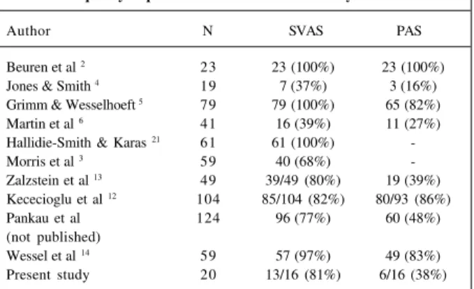
Table II - Cardiac defects in 20 patients with Williams-Beuren syndrome
Heart disease n (%)
SVAS Isolated (cases 8 and 10) 2/16 (19%)
Associated 11/16 (63%)
Pulmonary artery stenosis 4/11(36%) (cases 1, 7, 13, and 15)
Bicuspid aortic valve 3/11 (27%) (cases 4, 13 and 19)
Mitral valve prolapse 3/11 (27%) (cases 1, 3, and 15)
Aortic coarctation 2/11 (18%) (cases 10 and 20)
Thickened pulmonary valve 2/11 (18%) (cases 7 and 11)
Pulmonary valvular stenosis 1/11 (9%) (case 10)
Supravalvular pulmonary stenosis 1/11 (9%) (case 15)
Valvar aortic stenosis 1/11 (9%) (case 1)
Fixed subaortic stenosis 1/11 (9%) (case 19)
PAS Associated (cases 2, and 5) 2/16 (13%) Valvular pulmonary stenosis 1/2 (50%) (case 5)
Mitral valve prolapse 1/2 (50%) (case 2)
Isolated (case 18) 1/16 (13%) Mitral valve prolapse
Total 16/16 (100%)
enlarge the aorta with a bovine pericardial graft at the site of stenosis. All patients evolved well in the postoperative pe-riod with a reduction in the pressure gradient. Mean follow-up after surgery in 3 patients (cases 1, 3, and 19) was 5.7 years. The pressure gradient was normal after surgery in 2 patients (cases 1 and 3) in a mean follow-up of 8.4 years, wi-thout restenosis.
Two patients had aortic coarctation associated with supravalvular aortic stenosis (cases 10 and 20) with pres-sure gradients of 22 and 37 mmHg and with mild hemodyna-mic repercussions. Both patients were asymptomatic.
Table IV presents the mean pressure gradients and the follow-up period of patients with pulmonary aortic stenosis associated with supravalvular aortic stenosis (6/16=38%). Pulmonary artery branch stenosis and valvular pulmonary stenosis were diagnosed in case 5 at 9 months. Hemodynamic study, at 2 years, demonstrated multiple pulmonary artery stenoses, with stenosis in the origin and periphery of the right hypoplastic branch and the left branch the left ventricle and aorta were normal. Surgery was not indicated because the patient was compensated and the lesions were difficult to reach. Pressure gradient decreased spontaneously to 28 mmHg in a 6.6-year follow-up. Pulmo-nary artery stenosis in case 1 was corrected at 10 years, toge-ther with supravalvular aortic stenosis surgery. In the 5 remaining patients (cases 2, 5, 7, 13, and 15) stenosis resol-ved spontaneously even in cases 7 and 13 whose pulmo-nary artery stenosis was moderate.
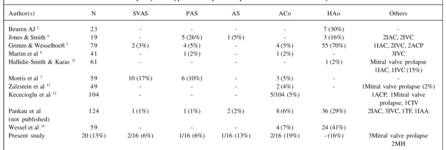
Table V - Supravalvular aortic stenosis and pulmonary artery frequency in patients with Williams-Beuren syndrome
Author N SVAS PAS
Beuren et al 2 23 23 (100%) 23 (100%)
Jones & Smith 4 19 7 (37%) 3 (16%)
Grimm & Wesselhoeft 5 79 79 (100%) 65 (82%)
Martin et al 6 41 16 (39%) 11 (27%)
Hallidie-Smith & Karas 21 61 61 (100%)
-Morris et al 3 59 40 (68%)
-Zalzstein et al 13 49 39/49 (80%) 19 (39%)
Kececioglu et al 12 104 85/104 (82%) 80/93 (86%)
Pankau et al 124 96 (77%) 60 (48%) (not published)
Wessel et al 14 59 57 (97%) 49 (83%)
Present study 20 13/16 (81%) 6/16 (38%)
*Modified from Wessel et al (1994).
Discussion
In the present study, heart disease was diagnosed in 81% of the patients, similar to that reported in the literature with incidences of 75 to 80% 11-13. Tables V and VI summarize
the types of heart diseases and their frequency in the present sample and in the main studies of literature.
Supravalvular aortic stenosis frequency (65%=13/20) in the present study was similar to that observed in the lite-rature 12,13. Pulmonary artery stenosis (38%) was the second
heart disease associated with supravalvular aortic stenosis, as in the studies in the literature (tab. V). Doppler echocar-diography is the examination of choice to detect supraval-vular aortic stenosis and other heart diseases 11-13.
Bicuspid aortic valve was seen in 19% of the patients from the present sample, whereas Hallidie-Smith and Karas21
observed this anomaly in 12% of their study patients with Williams-Beuren syndrome. However, the frequency of mitral valve prolapse (19%) in the present study was similar to that reported in the study. Other studies reported a lower incidence of mitral valve prolapse, ranging from 1% 12 to
2%13. In the present sample, the frequency of aortic
coarctation (13%) was greater than that reported in the lite-rature, which ranges from 2% 6,13 to 7% 14. Frequencies of
pulmonary valvular stenosis (1/16), supravalvular aortic stenosis (1/16), and aortic stenosis (1/16) in the present stu-dy were similar to those reported in the literature 4-6.
Fixed subaortic stenosis without hemodynamic reper-cussion was diagnosed in one of our patients. This abnor-mality was described in the literature by Narin et al 22 in an
8-year-old boy with Williams-Beuren syndrome.
In the present study, 1 patient (case 14) had bilateral renal artery stenosis in addition to supravalvular aortic ste-nosis and mitral valve prolapse. In the Zalzstein et al study13, of 8/11 of the patients with supravalvular aortic
ste-nosis and pulmonary artery steste-nosis, 1 patient had splenic artery stenosis and another patient had abdominal aortic stenosis. Arteriopathy in Williams-Beuren syndrome is ge-neralized and may involve any artery of the body due to elastin deletions 9,10.
Table IV – Pulmonary artery follow-up
Cases Initial Final Follow-up
gradient gradient period
1 - 0 11.8 years
2 26 0 10.3 years
5 92 28 6.9 years
7 65 0 13.5 years
13 59 0 6.3 years
15 24 0 9.6 years
Mean 44.3 4.7 9.7 years
Table III - Supravalvular aortic stenosis follow-up
Cases Initial Final Follow-up
Gradient Gradient Period
1 > 70 0 11.8 years
3 > 70 0 9.3 years
4 45 20 7.1 years
7 75 60 13.5 years
8 55 51 4.7 years
10 ... 25 6.8 years
11 15 0 8.4 years
12 6 ... ...
13 23 17 6.3 years
14 ... 30 13.3 years
15 19.5 0 9.6 years
19 > 70 14 2.2 years
20 94 0 9.3 years
Table VI - Frequency of the types of cardiopathies in patients with Williams-Beuren syndrome
Author(s) N SVAS PAS AS ACo HAo Others
Beuren AJ 2 23 - - - - 7 (30%)
-Jones & Smith 4 19 - 5 (26%) 1 (5%) - 3 (16%) 2IAC, 2IVC
Grimm & Wesselhoeft 5 79 2 (3%) 4 (5%) - 4 (5%) 55 (70%) 1IAC, 2IVC, 2ACP
Martin et al 6 41 - 1 (2%) - 1 (2%) - 3IVC
Hallidie-Smith & Karas 21 61 - - - - 1 (2%) Mitral valve prolapse
1IAC, 1IVC (15%)
Morris et al 3 59 10 (17%) 6 (10%) - 3 (5%) -
-Zalzstein et al 13 49 - - - 2 (4%) - 1Mitral valve prolapse (2%)
Kececioglu et al 12 104 - - - 5/104 (5%) 1ACP, 1Mitral valve
prolapse, 1CIV Pankau et al 124 1 (1%) 1 (1%) 2 (2%) 8 (6%) 36 (29%) 2IAC, 3IVC, 1TF, 1IAA (not published)
Wessel et al 14 59 - - - 4 (7%) 24 (41%)
Present study 20 (13%) 2/16 (6%) 1/16 (6%) 1/16 (13%) 2/16 (19%) - (16%) 3Mitral valve prolapse 2MH
Modified from Wessel, et al (1994). SVAS- supravalvular aortic stenosis; PS- pulmonary stenosis; AS- aortic stenosis; ACo- aortic coarctation; AoH- aortic hypoplasia; IAC- interatrial communication; IVC- interventricular communication; ACP- arterial channel persistence, MI- mitral insufficiency; TF- tetralogy of Fallot; AoAI- aortic arch interruption.
Table VII – Corrective surgery frequency in supravalvular aortic stenosis (SVAS)
Author SVAS Mean age
Zalzstein et al 13 12/39 (31%)
-Kececioglu et al 12 29/85 (34%) 11 years (2.5- 25)
Wessel et al 14 17/57 (32%)
-Present study 4/13 (31%) 5.7 years (19m-10 y) Great variability occurred in the pressure gradient of
patients with supravalvular aortic stenosis in the present study. These data were similar to those in the literature in which pressure gradient ranged from 0 to 110 mmHg in the study of Kececioglu et al 12, and 105 mmHg in the study of
Wessel et al 14.
In the present study, the patients with mild supravalvu-lar aortic stenosis, whose pressure gradients were <20mmHg, evolved with a reduction in the pressure gra-dient as reported in the literature. In the Wessel et al study14,
a mean pressure gradient of 9 mmHg remained unchanged in 32/45 of the patients with mild supravalvular aortic steno-sis during infancy, in a mean 13-year follow-up.
Zalzstein et al 13 subdivided the patients with
supra-valvular aortic stenosis into those with isolated supravalvu-lar aortic stenosis and those with supravalvusupravalvu-lar aortic ste-nosis associated with pulmonary artery steste-nosis. The first was present in 28/49 (57%) patients; in 17/28 (61%) of them, no worsening in stenosis occurred during a 6.3-year follow-up. However, 7/24 (29%) of the patients with initial pressure gradients higher than 30 mmHg evolved with an increase in pressure gradient and required corrective surgery of supra-valvular aortic stenosis. Eleven patients (11/49 = 22%) had supravalvular aortic stenosis and pulmonary artery steno-sis; 6 of them evolved with worsening of supravalvular aortic stenosis, whereas pulmonary artery stenosis either re-mained unchanged or even decreased. Five of them (5/6) underwent supravalvular aortic stenosis surgery.
In the Kececioglu et al study 12, supravalvular aortic
stenosis was classified into 3 groups according to the pres-sure gradient: 1) mild supravalvular aortic stenosis (0-30 mmHg) – 54/85 patients; 2) moderate supravalvular aortic stenosis (30-50 mmHg) – 12/85 patients, and 3) severe su-pravalvular aortic stenosis (50-110 mmHg) – 19/85 patients. The natural course of supravalvular aortic stenosis was as-sessed in 21 patients through hemodynamic study 11 years after the first catheterization, on average. The pressure
gra-dient in the aorta increased in 11 patients, and in 4 of them, the increase was >50 mmHg; the pressure gradient remained unchanged in 9/85 (11%), and decreased to 45 mmHg in 1/85 patients.
The frequency of patients undergoing supravalvular aortic stenosis surgery in the present study was similar to that reported in the literature (tab. VII). In the Kececioglu et al study 12, the mean postoperative follow-up was 8.6 years
in 29/85 patients with Williams-Beuren syndrome, under-going surgery to correct supravalvular aortic stenosis. Sur-gery resulted in a 30 mmHg decrease in the aortic pressure gradient in 22 patients.
We observed in the present sample that the severity of pulmonary artery stenosis decreased in childhood and adolescence as reported in the literature. Of the 6 patients with pulmonary artery stenosis, only 1 underwent surgical correction. The 5 remaining patients evolved with a sponta-neous reduction in the initial mean pressure gradient from 44.3 mmHg to 4.7 mmHg in a 9.7-year follow-up.
In the Zalzstein et al study 13, 8/49 of the patients had
isolated pulmonary stenosis, and 11/49 patients had pulmo-nary artery stenosis associated with supravalvular aortic stenosis. In 5/8 of the patients, no change in stenosis seve-rity occurred, and 1 patient underwent surgery at 12 years of age. In 6/11 patients with pulmonary aortic stenosis as-sociated with supravalvular aortic stenosis, pulmonary stenosis improved spontaneously in all cases.
pa-tients with Williams-Beuren syndrome in the Kececioglu et al study 12. The pressure gradient in the right ventricle was
increased in 55/80 patients, and it was mild or moderate in 75 patients, and severe in 5 patients. We observed a signifi-cant reduction in the pressure gradient of 14 patients.
In the Wessel et al study 14, the pressure gradient
as-sessed on hemodynamic study decreased spontaneously from a mean of 23 to 9.5 mmHg in 23/49 patients with pulmo-nary artery stenosis over 14 years. In adolescent and adults aged >33 years, the pressure gradient were lower than 20 mmHg in 22/23 patients.
In the present study, only 2 patients had aortic coarc-tation of mild hemodynamic repercussion. In the Kececio-glu et al study 12, 5% (5/104) had aortic coarctation, and all
patients underwent surgical correction. In the Wessel et al sample 14, aortic coarctation was also severe in 4/59 patients
(7%) who had it, 2 of them required surgery.
Cardiovascular anomalies caused by elastin deficien-cy are the most significant cause of morbidity and mortality in Williams-Beuren syndrome 11-23. Surgical correction of
severe supravalvular aortic stenosis must be indicated, once the patients with significant pressure gradients are at risk of sudden death 12,22. Kececioglu et al 12 estimated that
the risk of sudden death was 3% in a series of 104 patients with Williams-Beuren syndrome followed-up for 30 years. Pathologic findings observed by Bird et al. 23 in 10 patients
with Williams-Beuren syndrome who died suddenly were coronary artery stenosis and acute myocardial ischemia in 5 patients.
Immediate surgical results in patients with Williams-Beuren syndrome demonstrate low mortality and the long-term prospective studies revealed that most patients under-went surgery evolved well with stable pressure gradients and without restenosis.
An accurate cardiovascular evaluation is recommen-ded when diagnosis is performed in all patients with Wil-liams-Beuren syndrome, including electrocardiography and Doppler echocardiography. If the initial evaluation does not detect any cardiac abnormalities, a yearly follow-up should be performed to detect and prevent future complications.
1. Williams JCP, Barratt-Boyes BG, Lowe JB. Supravalvular aortic stenosis. Circulation 1961; 21: 1311-8.
2. Beuren AJ. Supravalvular aortic stenosis: a complex syndrome with and without mental retardation. Birth Defects: OAS VIII 1962; 5: 45-56.
3. Morris CA, Demsey AS, Leonard CO, Dilts C, Blackborn. Natural history of Wil-liams syndrome: physical characteristics. J Pediatr 1988; 113: 318-26. 4. Jones, KL, Smith, DW. The Williams elfin facies syndrome. J Pediatr 1975; 86: 718-23. 5. Grimm T, Wesselhoeft H. The genetic aspects of Williams-Beuren syndrome and the isolated form of the supravalvular aortic stenosis; investigation of 128 fami-lies. Z Kardiol 1980; 69: 168-72.
6. Martin NDT, Snodgrass GJAI, Cohen RD. Idiopathic infantile hypercalcemia: a continuing enigma. Arch Dis Child 1984; 59: 605-13.
7. Cortada X, Taysi K, Hartman AF. Familial Williams syndrome. Clin Genet 1980; 18: 173-6.
8. White RA, Preus M, Watters GV, Fraser FC. Familial occurrence of the Williams syndrome. J Pediatr 1977; 91: 614-6.
9. Lowery MC, Morris CA, Ewart A, et al. Strong correlation of elastin deletions, detected by FISH, with Williams syndrome: evaluation of 235 patients. Am J Hum Genet 1995; 57: 49-53.
10. Nickerson E, Greenberg F, Keating MT, McCaskill C, Shaffer LG. Deletions of the elastin gene at 7q11.23 occur in 90% of patients with Williams syndrome. Am J Hum Genet 1995; 56: 1156-61.
11. Conway JR, Noonan J, Marion, RW, Steeg CN. Myocardial infarction leading to sud-den death in the Williams syndrome: report of three cases. J Pediatr 1990; 117: 593-5. 12. Kececioglu D, Kotthoff S, Vogt J. Williams-Beurens syndrome: a 30-year
follow-up of natural and postoperative course. Eur Heart J 1993; 458-64.
References
13. Zalzstein E, Moes CAF, Musewe NN, Freedom RM. Spectrum of cardiovascular anomalies in Williams-Beuren syndrome. Pediatr Cardiol 1991; 12: 219-23. 14. Wessel A, Motz R, Pankau R, Bursch, JH. Arterial hypertension and blood
pres-sure profile in patients with Williams-Beuren syndrome. Z Kardiol 1997; 86: 251-7.
15. Sumboonnanonda A, Robinson BL, Gedroyc MWM, Saxton HM, Reidy JF, Haycock GB. Middle aortic syndrome: clinical and radiological findings. Arch Dis Child 1992; 67: 501-5.
16. Panayiotopoulos YP, Tyrrel MR, Koffman G, Reidy JF, Haycock GB, Taylor PR. Mid-aortic syndrome presenting in childhood. Br J Surg 1996; 83: 235-40. 17. Kawai M, Nishikawa T, Tanaka M, et al. An autopsied case of Williams syndrome
complicated by moyamoya disease. Acta Paediatr Jpn 1993; 35: 63-7. 18. Soper R, Chaloupka JC, Fayad PB, et al. Ischemic stroke and intracranial
multifo-cal cerebral arteriopathy in Williams syndrome. J Pediatr 1995; 126: 945-8. 19. Stumm M, Tönnies H, Wieacker PF. Molecular cytogeneic techniques for the
diagnosis of chromosomal abnormalities in childhood disease. Eur J Pediatr 1999; 158: 531-6.
20. Pinkel D, Straume T, Gray JW. Cytogenetic analysis using quantitative, high sen-sitivity fluorescence hybridization. Proc Natl Sci USA 1986; 83: 2934-8. 21. Hallidie-Smith KA, Karas S. Cardiac anomalies in Williams-Beuren syndrome.
Arch Dis Child 1988; 63: 809-13.
22. Narin N, Özyürek R, Bakiler AR, Parlar A, Arcasoy M, Köprübasi F. Williams syndrome and subaortic stenosis. Clin Genet 1993; 44: 223.