NT-Pro-BNP Levels and Their Response to Exercise in Patients with
Slow Coronary Flow
Mustafa Yurtdaş
1, İsmail Türkay Özcan
2, Ahmet Çamsarı
2, Dilek Çiçek
2, Lülüfer Tamer
3, Veli Gökhan Cin
2,
Oben Döven
2, Ali Sabri Seyis
4, Mehmet Necdet Akkuş
2Medisina Hospital, Department of Cardiology1, Van; University of Mersin, Department of Cardiology2, Mersin; University of Mersin, Department of Biochemistry3, Mersin; Medical Park Hospital, Department of Cardiology4, Mersin – Turkey
Mailing Address: Mustafa Yurtdaş •
Medisina Hospital, Department of Cardiology. Postal Code 65200, Van – Turkey
E-mail: [email protected]
Manucript received February 16, 2012; manuscript revised April 13, 2012; accepted May 23, 2012.
Abstract
Background: Natriuretic peptides are released by the heart in response to wall stress.
Objective: The NT-Pro-BNP concentrations in slow coronary flow (SCF) patients were assessed before and after the exercise test and compared with the values of healthy controls.
Methods: The study population was 34 patients with SCF [22 males (64.7%), aged 51.0±6.2 years], and 34 normal subjects with normal coronary arteries [21 males (61.8%), aged 53.2±6.6 years]. Coronary flow rates of all patients and control subjects were documented as Thrombolysis in Myocardial Infarction (TIMI) frame count. Blood samples were drawn at rest and after the exercise testing.
Results: The baseline NT-Pro-BNP concentrations of the SCF patients were higher than those of the control subjects (NT-Pro-BNP: 49.7±14.2 pg/mL vs. 25.3±4.6 pg/mL p<0.0001, respectively), and this difference increased after exercise test between the groups (NT-Pro-BNP: 69.5±18.6 pg/mL vs. 30.9±6.4 pg/mL p<0.0001). In SCF group after exercise, NT-Pro-BNP concentration in 15 patients with angina was higher than those without angina (76.8 ± 17.8 pg/mL vs. 63.8±17.5 pg/mL p=0.041). NT-Pro-BNP concentration in 11 patients with ST depression was also higher than those without ST depression (82.4 ± 17.3 pg/mL vs. 63.3 ± 16.1 pg/mL p=0.004). Median post-exercise increases in NT-Pro-BNP (∆ NT-NT-Pro-BNP) were higher in the SCF group than in the control group (∆ NT-NT-Pro-BNP: 19.8±7.7 pg/mL vs. 5.7±4.5 pg/mL p<0.0001).
Conclusion: The results of this study suggest that there may be an important pathophysiologic link between the severity of SCF (microvascular or epicardial coronary artery dysfunction) and the level of circulating NT-Pro-BNP in SCF patients. (Arq Bras Cardiol 2012;99(6):1115-1122)
Keywords: Coronary circulation; Exercise; Natriuretic Agents; Angina Pectoris.
in the heart. B-type natriuretic peptide (BNP), which has vasodilatory, diuretic, and renin-angiotensin-aldosterone antagonist effects, is synthesized as a 108-aminoacid-long prohormone termed proBNP. After secretion, this propeptide is divide into its biologically active BNP and the remaining NT-Pro-BNP. It is secreted mainly from the ventricular wall in response to increases in wall stress8,9. In the normal situation,
cardiac production and plasma concentration of NT-pro-BNP is very low. The most significant stimulant for cardiac secretion of NT-pro-BNP is stretch of cardiac myocytes10,11.
A great number of studies have revealed that the plasma level of BNP and the inactive amino-terminal fragment of its prohormone (N-terminal fragment of B-type natriuretic peptide pro-hormone [NT-pro-BNP]) elevate in a variety of right and left ventricular functional and structural abnormalities such as coronary artery disease (CAD), severe valve disease and heart failure12-17.
In the present study, we aimed to investigate the plasma NT-Pro-BNP concentrations of SCF patients before and post-exercise testing because, to the best of our knowledge, there
Introduction
Approximately 10–20% of patients with typical angina pectoris have normal coronary arteries, and most of these patients are syndrome X1,2. However, syndrome X is a
heterogeneous group that includes slow coronary flow (SCF)3.
SCF is characterized by late opacification of the epicardial coronary arteries without occlusive disease. Since its first description by Tambe et al4 in 1972, although endothelial and
vasomotor dysfunction, microvascular dysfunction, and disease of small coronary arteries have been convicted in SCF4-7, the
is no study that has evaluated NT-pro-BNP concentrations at rest and post-exercise in SCF patients.
Methods
Study population
Thirty-four patients with angiographically proven SCF but normal epicardial coronary arteries and thirty-four healthy subjects with angiographically normal coronary arteries without luminal irregularities as control group were selected from patients who had undergone coronary angiography because of typical and quasi-typical symptoms of angina. All control subjects with no history of cardiovascular disease and other systemic illness had normal echocardiographic and exercise studies. Patients who suffered from one of the following diseases or associated disorders were excluded: myocardial and/or valvular heart disease, systolic and/ or diastolic dysfunction, atrial disease or enlargement, cardiac arrhythmia such as atrial fibrillation, left ventricular hypertrophy and contraindication for exercise testing. After signed informed consent was obtained, all concomitant medication was stopped for at least five half lives before the test. The study was carried out according to the principles of the Declaration of Helsinki and approved by the Investigational Review Board of Mersin University, School of Medicine.
TIMI (Thrombolysis in Myocardial Infarction) Frame Count For the quantitative measurement of coronary blood flow, the time elapsed from the appearance of contrast agent until it reached the distal end of the coronary arteries in terms of cineframe count was considered to be the TIMI frame count18.
Thereafter, the final count was subtracted from the initial and the exact TIMI frame was calculated for the given artery. The cut-off values for the TIMI frame counts are taken from the study of Gibson et al18, in which 78 normal coronary arteries were
evaluated.18 Their cut-off values because of the length of time
needed for normal visualization of the coronary arteries were 36.2 ± 2.6 frames for the left anterior descending coronary artery (LAD), 22.2 ± 4.1 frames for the left circumflex coronary artery (LCX) and 20.4 ± 3 frames for the right coronary artery (RCA). They proposed that the TIMI frame count for the LAD was 1.7-fold higher than those of the LCX and RCA. In their study, the reason given for these differences was that the LAD is 1.5-fold longer than RCA and 1.6-fold longer than LCX anatomically. Therefore, the TIMI frame count was divided by 1.7 when the LAD was the case, for adjusted correction. The corrected cut-off value for the LAD was 21.1 ± 1.5 frames. Any values obtained above these thresholds were considered to be SCF in our study. The TIMI frame counting was undertaken separately by 2 cardiologists and in the case of conflict, the frames were referred to a third one. All TIMI frame counts were measured in matched projections using Medcon Telemedicine Technology (version 1.900, Israel).
Exercise Treadmill Testing
The 12-lead maximal effort exercise tests were performed using standard graduated treadmill protocols consistent with American Heart Association guidelines19, with all medications
discontinued for at least 5 half lives before evaluation. Patients were encouraged to give their maximal effort, but not to allow their angina to reach levels higher than previously experienced. The results were analyzed and reported using a computerized database (EXTRA; Mosby Publishers; Chicago, IL, USA)20. The
ST segment response considered was the most horizontal or downsloping ST-segment depression in any lead, except for aVR, during exercise or recovery. An abnormal response was
defined as ≥0.1 mV at 0.08 s after the J point of horizontal
or downsloping ST-segment depression and chest pain onset during exercise. Sampling time was determined according to study by Foote et al.15 Blood samples were drawn from the
forearm vein at rest and again at 1 minute post-exercise testing.
Analysis of NT-Pro-BNP levels
Resting and post-exercise blood samples were analyzed in batches for NT-pro-BNP using an electrochemiluminescent immunoassay (Roche diagnostics, Manheim GmbH, Germany) on an E-170 hormone autoanalyzer.
Statistical Analysis
Statistical analysis was performed using SPSS software package (Version 15.0, SPSS Inc, Chicago, IL, USA). Continuous variables were expressed as means ± SD. Categorical variables were expressed as percentage. Kolmogorow-Smirnov test was used to investigate whether the distribution of measurements was normal or not. To compare the two groups according to changes after the exercise in the variables that show normal distribution and according to measurements before and after exercise, independent t test was used. In contrast, for the variables that do not show normal distribution, Mann-Whitney U test was used. Also in the patients according to these measurements for comparison of that with chest pain and no pain; and also for comparison of that had ST segment depression and without ST depression, independent-t test was used. Paired-t test was used to determine the significance of changes observed before and after exercise in both patients and controls. Linear relations between measurements in both patients and controls were examined by Pearson’s correlation. All hypothesis testing was 2-tailed. A p-value of < 0.05 was considered significant.
Results
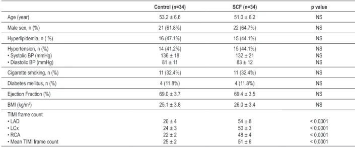
Clinical characteristics of control and patient groups were presented in Table 1. There was no statistically significant difference between two groups in respect to age, gender, hypertension, diabetes mellitus, hyperlipidemia and cigarette smoking. Besides, we found no significant difference between two groups in terms of body mass index (BMI), total cholesterol, LDL cholesterol, HDL-cholesterol and triglyceride levels. Systolic and diastolic blood pressures were similar in both groups measured during the exercise testing. Patients with SCF were detected to have significantly higher TIMI frame count for each major epicardial coronary artery compared to control subjects (Table 1).
NT-Pro-BNP (∆NT-Pro-BNP) were higher in the SCF group than in the control group (∆NT-Pro-BNP: 19.8 ± 7.7 pg/mL
vs. 5.7 ± 4.5 pg/mL; p<0.0001) (Table 2).
Exercise test was stopped because of chest pain and ST segment depression in 15 SCF patients. Of them, 11 had both chest pain and ST segment depression, 4 had only chest pain. Control subjects completed the whole protocol and none showed ST-segment changes, tachyarrhythmia or chest pain throughout the procedure. Peak exercise
heart rate, peak exercise systolic blood pressure and rate – pressure product were significantly higher in the control group than in the patient group. The exercise parameters of both groups are summarized in Table 3. In SCF group, NT-Pro-BNP concentrations in 15 patients with angina was higher than those without angina after exercise (76.8 ± 17.8 pg/mL vs. 63.8±17.5 pg/mL; p=0.041) (Figure 1). NT-Pro-BNP concentration in 11 patients with ST segment depression was also higher than those without ST segment Table 1 - Comparison of baseline clinical characteristics, and TIMI frame counts of SCF patients and control subjects
Control (n=34) SCF (n=34) p value
Age (year) 53.2 ± 6.6 51.0 ± 6.2 NS
Male sex, n (%) 21 (61.8%) 22 (64.7%) NS
Hyperlipidemia, n ( %) 16 (47.1%) 15 (44.1%) NS
Hypertension, n (%)
• Systolic BP (mmHg) • Diastolic BP (mmHg)
14 (41.2%) 136 ± 18
81 ± 11
15 (44.1%) 132 ± 21
83 ± 12
NS NS NS
Cigarette smoking, n (%) 11 (32.4%) 11 (32.4%) NS
Diabetes mellitus, n (%) 4 (11.8%) 4 (11.8%) NS
Ejection Fraction (%) 69.0 ± 3.7 69.4 ± 3.5 NS
BMI (kg/m2) 25.1 ± 3.8 26.0 ± 3.4 NS
TIMI frame count • LAD • LCx • RCA
• Mean TIMI frame count
26 ± 4 24 ± 3 22 ± 2 25 ± 2
54 ± 8 50 ± 3 48 ± 4 51 ± 6
< 0.0001 < 0.0001 < 0.0001 < 0.0001
NS: non-signiicant; TIMI: Thrombolysis In Myocardial Infarction; BP: Blood Pressure; BMI: Body mass index; LAD: Left anterior descending; LCx: Left circumlex; RCA: Right coronary artery
Table 2 - Baseline and post-exercise plasma NT-Pro-BNP concentrations in SCF and control groups
Controls (n=34) SCF (n=34) p value
Baseline NT-Pro-BNP (pg/mL) 25.3 ± 4.6 49.7 ± 14.2 < 0.0001
Post-exercise NT-Pro-BNP (pg/mL) 30.9 ± 6.4 69.5 ± 18.6 < 0.0001
Δ NT-Pro-BNP (pg/mL) 5.7 ± 4.5 19.8 ± 7.7 < 0.0001
∆: median post-exercise increases
Table 3 - Exercise parameters of SCF and control groups
Controls (n=34) SCF (n=34) p value
Baseline heart rate (beats/min) 78 ± 6 77 ± 9 NS
Peak exercise heart rate (beats/min) 173 ± 6 159 ± 8 < 0.0001
Peak systolic blood pressure (mmHg) 188 ± 7 178 ± 11 < 0.0001
Rate-pressure product
[(mmHg x beats/min)] x 102 327 ± 18 284 ± 25 < 0.0001
Angina, n (%) – 15 (44%) −
ST segment depression, n (%) – 11 (32%) –
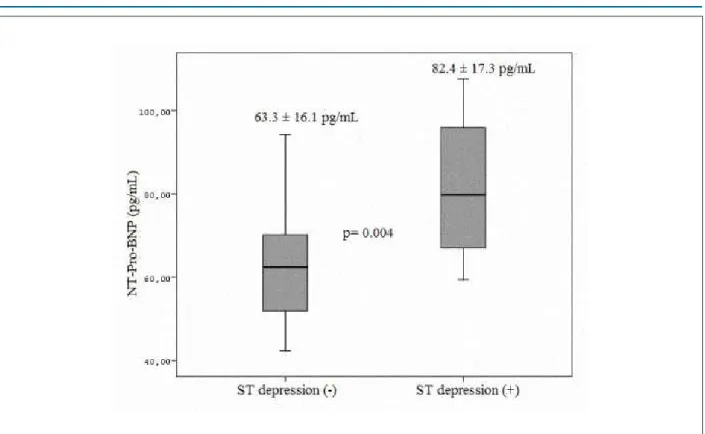
depression after exercise (82.4 ± 17.3 pg/mL vs. 63.3 ± 16.1 pg/mL; p=0.004) (Figure 2). In addition, NT-Pro-BNP value in 11 patients with both angina and ST segment depression was greater than those with only angina after exercise (82.4 ± 17.3 pg/mL vs. 61.2 ± 7.4 pg/mL; p=0.006). Only the exercise rate-pressure product among exercise testing parameters was negatively correlated with post-exercise NT-Pro-BNP in patient group (Figure 3). There was no correlation between exercise testing parameters and NT-Pro-BNP levels in both groups. The mean TIMI frame count was 51.3 ± 6.1 in the SCF patients. The mean TIMI frame count was not correlated with baseline and/or post-exercise NT-Pro-BNP concentrations.
Discussion
The results of our study have three main findings; 1) NT-Pro-BNP plasma concentration was higher in patients with SCF than in a matched group of control subjects, 2) The differences in the NT-Pro-BNP concentrations between the two groups became greater after the exercise treadmill test, as a result of a significant increase in NT-Pro-BNP in patients with SCF, 3) In SCF group, NT-Pro-BNP concentrations in patients with angina and ST segment depression were higher than those without angina and ST segment depression after exercise test.
SCF is an angiographic finding defined as the slow movement of contrast throughout the coronary lumen in
the absence of epicardial coronary stenosis. Although the underlying mechanism is not exactly known, many authors state that SCF is closely related to the microvascular and macrovascular disease, and has a dynamic process1-7,21-23.
Mosseriet al6 studied six patients who suffered from angina
pectoris but had angiographically patent major coronary arteries, and they concluded that right endomyocardial biopsy revealed pathologic small coronary arteries with fibromuscular hyperplasia, hypertrophy of the media, myointimal proliferation, and endothelial degeneration. Additionally, Mangieri et al7 evaluated left ventricular biopsy
samples of 10 patients with SCF, none of whom had a cardiac or systemic illness, and found endothelial thickening due to cell edema, capillary damage, and decreased luminal diameter. However, Van Lierde et al21 have shown
that extremely slow blood flow velocity confirmed by intracoronary Doppler measurements in patients with SCF, but coronary flow reserve and coronary blood flow were within the normal range, and these findings suggest that SCF may not always be a microvascular disease. They speculated that SCF may be caused by epicardial artery disease such as coronary ectasia. Some investigators have recently demonstrated that the intima+media area was thickened diffusely throughout the given coronary artery in patients with SCF by using intravascular ultrasound. They have shown the presence of diffuse early atherosclerosis which does not cause luminal irregularities in the coronary angiography of this group of patients22,23. As a result, SCF
Figure 2 - NT-Pro-BNP concentrations in patients with ST depression (-) and ST depression (+)
probably prepares a suitable environment for the myocyte stretch due to its underlying pathophysiology. In a previous study, Foote et al15 have compared 74 CAD patients with
21 healthy volunteers (with normal left ventricular functions) in terms of resting levels of NT-Pro-BNP and BNP. They found that patients with known CAD had higher NT-Pro-BNP levels than normal subject without CAD at rest. Again, in other study, it was showed that NT-Pro-BNP levels were > 4 times higher in patients with CAD than in control subjects16.
Another crucial stimulus for cardiac NT-Pro-BNP secretion is hypoxia, which, independently of stretch, may also induce peptide release24. Indeed, Beltrame et al25 have revealed that
resting coronary sinus oxygen saturation was low compared with control subjects, reflecting an increased resting coronary vasomotor tone in coronary resistance vessels in patients with SCF. This information may be important to explain why SCF patients have higher NT-Pro-BNP levels than control subjects at rest. So far, an association between the SCF and the levels of NT-Pro-BNP has been showed in the only one study, in which NT-Pro-BNP levels has been significantly higher in patients with SCF than normal control subjects26, and they
have concluded that this association could be suggestive of a low grade chronic inflammation in SCF patients. Thus, in the pre-exercise state, presence of microvascular dysfunction, epicardial coronary artery disease, hypoxia and chronic inflammation may act together on increased NT-Pro-BNP concentration in the SCF patients.
One of the important findings observed in our study was increased levels of NT-Pro-BNP after exercise in SCF patients. The effects of adrenergic stimulation, such as exercise, on the coronary microvascular tone are controversial. Physical exercise stress induces only a 10% increase in plasma NT-Pro-BNP concentration in the normal conditions15. But, in pathological
conditions such as endothelial dysfunction, exercise may give rise to marked increases in NT-Pro-BNP level. Increased sympathetic stimulation may cause abnormal microvascular constriction27 and promotes coronary flow indirectly
through the metabolic vasodilation secondary to an increase in heart rate and myocardial contractility28 and, in part, to
endothelium-mediated vasodilation29. On the other hand, it
may have both direct vasoconstrictor and vasodilator effects through α- and β- receptor stimulation, respectively29. The
net effect likely depending on the pathophysiologic state of the small coronary arteries. The endothelial dysfunction in the small coronary arteries (increased ET-1 concentrations) in patients with angina pectoris and normal coronary arteriography may increase coronary resistance and hence lead to myocardial ischemia 22,30,31. Thereafter, ischemia
may give rise to transient left ventricle (LV) systolic and diastolic dysfunction, and may also cause an increase in LV end-diastolic wall tension, thus leading to an extreme elevation of NT-Pro-BNP levels. In addition, some hormones such as angiotensin II, catecholamines, and endothelin-1 may further magnify secretion of NT-ProBNP through the paracrine and possibly endocrine mechanisms10,11,14.
Previous studies22,31 have shown that endothelin-1 (ET-1) is
higher and NO concentration is lower in patients with SCF than in a matched group of control subjects with normal coronary flow, and that endothelial dysfunction could be of
great importance in the etiopathogenesis of SCF. In line with these data, it is reasonable to state that endocrine activation and ischemia, through the mechanisms discussed above, could be a major triggering factor for the elevated NT-Pro-BNP levels in patients with SCF after exercise testing.
Another important finding in our study was that patients with angina and ST segment depression had higher NT-Pro-BNP concentrations than those without angina and ST segment depression after exercise test in SCF patients. Furthermore, although there is no correlation between the mean TIMI frame count and NT-Pro-BNP concentrations in both groups, we found a negative correlation between the exercise rate-pressure product and post-exercise NT-Pro-BNP concentrations in SCF group. Myocardial oxygen consumption is correlated with the rate-pressure product (heart rate x systolic blood pressure). Hence, inability to supply oxygen to myocardium, when demand is high, may result in severe cardiovascular events such as transient myocardial ischemia, leading to increased NT-Pro-BNP levels. Until recently, one study has examined the effects of exercise on the levels of NT-Pro-BNP and/or BNP in patients with CAD, in which median post-exercise increases in NT-Pro-BNP and BNP were higher in the ischemic group than in the non-ischemic group15, a finding consistent with
the results of the present study. Ndrepepa et al16 have
shown that NT-Pro-BNP concentrations are high across the entire spectrum of CAD and parallel the clinical or angiographic severity of CAD. Similarly, Sabatine et al17
also determined that transient myocardial ischemia was associated with an immediate rise in circulation BNP levels, to lesser extent, NT-Pro-BNP levels, and that the degree of rise was proportional to the severity of ischemia. Based on these notions, it is generally considered that the severity of ischemia caused by exercise may closely be related with increased ventricular wall stress or damage, thus, further contributing to inducing NT-Pro-BNP in SCF patients.
Limitations
Some of the main limitations of this study were that the overall sample size is small and, hemodynamic parameters such as cardiac output, pulmonary capillary wedge pressure, left ventricular end-diastolic pressure and right atrial pressure were not measured due to ethical concerns. Additionally, another limitation was that the levels of atrial natriuretic peptide (ANP) could not be measured simultaneously with NT-Pro-BNP levels. Further studies with larger samples are required to confirm our findings.
Conclusion
NT-Pro-BNP in this group of patients. The present study may be of great value for the first time to demonstrate such an pathophysiologic association.
Potential Conflict of Interest
There is no potential conflict of interest.
Sources of Funding
There were no external funding sources for this study.
Study Association
This study is not associated with any post-graduation program.
References
1. Kemp HG, Kronmal RA, Vlietstra RE, Frye RL. Seven year survival of patients with normal or near normal coronary arteriograms: a CASS registry study. J Am Coll
Cardiol. 1986;7(3):479-83.
2. Kemp HG Jr. Left ventricular function in patients with the anginal syndrome and normal coronary arteriograms. Am J Cardiol. 1973;32(3):375-6.
3. Voelker W, Euchaner U, Dittmann H, Karsch KR. Long-term clinical course of patients with angina and angiographically normal coronary arteries. Clin Cardiol. 1991;14(4):4307-11.
4. Tambe AA, Demany MA, Zimmerman HA, Mascarenhas E. Angina pectoris and slow flow velocity of dye in coronary arteries: a new angiographic finding. Am
Heart J. 1972;84(1):66-71.
5. Gunes Y, Gumrukcuoglu HA, Akdag S, Simsek H, Sahin M, Tuncer M. Vascular endothelial function in patients with slow coronary flow and the effects of Nebivolol. Arq Bras Cardiol. 2011;97(4):275-80.
6. Mosseri M, Yarom R, Gotsman MS, Hasin Y. Histologic evidence for small vessel coronary artery disease in patients with angina pectoris and patent large coronary arteries. Circulation. 1986;74(5):964-72.
7. Mangieri E, Macchiarelli G, Ciavolella M, Barilla F, Avella A, Martinotti A, et al. Slow coronary flow: clinical and histopathological features in patients with otherwise normal epicardial coronary arteries. Cathet Cardiovasc Diagn. 1996;37(4):375-81.
8. Munagala VK, Burnett JC, Redfield MM. The natriuretic peptides in
cardiovascular medicine. Curr Probl Cardiol. 2004;29(12):707-69.
9. Yeo KT, Lee HK, Wong KC, Foote RS. Can exercise-induced changes in B-type natriuretic peptides be used to detect cardiac ischemia? J Card Fail.
2005;11(5 Suppl):S59-64.
10. Kuwahara K, Saito Y, Ogawa Y, Tamura N, Ishikawa M, Harada M, et al. Interaction of myocytes and nonmyocytes is necessary for mechanical stretch
to induce ANP/BNP production in cardiocyte culture. J Cardiovasc Pharmacol.
1998;31(Suppl 1):S357-9.
11. Bruneau BG, Piazza LA, De Bold AJ. BNP gene expression is specifically modulated by stretch and ET-1 in a new model of isolated rat atria. Am J
Physiol Heart Circ Physiol. 1997;273(6 Pt 2):H2678-86.
12. Yasue H, Yoshimura M, Sumida H, Kikuta K, Kugiyama K, Jougasaki M, et
al. Localization and mechanism of secretion of B-type natriuretic peptide in comparison with those of A-type natriuretic peptide in normal subjects and patients with heart failure. Circulation. 1994;90(1):195-203.
13. Ikeda T, Matsuda K, Itoh H, Shirakami G, Miyamoto Y, Yoshimasa T, et al. Plasma levels of brain and atrial natriuretic peptides elevate in proportion to left ventricular end systolic wall stress in patients with aortic stenosis.
Am Heart J. 1997;133(3):307-14.
14. Magga J, Marttila M, Mantymaa P, Vuolteenaho O, Ruskoaho H. Brain
natriuretic peptide in plasma, atria, and ventricles of vasopressin a n d p h e n y l e p h r i n e - i n f u s e d c o n s c i o u s r a t s . E n d o c r i n o l o g y. 1994;134(6):2505-15.
15. Foote RS, Pearlman JD, Siegel AH, Yeo KT. Detection of Exercise-Induced
Ischemia by Changes in B-type natriuretic peptides. J Am Coll Cardiol.
2004;44(10):1980-7.
16. Ndrepepa G, Braun S, Mehilli J, von Beckerath N, Vogt W, Schomig A, et
al. Plasma levels of n-terminal pro-brain natriuretic peptide in patients with coronary artery disease and relation to clinical presentation, angiographic
severity, and left ventricular ejection fraction. Am J Cardiol. 2005;95(5):553-7.
17. Sabatine MS, Morrow DA, de Lemos JA, Omland T, Desai MY, Tansyevic N, et al.
Acute changes in circulating natriuretic peptide levels in relation to myocardial
ischemia. J Am Coll Cardiol. 2004;44(10):1988-95.
18. Gibson CM, Cannon CP, Daley WL, Dodge JT Jr, Alexandr B Jr, Marble SJ, et
al. TIMI frame count: a quantitative method of assessing coronary artery flow. Circulation. 1996;93(5):879-88.
19. Fletcher GF, Balady G, Froelicher VF, Hartley LH, Haskell WL, Pollock ML. Exercise standards: a statement for health care professionals from the American Heart Association Writing Group. Circulation. 1995;91(2):580-615.
20. Shue P, Froelicher V. Extra: an expert system for exercise reporting. J Noninvasive
Test. 1998;II-4:21-7.
21. Van Lierde J, Vrolix M, Sionis D, De Geest H, Piessens J. Lack of evidence for small
vessel disease in a patient with ‘slow dye progression’ in the coronary arteries. Cathet Cardiovasc Diagn. 1991;23(2):117-20.
22. Pekdemir H, Polat G, Cin VG, Camsari A, Cicek D, Akkus MN, et al. Elevated plasma endothelin-1 levels in coronary sinus during rapid right atrial pacing in patients with slow coronary flow. Int J Cardiol. 2004;97(1):35-41.
23. Cin VG, Pekdemir H, Camsari A, Cicek D, Akkus MN, Parmaksyz T, et al.
Diffuse intimal thickening of coronary arteries in slow coronary flow. Jpn Heart J. 2003;44(6):907-19.
24. Hopkins WE, Chen Z, Fukagawa NK, Hall C, Knot HJ, LeWinter MM. Increased
atrial and brain natriuretic peptides in adults with cyanotic congenital heart disease: enhanced understanding of the relationship between hypoxia and natriuretic peptide secretion. Circulation. 2004;109(23):2872-7.
25. Beltrame JF, Limaye SB, Wuttke RD, Horowitz JD. Coronary hemodynamic
and metabolic studies of the coronary slow flow phenomenon. Am Heart J.
2003;146(1):84-90.
26. Madak N, Nazlı Y, Mergen H, Aysel S, Kandaz M, Yanik E, et al. Acute phase
reactants in patients with coronary slow flow phenomenon. Anadolu Kardiyol Derg. 2010;10(5):416-20.
27. Cordero DL, Cagin NA, Natelson BH. Neurocardiology update: Role of the nervous system in coronary vasomotion. Cardiovasc Res. 1995;29(3):319-28.
28. Feigl EO. Adrenergic control of transmural coronary blood flow. Basic Res Cardiol. 1990;85 Suppl I:167-76.
29. Zehier AM, Drexler H, Wollschlaeger H, Saurbier B, Just H. Coronary
vasomotion in response to sympathetic stimulation in humans: Importance of the functional integrity of the endothelium. J Am Coll Cardiol.
1989;14(5):1181-90.
30. Kaski JC, Cox ID, Crook JR, Salomone OA, Fredericks S, Hann C, et al. Differential
plasma endothelin concentrations in subgroups of patients with angina and angiographically normal coronary arteries: Coronary Artery Disease Research
Group. Am Heart J. 1998;136(3):412-7.
31. Camsari A, Pekdemir H, Cicek D, Polat G, Akkus MN, Doven O, et al. Endothelin-1 and nitric oxide concentrations and their response to exercise in